. 2008 Sep 26;283(39):26461-7.
doi: 10.1074/jbc.M801625200. Epub 2008 Jul 29.

RIBEYE recruits Munc119, a mammalian ortholog of the Caenorhabditis elegans protein unc119, to synaptic ribbons of photoreceptor synapses

Affiliations

RIBEYE recruits Munc119, a mammalian ortholog of the Caenorhabditis elegans protein unc119, to synaptic ribbons of photoreceptor synapses

Kannan Alpadi et al. J Biol Chem. .

Abstract

Munc119 (also denoted as RG4) is a mammalian ortholog of the Caenorhabditis elegans protein unc119 and is essential for vision and synaptic transmission at photoreceptor ribbon synapses by unknown molecular mechanisms. Munc119/RG4 is related to the prenyl-binding protein PrBP/delta and expressed at high levels in photoreceptor ribbon synapses. Synaptic ribbons are presynaptic specializations in the active zone of these tonically active synapses and contain RIBEYE as a unique and major component. In the present study, we identified Munc119 as a RIBEYE-interacting protein at photoreceptor ribbon synapses using five independent approaches. The PrBP/delta homology domain of Munc119 is essential for the interaction with the NADH binding region of RIBEYE(B) domain. But RIBEYE-Munc119 interaction does not depend on NADH binding. A RIBEYE point mutant (RE(B)E844Q) that no longer interacted with Munc119 still bound NADH, arguing that binding of Munc119 and NADH to RIBEYE are independent from each other. Our data indicate that Munc119 is a synaptic ribbon-associated component. We show that Munc119 can be recruited to synaptic ribbons via its interaction with RIBEYE. Our data suggest that the RIBEYE-Munc119 interaction is essential for synaptic transmission at the photoreceptor ribbon synapse.


Figures

FIGURE 1.
FIGURE 1.
Interaction of RIBEYE(B) and RIBEYE(AB) with Munc119 in the YTH system. A, amino acid sequence of bovine Munc119. The proline-rich domain (PRD, aa1–77) is colored in blue, the PrBP/δ-homology domain of Munc119 (aa78–240) colored in green. The boxed lysine indicates the site of a premature stop mutation that causes cone rod dystrophy in a human patient (5). The amino acids methionine 1 (M1), lysine 92 (K92), and isoleucine 93 (I93), which are underlined in yellow indicate the beginning of the reading frames of three independently obtained Munc119 YTH prey clones. The amino acid sequence of Munc119 obtained in our YTH screen is identical to the Munc119 sequence deposited at GenBank™ (accession BC103449.1). B, schematic domain structures of RIBEYE and Munc119. RIBEYE contains of a large N-terminal A-domain and a C-terminal B-domain. The B-domain of RIBEYE contains the NAD(H) binding subdomain (NBD, depicted in yellow) and the substrate binding subdomain (SBD, depicted in red). C, RIBEYE(B) interacts with the PrBP/δ-homology domain of Munc119 in YTH. Summary plates of YTH analyses obtained with the indicated bait and prey plasmids. For convenience, experimental bait-prey pairs are underlayered in color (green in the case of interacting bait-prey pairs; yellow in the case of non-interacting bait-prey-pairs; control matings are non-colored). D, RIBEYE(B) and also full-length RIBEYE (RIBEYE(AB) interact with Munc119. The interaction is mediated via the NBD of RIBEYE and the PrBP/δ homology domain of Munc119, Munc119(78–240) (C; matings 6, 12–13). Mating 22 in C and mating 6 in D denote an unrelated positive control mating (CtIP). pSE1111 is an irrelevant prey vector, and pSE1112 an irrelevant bait vector (22).
FIGURE 2.
FIGURE 2.
RIBEYE(B) specifically interacts with Munc119 in fusion protein pull-down assays. Pull-down analyses of RIBEYE(B)/Munc119 complexes using bacterially expressed fusion proteins. In A, pull-down experiments were analyzed by Coomassie Blue-stained polyacrylamide gel after SDS-PAGE. In B, by Western blot analyses with the indicated antibodies. A and B, lanes 1–4 show the indicated purified fusion proteins (input fractions). All input lanes, except for lane 4, represent 50% of the input fraction. Lane 4 represents 25% of the input fraction. In lanes 5–8, 100% was loaded. GST-tagged fusion proteins were used as immobilized bait proteins and MBP-tagged proteins as soluble prey proteins. Only Munc119-GST pulled-down RE(B)-MBP (lane 8) but not GST alone (lane 6). Neither GST alone nor Munc119-GST pulled-down MBP alone (lanes 5 and 7). The asterisks in lanes 3, 7, and 8 of A label a break-down product of Munc119-GST. SDS-PAGE clearly demonstrated that Munc119-GST does not pull-down MBP alone (A). To further exclude that any MBP is nonspecifically pulled-down by Munc119-GST, we also analyzed the results of the pull-down assays by Western blotting with anti-MBP antibodies. B, Western blot analyses with anti-MBP antibodies (B) clearly show that only RE(B)-MBP (lane 8) but not MBP alone (lane 7) is pulled-down by Munc119-GST. GST alone does not pull-down RE(B)-MBP as well as MBP alone as shown by Western blotting with antibodies against MBP (Ba) demonstrating the specificity of the interaction and completely confirming the results in A. In Bb, the same blot as analyzed in Ba was reprobed (after stripping) with antibodies against GST to show equal loading of the bait proteins. Abbreviations: CB, Coomassie Blue.
FIGURE 3.
FIGURE 3.
RIBEYE interacts with Munc119 in transfected COS cells. COS cells were co-transfected either with Munc119-GSTpEBG and RE(B)-EGFP (experimental assays) or with empty GSTpEBG and RE(B)-EGFP (control assays) using lipofection. Glutathione beads were added to the respective cell lysates. Proteins bound to the glutathione beads (lanes 3 and 4) were analyzed via Western blotting with antibodies against GST and EGFP. Munc119-GST pulled-down RIBEYE(B)-EGFP (lane 3) but not GST alone (lane 4) demonstrating the specific interaction between Munc119 and RIBEYE(B). Lanes 1–2 show the respective input fractions (10% of total input); Lanes 3 and 4 show 100% of the pulled-down proteins. In b, the same blot as shown in a was reprobed (after stripping) with antibodies against GST to show equal loading of the samples.
FIGURE 4.
FIGURE 4.
Co-immunoprecipitation of RIBEYE and Munc119 from R28 retinal precursor cells. R28 retinal progenitor cells endogenously express soluble Munc119 and RIBEYE, which can be readily solubilized from R28 cells by Triton X-100 lysis as described under “Experimental Procedures.” Munc119 was co-immunoprecipitated by antibodies against RIBEYE from extracts of R28 retinal progenitor cells (lane 2). Immunoprecipitated Munc119 is indicated by an arrowhead in lane 2. The RIBEYE preimmune serum did not co-immunoprecipitate Munc119 (lane 3) demonstrating the specificity of the co-immunoprecipitation. Lane 1 shows the input fraction (5% of total input); all of the protein A-beads with the immunoprecipitated proteins (100%) were loaded on the gel (lanes 2 and 3). Asterisks indicate the immunoglobulin heavy chains.
FIGURE 5.
FIGURE 5.
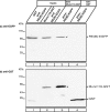
Co-immunoprecipitation of RIBEYE and Munc119 from the bovine retina. Co-immunoprecipitation of RIBEYE and Munc119 from bovine retina. In A, RIBEYE immune serum and RIBEYE preimmune serum were tested for their capability to co-immunoprecipitate Munc119. Munc119 is co-immunoprecipitated by RIBEYE immune serum (lane 2, Aa) but not by RIBEYE preimmune serum (lane 3, A). Ab, shows the same blot as in Aa but reprobed with anti-RIBEYE antibodies. This blot shows the presence of RIBEYE precipitated by the immune serum (lane 2) but not by the preimmune serum (lane 3). Asterisks indicate the immunoglobulin heavy chains. Lane 1 shows the input fraction (2% of total input). The loaded 2% input fraction corresponds roughly to 200 μg of total proteins (in a volume of ≈20 μl). Considerably more input fraction could not be loaded on the gel for volume reasons and also not to overload the gel. Furthermore, synaptic ribbons are mechanically stable, Triton X-100-insoluble structures, which can only be extracted to a certain extent from the bovine retina by the combination of mechanical and chemical lysis. Therefore, the RIBEYE immunosignal is weak in the input fractions. RIBEYE is highly enriched in the experimental immunoprecipitates (lane 2) but absent in the control immunoprecipitates (lane 3). Asterisks indicate the immunoglobulin heavy chains. 100% of the protein A-beads containing the immunoprecipitated proteins were loaded on the gel for experimental and control immunoprecipitations (lanes 2 and 3). In B, Munc119 immune serum and Munc119 preimmune serum were tested for their capability to co-immunoprecipitate RIBEYE. RIBEYE is co-immunoprecipitated by Munc119 immune serum (lane 2, Ba) but not by Munc119 preimmune serum (lane 3, Ba). Bb shows the same blot as in Ba but reprobed with anti-Munc119. This blot shows the presence of Munc119 immunoprecipitated by the immune serum but not by the preimmune serume. Asterisks indicate the immunoglobulin heavy chains.
FIGURE 6.
FIGURE 6.
Binding of Munc119 to RIBEYE(B) is independent of NADH binding. Summary plates of YTH analyses obtained with the indicated bait and prey plasmids. For convenience, experimental bait-prey pairs are underlayered in color (green in the case of interacting bait-prey pairs; control matings are non-colored). The NADH binding deficient RIBEYE point mutant RE(B)G730A (mating 2) interacts with Munc119 in YTH indicating that NADH binding to RIBEYE is not essential for the binding of Munc119.
FIGURE 7.
FIGURE 7.
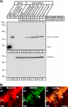
Purified synaptic ribbons specifically recruit Munc119. A, binding of Munc119 fusion protein to synaptic ribbons. 20 μg of purified synaptic ribbons were tested for their capability to bind soluble Munc119 fusion protein at the indicated concentrations. GST alone was used as control protein. Purified ribbons specifically bound Munc119-GST but not GST (A). For GST (lane 1) and Munc119-GST (lane 2) 10% were loaded as input; for the synaptic ribbon (lane 3) 100% were loaded as input. The two depicted blots show representative examples of four different experiments, which all showed the same result. The lower blot is stripped and reprobed with antibodies against RIBEYE to show equal loading of ribbons. A quantitative analysis of binding of Munc119 to synaptic ribbons is given in supplemental Fig. S5. B, Munc119 co-localizes with synaptic ribbons. Immunolabeling of the outer plexiform layer of the bovine retina that contain photoreceptor ribbon synapses with polyclonal antibodies against Munc119 and monoclonal antibodies against RIBEYE(B)/CtBP2. Strong immunosignals of Munc119 were found at synaptic ribbons and in close vicinity to synaptic ribbons. Abbreviations: ONL, outer nuclear layer; OPL, outer plexiform layer; INL, inner nuclear layer. Scale bar:10 μm.

Similar articles

Cited by

References

    1. Higashide, T., Murakami, A., McLaren, M. J., and Inana, G. (1996) J. Biol. Chem. 271 1797–1804 - PubMed
    1. Higashide, T., McLaren, M. J., and Inana, G. (1998) Invest. Ophthalmol. Vis. Sci. 39 690–698 - PubMed
    1. Swanson, D. A., Chang, J. T., Campochiara, P. A., Zack, D. J., and Valle, D. (1998) Investig. Ophthalmol. Vis. Sci. 39 2085–2094 - PubMed
    1. Li, L., Florio, S. K., Pettenati, M. J., Rao, N., Beavo, J. A., and Baehr, W. (1998) Genomics 49 76–82 - PubMed
    1. Kobayashi, A., Higashide, T., Hamasaki, D., Kubota, S., Sakuma, H., An, W., Fujimaki, T., McLaren, M. J., Weleber, R. G., and Inana, G. (2000) Investig. Ophthalmol. Vis. Sci. 11 3268–3277 - PubMed

Publication types

MeSH terms

Associated data

LinkOut - more resources