

Umbilical cord blood regulatory T-cell expansion and functional effects of tumor necrosis factor receptor family members OX40 and 4-1BB expressed on artificial antigen-presenting cells
- PMID: 18645038
- PMCID: PMC2556620
- DOI: 10.1182/blood-2008-01-132951
Umbilical cord blood regulatory T-cell expansion and functional effects of tumor necrosis factor receptor family members OX40 and 4-1BB expressed on artificial antigen-presenting cells
Abstract
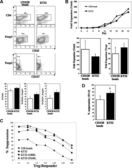
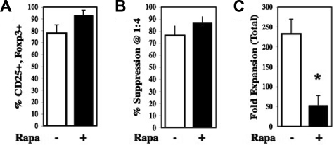
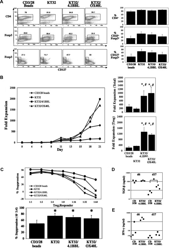
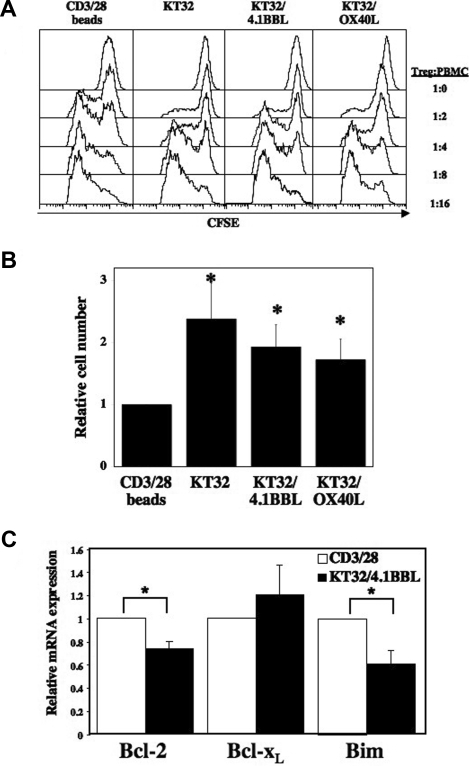
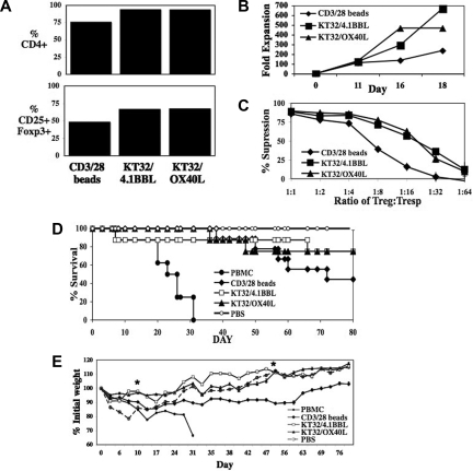
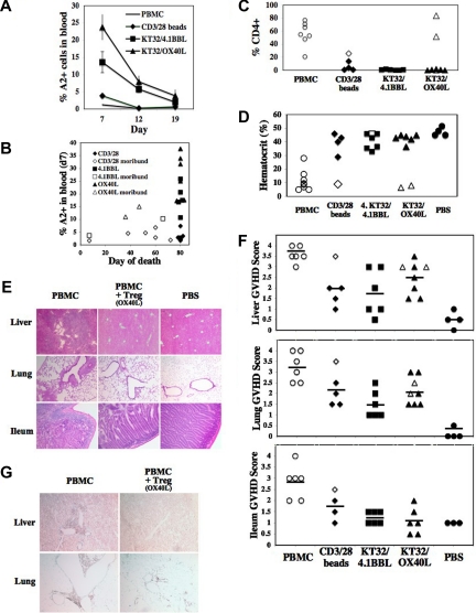
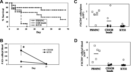
Previously, we showed that human umbilical cord blood (UCB) regulatory T cells (Tregs) could be expanded approximately 100-fold using anti-CD3/28 monoclonal antibody (mAb)-coated beads to provide T-cell receptor and costimulatory signals. Because Treg numbers from a single UCB unit are limited, we explored the use of cell-based artificial antigen-presenting cells (aAPCs) preloaded with anti-CD3/28 mAbs to achieve higher levels of Treg expansion. Compared with beads, aAPCs had similar expansion properties while significantly increasing transforming growth factor beta (TGF-beta) secretion and the potency of Treg suppressor function. aAPCs modified to coexpress OX40L or 4-1BBL expanded UCB Tregs to a significantly greater extent than bead- or nonmodified aAPC cultures, reaching mean expansion levels exceeding 1250-fold. Despite the high expansion and in contrast to studies using other Treg sources, neither OX40 nor 4-1BB signaling of UCB Tregs reduced in vitro suppression. UCB Tregs expanded with 4-1BBL expressing aAPCs had decreased levels of proapoptotic bim. UCB Tregs expanded with nonmodified or modified aAPCs versus beads resulted in higher survival associated with increased Treg persistence in a xeno-geneic graft-versus-host disease lethality model. These data offer a novel approach for UCB Treg expansion using aAPCs, including those coexpressing OX40L or 4-1BBL.
Figures
Similar articles
-
Optimization of cGMP purification and expansion of umbilical cord blood-derived T-regulatory cells in support of first-in-human clinical trials.Cytotherapy. 2017 Feb;19(2):250-262. doi: 10.1016/j.jcyt.2016.10.011. Epub 2016 Nov 22. Cytotherapy. 2017. PMID: 27887864 Free PMC article.
-
Ex vivo expansion of CD4+CD25+FoxP3+ T regulatory cells based on synergy between IL-2 and 4-1BB signaling.J Immunol. 2007 Dec 1;179(11):7295-304. doi: 10.4049/jimmunol.179.11.7295. J Immunol. 2007. PMID: 18025172
-
4-1BB is superior to CD28 costimulation for generating CD8+ cytotoxic lymphocytes for adoptive immunotherapy.J Immunol. 2007 Oct 1;179(7):4910-8. doi: 10.4049/jimmunol.179.7.4910. J Immunol. 2007. PMID: 17878391 Free PMC article.
-
A comprehensive review on the role of co-signaling receptors and Treg homeostasis in autoimmunity and tumor immunity.J Autoimmun. 2018 Dec;95:77-99. doi: 10.1016/j.jaut.2018.08.007. Epub 2018 Aug 31. J Autoimmun. 2018. PMID: 30174217 Free PMC article. Review.
-
Immune regulation and control of regulatory T cells by OX40 and 4-1BB.Cytokine Growth Factor Rev. 2008 Jun-Aug;19(3-4):253-62. doi: 10.1016/j.cytogfr.2008.04.003. Epub 2008 May 27. Cytokine Growth Factor Rev. 2008. PMID: 18508403 Free PMC article. Review.
Cited by
-
Promoting transplantation tolerance; adoptive regulatory T cell therapy.Clin Exp Immunol. 2013 May;172(2):158-68. doi: 10.1111/cei.12052. Clin Exp Immunol. 2013. PMID: 23574313 Free PMC article. Review.
-
miR-146b antagomir-treated human Tregs acquire increased GVHD inhibitory potency.Blood. 2016 Sep 8;128(10):1424-35. doi: 10.1182/blood-2016-05-714535. Epub 2016 Aug 2. Blood. 2016. PMID: 27485827 Free PMC article.
-
Beyond FOXP3: a 20-year journey unravelling human regulatory T-cell heterogeneity.Front Immunol. 2024 Jan 12;14:1321228. doi: 10.3389/fimmu.2023.1321228. eCollection 2023. Front Immunol. 2024. PMID: 38283365 Free PMC article. Review.
-
GVHD-associated, inflammasome-mediated loss of function in adoptively transferred myeloid-derived suppressor cells.Blood. 2015 Sep 24;126(13):1621-8. doi: 10.1182/blood-2015-03-634691. Epub 2015 Aug 11. Blood. 2015. PMID: 26265697 Free PMC article.
-
Immune regulatory cells in umbilical cord blood: T regulatory cells and mesenchymal stromal cells.Br J Haematol. 2009 Oct;147(2):200-6. doi: 10.1111/j.1365-2141.2009.07781.x. Br J Haematol. 2009. PMID: 19796269 Free PMC article. Review.
References
-
- Welniak LA, Blazar BR, Murphy WJ. Immunobiology of allogeneic hematopoietic stem cell transplantation. Annu Rev Immunol. 2007;25:139–170. - PubMed
-
- Sakaguchi S, Sakaguchi N, Asano M, Itoh M, Toda M. Immunologic self-tolerance maintained by activated T cells expressing IL-2 receptor alpha-chains (CD25). Breakdown of a single mechanism of self-tolerance causes various autoimmune diseases. J Immunol. 1995;155:1151–1164. - PubMed
-
- Sakaguchi S, Ono M, Setoguchi R, et al. Foxp3+ CD25+ CD4+ natural regulatory T cells in dominant self-tolerance and autoimmune disease. Immunol Rev. 2006;212:8–27. - PubMed
-
- Shevach EM, DiPaolo RA, Andersson J, Zhao DM, Stephens GL, Thornton AM. The lifestyle of naturally occurring CD4+ CD25+ Foxp3+ regulatory T cells. Immunol Rev. 2006;212:60–73. - PubMed
Publication types
MeSH terms
Substances
Grants and funding
LinkOut - more resources
Full Text Sources
Other Literature Sources
Research Materials
