Mechanisms of cytomegalovirus-accelerated vascular disease: induction of paracrine factors that promote angiogenesis and wound healing
- PMID: 18637518
- PMCID: PMC2699259
- DOI: 10.1007/978-3-540-77349-8_22
Mechanisms of cytomegalovirus-accelerated vascular disease: induction of paracrine factors that promote angiogenesis and wound healing
Abstract
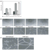
Human cytomegalovirus (HCMV) is associated with the acceleration of a number of vascular diseases such as atherosclerosis, restenosis, and transplant vascular sclerosis (TVS). All of these diseases are the result of either mechanical or immune-mediated injury followed by inflammation and subsequent smooth muscle cell (SMC) migration from the vessel media to the intima and proliferation that culminates in vessel narrowing. A number of epidemiological and animal studies have demonstrated that CMV significantly accelerates TVS and chronic rejection (CR) in solid organ allografts. In addition, treatment of human recipients and animals alike with the antiviral drug ganciclovir results in prolonged survival of the allograft, indicating that CMV replication is a requirement for acceleration of disease. However, although virus persists in the allograft throughout the course of disease, the number of directly infected cells does not account for the global effects that the virus has on the acceleration of TVS and CR. Recent investigations of up- and downregulated cellular genes in infected allografts in comparison to native heart has demonstrated that rat CMV (RCMV) upregulates genes involved in wound healing (WH) and angiogenesis (AG). Consistent with this result, we have found that supernatants from HCMV-infected cells (HCMV secretome) induce WH and AG using in vitro models. Taken together, these findings suggest that one mechanism for HCMV acceleration of TVS is mediated through induction of secreted cytokines and growth factors from virus-infected cells that promote WH and AG in the allograft, resulting in the acceleration of TVS. We review here the ability of CMV infection to alter the local environment by producing cellular factors that act in a paracrine fashion to enhance WH and AG processes associated with the development of vascular disease, which accelerates chronic allograft rejection.
Figures


Similar articles
-
Human cytomegalovirus secretome contains factors that induce angiogenesis and wound healing.J Virol. 2008 Jul;82(13):6524-35. doi: 10.1128/JVI.00502-08. Epub 2008 Apr 30. J Virol. 2008. PMID: 18448536 Free PMC article.
-
The role of angiogenic and wound repair factors during CMV-accelerated transplant vascular sclerosis in rat cardiac transplants.Am J Transplant. 2008 Feb;8(2):277-87. doi: 10.1111/j.1600-6143.2007.02062.x. Epub 2007 Dec 18. Am J Transplant. 2008. PMID: 18093265
-
Cytomegalovirus latency promotes cardiac lymphoid neogenesis and accelerated allograft rejection in CMV naïve recipients.Am J Transplant. 2011 Jan;11(1):45-55. doi: 10.1111/j.1600-6143.2010.03365.x. Am J Transplant. 2011. PMID: 21199347 Free PMC article.
-
The role of cytomegalovirus in angiogenesis.Virus Res. 2011 May;157(2):204-11. doi: 10.1016/j.virusres.2010.09.011. Epub 2010 Oct 1. Virus Res. 2011. PMID: 20869406 Free PMC article. Review.
-
Cytomegalovirus infection and cardiac allograft vasculopathy.Transpl Infect Dis. 1999 Jun;1(2):115-26. doi: 10.1034/j.1399-3062.1999.010205.x. Transpl Infect Dis. 1999. PMID: 11428979 Review.
Cited by
-
Human CMV infection of endothelial cells induces an angiogenic response through viral binding to EGF receptor and beta1 and beta3 integrins.Proc Natl Acad Sci U S A. 2008 Apr 8;105(14):5531-6. doi: 10.1073/pnas.0800037105. Epub 2008 Mar 28. Proc Natl Acad Sci U S A. 2008. PMID: 18375753 Free PMC article.
-
A systemic network triggered by human cytomegalovirus entry.Adv Virol. 2011;2011:262080. doi: 10.1155/2011/262080. Epub 2011 May 15. Adv Virol. 2011. PMID: 22312338 Free PMC article.
-
Human cytomegalovirus glycoprotein UL141 targets the TRAIL death receptors to thwart host innate antiviral defenses.Cell Host Microbe. 2013 Mar 13;13(3):324-35. doi: 10.1016/j.chom.2013.02.003. Cell Host Microbe. 2013. PMID: 23498957 Free PMC article.
-
Cytomegalovirus microRNA expression is tissue specific and is associated with persistence.J Virol. 2011 Jan;85(1):378-89. doi: 10.1128/JVI.01900-10. Epub 2010 Oct 27. J Virol. 2011. PMID: 20980502 Free PMC article.
-
Lymphotropic herpesvirus DNA detection in patients with active CMV infection - a possible role in the course of CMV infection after hematopoietic stem cell transplantation.Med Sci Monit. 2011 Aug;17(8):CR432-441. doi: 10.12659/msm.881904. Med Sci Monit. 2011. PMID: 21804462 Free PMC article.
References
-
- Almeida GD, Porada CD, St Jeor S, Ascensao JL. Human cytomegalovirus alters interleukin-6 production by endothelial cells. Blood. 1994;83:370–376. - PubMed
-
- Almeida-Porada G, Porada CD, Shanley JD, Ascensao JL. Altered production of GM-CSF and IL-8 in cytomegalovirus-infected, IL-1- primed umbilical cord endothelial cells. Exp Hematol. 1997;25:1278–1285. - PubMed
-
- Auerbach R, Lewis R, Shinners B, Kubai L, Akhtar N. Angiogenesis assays: a critical overview. Clin Chem. 2003;49:32–40. - PubMed
-
- Billingham ME. Histopathology of Graft Coronary Disease. J Heart Lung Transplant. 1992;11:S38–S44. - PubMed
-
- Bouis D, Kusumanto Y, Meijer C, Mulder NH, Hospers GA. A review on pro- and anti-angiogenic factors as targets of clinical intervention. Pharmacol Res. 2006;53:89–103. - PubMed
Publication types
MeSH terms
Substances
Grants and funding
- R01 HL071695-04/HL/NHLBI NIH HHS/United States
- R01 HL083194/HL/NHLBI NIH HHS/United States
- R01 HL071695/HL/NHLBI NIH HHS/United States
- AI21640/AI/NIAID NIH HHS/United States
- R01 HL066238/HL/NHLBI NIH HHS/United States
- R01 HL088603-01A1/HL/NHLBI NIH HHS/United States
- R01 HL066238-01/HL/NHLBI NIH HHS/United States
- R37 AI021640/AI/NIAID NIH HHS/United States
- R01 AI021640/AI/NIAID NIH HHS/United States
- R01 AI021640-23/AI/NIAID NIH HHS/United States
- R01 HL083194-04/HL/NHLBI NIH HHS/United States
- HL71695/HL/NHLBI NIH HHS/United States
- R01 HL065754-08/HL/NHLBI NIH HHS/United States
- R01 HL085451/HL/NHLBI NIH HHS/United States
- HL083194/HL/NHLBI NIH HHS/United States
- R01 HL088603/HL/NHLBI NIH HHS/United States
- HL65754/HL/NHLBI NIH HHS/United States
- HL 66238-01/HL/NHLBI NIH HHS/United States
- R01 HL065754/HL/NHLBI NIH HHS/United States
LinkOut - more resources
Full Text Sources
Other Literature Sources
Medical

