. 2008 Sep;28(18):5668-86.
doi: 10.1128/MCB.00418-08. Epub 2008 Jul 14.

The protein kinase C-responsive inhibitory domain of CARD11 functions in NF-kappaB activation to regulate the association of multiple signaling cofactors that differentially depend on Bcl10 and MALT1 for association

Affiliations

The protein kinase C-responsive inhibitory domain of CARD11 functions in NF-kappaB activation to regulate the association of multiple signaling cofactors that differentially depend on Bcl10 and MALT1 for association

Ryan R McCully et al. Mol Cell Biol. 2008 Sep.

Abstract

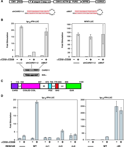
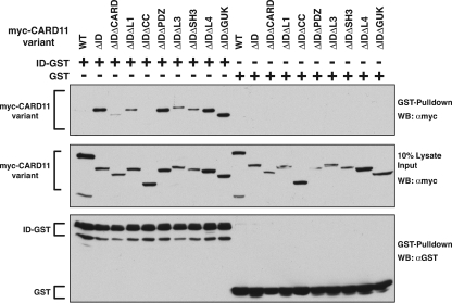
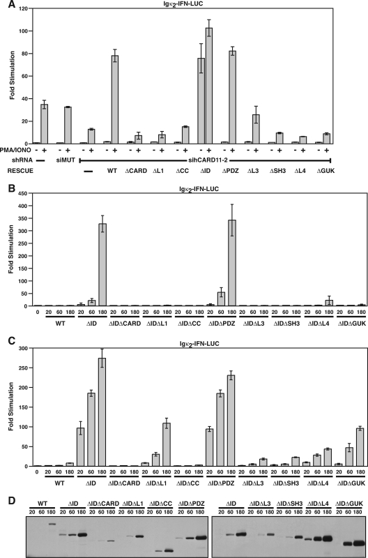
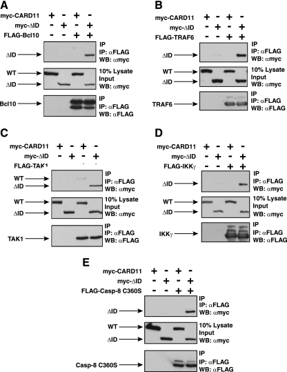
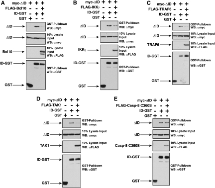
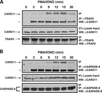
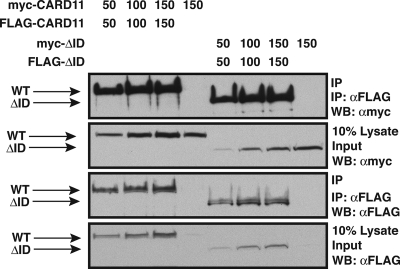
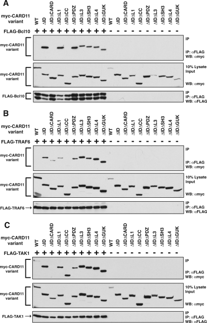
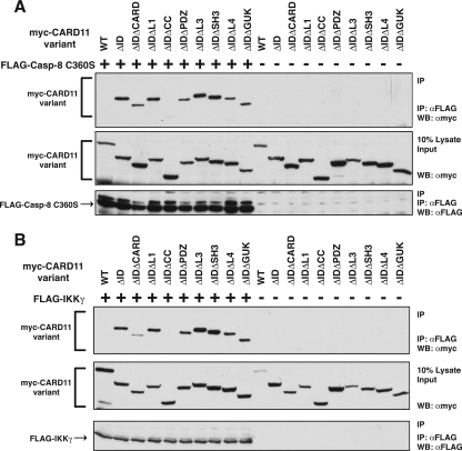
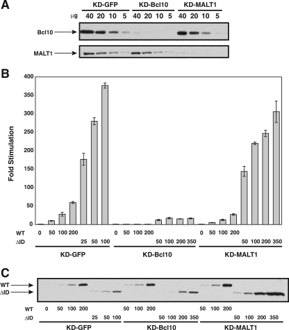
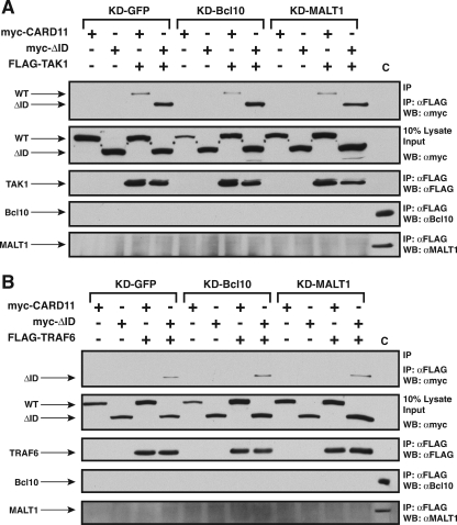
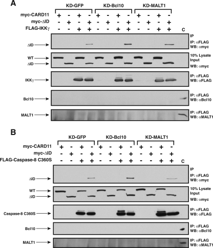
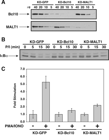
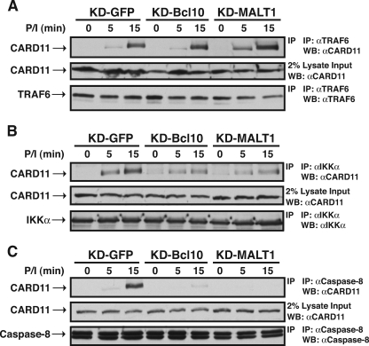
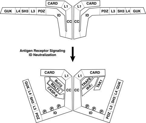
The activation of NF-kappaB by T-cell receptor (TCR) signaling is critical for T-cell activation during the adaptive immune response. CARD11 is a multidomain adapter that is required for TCR signaling to the IkappaB kinase (IKK) complex. During TCR signaling, the region in CARD11 between the coiled-coil and PDZ domains is phosphorylated by protein kinase Ctheta (PKCtheta) in a required step in NF-kappaB activation. In this report, we demonstrate that this region functions as an inhibitory domain (ID) that controls the association of CARD11 with multiple signaling cofactors, including Bcl10, TRAF6, TAK1, IKKgamma, and caspase-8, through an interaction that requires both the caspase recruitment domain (CARD) and the coiled-coil domain. Consistent with the ID-mediated control of their association, we demonstrate that TRAF6 and caspase-8 associate with CARD11 in T cells in a signal-inducible manner. Using an RNA interference rescue assay, we demonstrate that the CARD, linker 1, coiled-coil, linker 3, SH3, linker 4, and GUK domains are each required for TCR signaling to NF-kappaB downstream of ID neutralization. Requirements for the CARD, linker 1, and coiled-coil domains in signaling are consistent with their roles in the association of CARD11 with Bcl10, TRAF6, TAK1, caspase-8, and IKKgamma. Using Bcl10- and MALT1-deficient cells, we show that CARD11 can recruit signaling cofactors independently of one another in a signal-inducible manner.


Figures

FIG. 1.
FIG. 1.
The linker domains in CARD11 are required for regulated TCR signaling to NF-κB. (A) Schematic of lentiviral constructs. Following integration, the human H1 RNA promoter drives expression of an shRNA, and the CMV enhancer/chicken β-actin promoter fusion drives expression of the puromycin resistance gene (PURO). The virus contains a flap sequence (F) for increased nuclear translocation efficiency and a Woodchuck hepatitis virus posttranscriptional regulatory element (WPRE) for enhanced expression. The sequences of the sihCARD11-2 and siMUT shRNAs are depicted. (B) Jurkat T-cell pools infected with viruses expressing no hairpin (-), the sihCARD11-2 hairpin, or the siMUT hairpin were transfected with 200 ng of pCSK-LacZ and 2,800 ng of either Igκ2-IFN-LUC or NFAT-LUC and stimulated with anti-CD3/anti-CD28 cross-linking as indicated. The lower panels are Western blots of total cell extracts of the infected pools probed with antibodies to CARD11 or IKKα. (C) Schematic of the domain structure of CARD11. The numbers indicate the amino acid position according to Pomerantz et al. (44). (D) The Jurkat T-cell pool stably expressing the sihCARD11-2 shRNA was transfected with 200 ng of pCSK-LacZ and 1,800 ng of Igκ2-IFN-LUC in the absence or presence of 200 ng of the indicated CARD11 variant expression construct. Cells were stimulated with anti-CD3/anti-CD28 cross-linking as indicated. The bars represent the mean values of triplicate samples, and error bars indicate the standard deviation. WT, wild type; α, anti.
FIG. 2.
FIG. 2.
Determinants of ID association with the CARD11 ΔID variant. (A) HEK293T cells were transfected with expression vectors for the indicated CARD11 variants and 150 ng of expression vector for either the ID-GST fusion (pEBB-ID-GST) or GST (pEBG). Glutathione-agarose precipitations were performed as described in Materials and Methods. To achieve approximately equivalent expression of each CARD11 variant, the following amounts of each expression vector were used (in ng): wild type (WT), 50; ΔID, 50; ΔIDΔCARD, 1,000; ΔIDΔL1, 150; ΔIDΔCC, 75; ΔIDΔPDZ, 100; ΔIDΔL3, 150; ΔIDΔSH3, 150; ΔIDΔL4, 100; ΔIDΔGUK, 100. Western blotting (WB) with the indicated antibodies was performed to develop the contents of the precipitate (top and bottom panels) and the lysate input (middle panel). α, anti.
FIG. 3.
FIG. 3.
CARD11 domain requirements for NF-κB activation by PMA/ionomycin treatment and by the CARD11 ΔID variant. (A) Jurkat T cell pools stably expressing either no hairpin (−), the siMUT shRNA, or the sihCARD11-2 shRNA were transfected with 200 ng of pCSK-LacZ and 1,800 ng of Igκ2-IFN-LUC in the absence or presence of 200 ng of the indicated CARD11 variant expression construct. Cells were stimulated with PMA and ionomycin as indicated. (B) Wild-type Jurkat T cells were transfected with 200 ng of pCSK-LacZ and 2,500 ng of Igκ2-IFN-LUC in the presence of the indicated amounts (in ng) of expression vectors for the indicated CARD11 variants. (C) HEK293T cells were transfected with 6 ng of pCSK-LacZ and 20 ng of Igκ2-IFN-LUC in the presence of the indicated amounts (in ng) of expression vectors for the indicated CARD11 variants. (D) The lysates from samples in panel C were probed by Western blotting using anti-myc primary antibody to indicate the relative expression level of each variant. The bars represent the mean values of triplicate samples, and error bars indicate the standard deviations. WT, wild type.
FIG. 4.
FIG. 4.
The ID regulates the ability of CARD11 to associate with Bcl10, TRAF6, TAK1, IKKγ, and caspase-8. HEK293T cells were transfected with 50 to 150 ng of the expression vector for wild-type CARD11 or the ΔID in the absence and presence of vectors encoding FLAG-tagged versions of Bcl10 (100 ng), TRAF6 (200 ng), TAK1 (200 ng), IKKγ (1,000 ng), or caspase-8 C360S (1,000 ng), as indicated. Anti-FLAG immunoprecipitations were performed as described in Materials and Methods. In each panel, Western blotting (WB) with the indicated antibodies (at right) was performed to develop the contents of the immunoprecipitate (top and bottom) and the lysate input (middle). WT, wild type; α, anti; IP, immunoprecipitation.
FIG. 5.
FIG. 5.
Bcl10, TRAF6, TAK1, and IKKγ compete with the ID in trans for binding to the ΔID variant. HEK293T cells were transfected with expression vectors for the ΔID and either the ID-GST fusion or GST in the absence and presence of vectors encoding Bcl10 (A), IKKγ (B), TRAF6 (C), TAK1 (D), or caspase-8 C360S (E). Glutathione-agarose precipitations were performed as described in Materials and Methods. To achieve approximately equivalent expression of the ΔID and the ID-GST in the absence and presence of each signaling cofactor, the following amount of each expression vector was used (in ng): ΔID, 50 to 100; ID-GST, 100 to 300; GST, 50; Bcl10, 150; IKKγ, 300; TRAF6, 300; TAK1, 100; or caspase-8 C360S, 300. The panels show the contents of the precipitate and lysate input, developed with anti-myc, anti-FLAG, or anti-GST primary antibody, as indicated. α, anti; WB, Western blotting.
FIG. 6.
FIG. 6.
Signal-induced association of CARD11 with TRAF6 and caspase-8 in T cells. Jurkat T cells were stimulated with a time course of PMA/ionomycin as indicated. Anti-TRAF6 or anti-caspase-8 immunoprecipitations were performed as described in Materials and Methods. In each panel, Western blotting (WB) with the indicated antibodies (at right) was performed to develop the contents of the immunoprecipitate (top and bottom) and the lysate input (middle). The asterisk indicates an undefined inducible band in the immunoprecipitate that is reactive to the anti-CARD11 primary antibody. α, anti; IP, immunoprecipitation.
FIG. 7.
FIG. 7.
The ID does not determine whether CARD11 can self-associate. HEK293T cells were transfected with the indicated amounts (in ng) of expression vectors for wild-type CARD11 or the ΔID variant as either myc-tagged or FLAG-tagged fusions. Anti-FLAG immunoprecipitations were performed as described in Materials and Methods. The panels show the contents of the immunoprecipitate and the lysate input, developed with either anti-myc or anti-FLAG primary antibody, as indicated. WT, wild type; α, anti; IP, immunoprecipitation; WB, Western blotting.
FIG. 8.
FIG. 8.
Determinants of the association of the ΔID with Bcl10, TRAF6, and TAK1. (A) HEK293T cells were transfected with expression vectors for the indicated CARD11 variants in the absence (−) or presence (+) of the expression vector for FLAG-tagged Bcl10. Anti-FLAG immunoprecipitations were performed as described in Materials and Methods. To achieve approximately equivalent expression of each CARD11 variant, the following amount of each expression vector was used (in ng): wild type (WT), 100; ΔID, 100; ΔIDΔCARD, 1,000; ΔIDΔL1, 100; ΔIDΔCC, 100; ΔIDΔPDZ, 150; ΔIDΔL3, 125; ΔIDΔSH3, 150; ΔIDΔL4, 50; ΔIDΔGUK, 50. To achieve approximately equivalent expression of Bcl10 in each sample, 100 ng of Bcl10 expression vector was used in each sample except that 500 ng was used in the ΔIDΔCARD sample and 300 ng was used in the ΔIDΔPDZ sample. Western blotting (WB) with the indicated antibodies (at right) was performed to develop the contents of the immunoprecipitate (top and bottom panels) and the lysate input (middle panel). (B) Anti-FLAG immunoprecipitations were performed as described in panel A except that 200 ng of the expression vector for FLAG-tagged TRAF6 was used in all indicated samples (+). The following amount (in ng) of expression vector for each CARD11 variant was used: WT, 200; ΔID, 200; ΔIDΔCARD, 2,000; ΔIDΔL1, 200; ΔIDΔCC, 90; ΔIDΔPDZ, 300; ΔIDΔL3, 250; ΔIDΔSH3, 300; ΔIDΔL4, 100; ΔIDΔGUK, 100. (C) Anti-FLAG immunoprecipitations were performed as described in panel A except that 200 ng of the expression vector for FLAG-tagged TAK1 was used in all indicated samples (+). The following amount (in ng) of expression vector for each CARD11 variant was used: WT, 100; ΔID, 100; ΔIDΔCARD, 1,000; ΔIDΔL1, 100; ΔIDΔCC, 100; ΔIDΔPDZ, 150; ΔIDΔL3, 175; ΔIDΔSH3, 150; ΔIDΔL4, 50; ΔIDΔGUK, 50. α, anti; IP, immunoprecipitation.
FIG. 9.
FIG. 9.
Determinants of the association of the ΔID with caspase-8 C360S and IKKγ. (A) Anti-FLAG immunoprecipitations were performed as described in the legend of Fig. 8A except that 1,000 ng of the expression vector for FLAG-tagged caspase-8 C360S was used in all indicated samples (+). The following amount (in ng) of expression vector for each CARD11 variant was used: wild type (WT), 100; ΔID, 100; ΔIDΔCARD, 1,000; ΔIDΔL1, 100; ΔIDΔCC, 50; ΔIDΔPDZ, 150; ΔIDΔL3, 125; ΔIDΔSH3, 150; ΔIDΔL4, 50; ΔIDΔGUK, 50. (B) Anti-FLAG immunoprecipitations were performed as described in the legend of Fig. 8A except that 800 ng of the expression vector for FLAG-tagged IKKγ was used in all indicated samples (+). The following amount (in ng) of expression vector for each CARD11 variant was used: WT, 120; ΔID, 120; ΔIDΔCARD, 1,200; ΔIDΔL1, 120; ΔIDΔCC, 120; ΔIDΔPDZ, 180; ΔIDΔL3, 210; ΔIDΔSH3, 180; ΔIDΔL4, 60; ΔIDΔGUK, 60. α, anti; IP, immunoprecipitation; WB, Western blotting.
FIG. 10.
FIG. 10.
Stable knockdown of Bcl10 and MALT1 in HEK293T cell lines. HEK293T cells were infected with lentiviruses expressing shRNAs that target either Bcl10, MALT1, or GFP as a control, as described in Materials and Methods, to establish the KD-GFP, KD-Bcl10, and KD-MALT1 cell lines. (A) The indicated amounts (in μg) of stably infected cell lysates were assayed by Western blotting for Bcl10 and MALT1 protein levels using anti-Bcl10 and anti-MALT1 primary antibodies. (B) The KD-GFP, KD-Bcl10, and KD-MALT1 cell lines were transfected with 6 ng of pCSK-LacZ, 20 ng of Igκ2-IFN-LUC, and the indicated amounts (in ng) of expression vectors for myc-tagged wild-type CARD11 or the ΔID variant. Stimulation was determined as described in Materials and Methods. (C) The lysates from samples in panel B were probed by Western blotting using anti-myc primary antibody to indicate the relative expression level of each variant. The bars represent the mean values of triplicate samples, and error bars indicate the standard deviations. WT, wild type.
FIG. 11.
FIG. 11.
Effect of Bcl10 and MALT1 knockdown on the association of the ΔID with TAK1 and TRAF6 in HEK293T cells. The KD-GFP, KD-Bcl10, and KD-MALT1 cell lines were transfected with expression vectors for the wild-type CARD11 or the ΔID in the absence (−) or presence (+) of expression vectors for FLAG-tagged TAK1 (A) or TRAF6 (B). Anti-FLAG immunoprecipitations were performed as described in Materials and Methods. Where indicated, transfections included 100 ng of expression vector for wild-type CARD11 and 200 ng of expression vector for either TAK1 or TRAF6. To achieve approximately equivalent expression of the ΔID variant in each cell line, 200 ng of ΔID expression vector was used in the KD-GFP line, and 400 ng was used in the KD-Bcl10 and KD-MALT1 lines. The panels show the contents of the immunoprecipitate and lysate input developed with the indicated primary antibodies. In the control lane (C), the equivalent of 5% of the lysate input from the KD-GFP sample was resolved so as to provide a positive control for the Western blot (WB) analysis of the immunoprecipitates for the presence of Bcl10 and MALT1. WT, wild type; α, anti; IP, immunoprecipitation.
FIG. 12.
FIG. 12.
Effect of Bcl10 and MALT1 knockdown on the association of the ΔID with IKKγ and caspase-8 C360S in HEK293T cells. The KD-GFP, KD-Bcl10, and KD-MALT1 cell lines were transfected with expression vectors for the wild-type CARD11 or the ΔID in the absence (−) or presence (+) of expression vectors for FLAG-tagged IKKγ or caspase-8. Anti-FLAG immunoprecipitations were performed as described in Materials and Methods. In panel A, transfections included 800 ng of expression vector for IKKγ where indicated. To achieve approximately equivalent expression of wild-type and ΔID CARD11 variants in each cell line, the following amounts were used, from left to right (in ng): 100 WT, 200 ΔID, 100 WT, and 200 ΔID (for the KD-GFP cells); 200 WT, 400 ΔID, 100 WT, and 400 ΔID (for KD-Bcl10 cells); 200 WT, 400 ΔID, 100 WT, and 400 ΔID (for KD-MALT1 cells). In panel B, where indicated, transfections included 100 ng of expression vector for wild-type CARD11 and 1,000 ng of expression vector for caspase-8. To achieve approximately equivalent expression of the ΔID variant in each cell line, 200 ng of ΔID expression vector was used in the KD-GFP line, and 400 ng was used in the KD-Bcl10 and KD-MALT1 lines. The panels show the contents of the immunoprecipitate and lysate input developed with the indicated primary antibodies. In the control lane (C), the equivalent of 5% of the lysate input from the KD-GFP sample was resolved so as to provide a positive control for the Western blot (WB) analysis of the immunoprecipitates for the presence of Bcl10 and MALT1. WT, wild type; α, anti; IP, immunoprecipitation.
FIG. 13.
FIG. 13.
Stable knockdown of Bcl10 and MALT1 in Jurkat T cell lines. Jurkat T cells were infected with lentiviruses expressing shRNAs that target either Bcl10, MALT1, or GFP as a control, as described in Materials and Methods, to establish the KD-GFP, KD-Bcl10, and KD-MALT1 cell lines. (A) The indicated amounts (in μg) of stably infected cell lysates were assayed by Western blotting for Bcl10 and MALT1 protein levels using anti-Bcl10 and anti-MALT1 primary antibodies. (B) The KD-GFP, KD-Bcl10, and KD-MALT1 cell lines were stimulated with PMA (50 ng/ml) and ionomycin (1 μM) (P/I) for the indicated times. Lysates were assayed by Western blotting for IκBα degradation using anti-IκBα primary antibody. (C) The KD-GFP, KD-Bcl10, and KD-MALT1 cell lines were transfected with 200 ng of pCSK-LacZ, 1,500 ng of Igκ2-IFN-LUC, and 1,300 ng of pBSKII+ and stimulated with 50 ng/ml PMA and 1 μM ionomycin for 1 h. Stimulation was determined as described in Materials and Methods. The bars represent the mean values of triplicate samples, and error bars indicate the standard deviations.
FIG. 14.
FIG. 14.
Effect of Bcl10 and MALT1 knockdown on the inducible association of CARD11 with cofactors in Jurkat T cells. The KD-GFP, KD-Bcl10, and KD-MALT1 cell lines were stimulated with a time course of PMA-ionomycin (P/I) as indicated. Anti-TRAF6 (A), anti-IKKα (B), and anti-caspase-8 (C) immunoprecipitations were performed as described in Materials and Methods. In each panel, Western blotting (WB) with the indicated antibodies (at right) was performed to develop the contents of the immunoprecipitate (top and bottom) and the lysate input (middle). α, anti; IP, immunoprecipitation.
FIG. 15.
FIG. 15.
Model of the signal-induced conversion of CARD11 to an active signaling scaffold. Prior to TCR engagement, the ID of CARD11 interacts with the CARD and the coiled-coil (CC) domain to prevent the association of signaling cofactors. Although it is depicted here as an intramolecular interaction in the inhibited state, it is also possible that the ID interacts with the CARD and the coiled-coil domain of another CARD11 molecule in the oligomer. TCR signaling leads to the neutralization of ID activity, in part through the phosphorylation of the ID by PKCθ. Once the ID is neutralized, signaling cofactors can be recruited to the N-terminal portion of CARD11. The model indicates which domains of CARD11 are required for the association with the cofactors analyzed. TAK1 requires only the CARD, while Bcl10 requires the CARD and coiled-coil domain. TRAF6 requires the CARD and the L1 and coiled-coil domains, while caspase-8 and IKKγ each require the CARD and the coiled-coil domain. MALT1 is presumably recruited through interactions with Bcl10. Caspase-8 (CASP-8) requires both Bcl10 and MALT1 to associate with CARD11 during signaling. The association of TAK1, TRAF6, and IKKγ with activated CARD11 can occur in a Bcl10-independent and MALT1-independent manner. The ID does not appear to influence whether CARD11 oligomerizes, suggesting that oligomerization occurs both before and after signaling through a region of the coiled-coil domain that is not targeted by the ID. The depicted interactions between CARD11 and signaling cofactors may be direct or may require other proteins to bridge the associations.

Similar articles

Cited by

References

    1. Akagi, T., M. Motegi, A. Tamura, R. Suzuki, Y. Hosokawa, H. Suzuki, H. Ota, S. Nakamura, Y. Morishima, M. Taniwaki, and M. Seto. 1999. A novel gene, MALT1 at 18q21, is involved in t(11;18) (q21;q21) found in low-grade B-cell lymphoma of mucosa-associated lymphoid tissue. Oncogene 185785-5794. - PubMed
    1. Bell, S., K. Degitz, M. Quirling, N. Jilg, S. Page, and K. Brand. 2003. Involvement of NF-κB signalling in skin physiology and disease. Cell Signal. 151-7. - PubMed
    1. Bertin, J., L. Wang, Y. Guo, M. D. Jacobson, J. L. Poyet, S. M. Srinivasula, S. Merriam, P. S. DiStefano, and E. S. Alnemri. 2001. CARD11 and CARD14 are novel caspase recruitment domain (CARD)/membrane-associated guanylate kinase (MAGUK) family members that interact with BCL10 and activate NF-κB. J. Biol. Chem. 27611877-11882. - PubMed
    1. Bidere, N., A. L. Snow, K. Sakai, L. Zheng, and M. J. Lenardo. 2006. Caspase-8 regulation by direct interaction with TRAF6 in T cell receptor-induced NF-κB activation. Curr. Biol. 161666-1671. - PubMed
    1. Cannons, J. L., L. J. Yu, B. Hill, L. A. Mijares, D. Dombroski, K. E. Nichols, A. Antonellis, G. A. Koretzky, K. Gardner, and P. L. Schwartzberg. 2004. SAP regulates TH2 differentiation and PKC-θ-mediated activation of NF-κB1. Immunity 21693-706. - PubMed

Publication types

MeSH terms

Substances

LinkOut - more resources