

Syntaxin 1A interaction with the dopamine transporter promotes amphetamine-induced dopamine efflux
- PMID: 18617632
- PMCID: PMC2728020
- DOI: 10.1124/mol.108.048447
Syntaxin 1A interaction with the dopamine transporter promotes amphetamine-induced dopamine efflux
Abstract
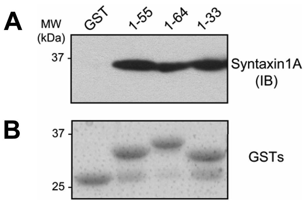
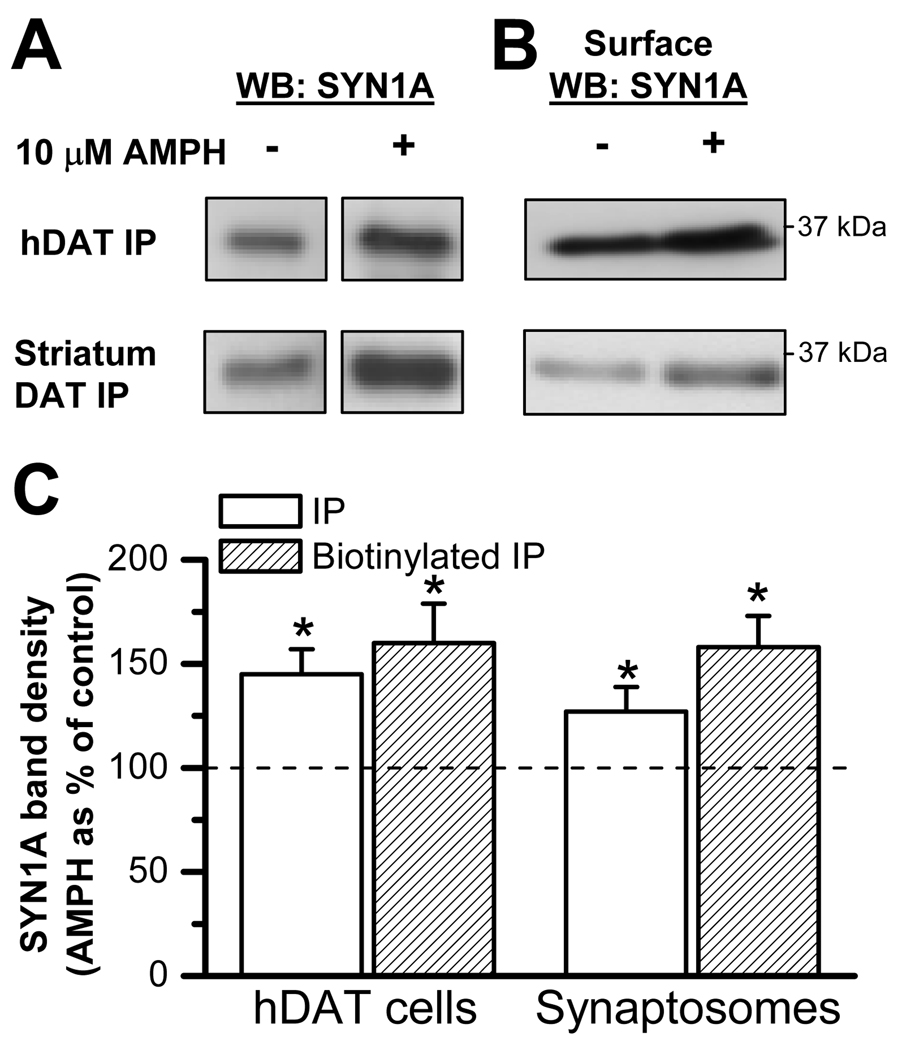
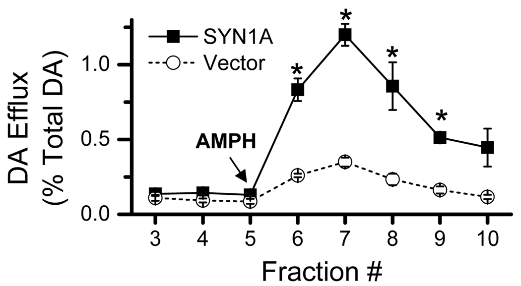
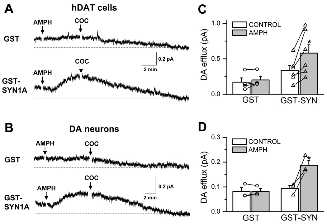
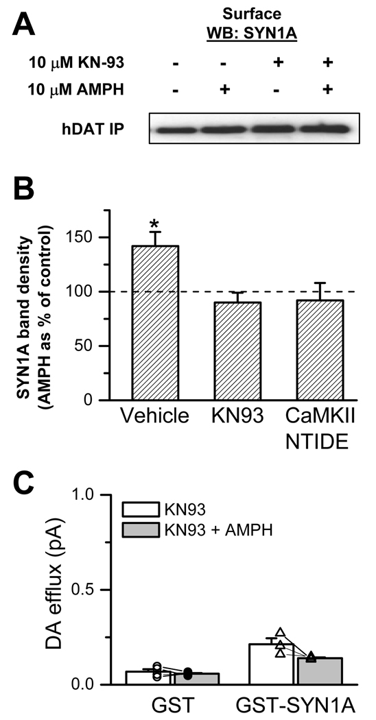
The soluble N-ethylmaleimide-sensitive factor attachment protein receptor protein syntaxin 1A (SYN1A) interacts with and regulates the function of transmembrane proteins, including ion channels and neurotransmitter transporters. Here, we define the first 33 amino acids of the N terminus of the dopamine (DA) transporter (DAT) as the site of direct interaction with SYN1A. Amphetamine (AMPH) increases the association of SYN1A with human DAT (hDAT) in a heterologous expression system (hDAT cells) and with native DAT in murine striatal synaptosomes. Immunoprecipitation of DAT from the biotinylated fraction shows that the AMPH-induced increase in DAT/SYN1A association occurs at the plasma membrane. In a superfusion assay of DA efflux, cells overexpressing SYN1A exhibited significantly greater AMPH-induced DA release with respect to control cells. By combining the patch-clamp technique with amperometry, we measured DA release under voltage clamp. At -60 mV, a physiological resting potential, AMPH did not induce DA efflux in hDAT cells and DA neurons. In contrast, perfusion of exogenous SYN1A (3 microM) into the cell with the whole-cell pipette enabled AMPH-induced DA efflux at -60 mV in both hDAT cells and DA neurons. It has been shown recently that Ca2+/calmodulin-dependent protein kinase II (CaMKII) is activated by AMPH and regulates AMPH-induced DA efflux. Here, we show that AMPH-induced association between DAT and SYN1A requires CaMKII activity and that inhibition of CaMKII blocks the ability of exogenous SYN1A to promote DA efflux. These data suggest that AMPH activation of CaMKII supports DAT/SYN1A association, resulting in a mode of DAT capable of DA efflux.
Figures
Similar articles
-
Amphetamine induces a calcium/calmodulin-dependent protein kinase II-dependent reduction in norepinephrine transporter surface expression linked to changes in syntaxin 1A/transporter complexes.Mol Pharmacol. 2007 Jan;71(1):230-9. doi: 10.1124/mol.106.026690. Epub 2006 Oct 10. Mol Pharmacol. 2007. PMID: 17032905
-
Amphetamine-induced dopamine efflux. A voltage-sensitive and intracellular Na+-dependent mechanism.J Biol Chem. 2003 Apr 4;278(14):12070-7. doi: 10.1074/jbc.M212815200. Epub 2003 Jan 29. J Biol Chem. 2003. PMID: 12556446
-
Membrane-permeable C-terminal dopamine transporter peptides attenuate amphetamine-evoked dopamine release.J Biol Chem. 2013 Sep 20;288(38):27534-27544. doi: 10.1074/jbc.M112.441295. Epub 2013 Jul 24. J Biol Chem. 2013. PMID: 23884410 Free PMC article.
-
Phosphorylation of the Amino Terminus of the Dopamine Transporter: Regulatory Mechanisms and Implications for Amphetamine Action.Adv Pharmacol. 2018;82:205-234. doi: 10.1016/bs.apha.2017.09.002. Epub 2017 Oct 25. Adv Pharmacol. 2018. PMID: 29413521 Free PMC article. Review.
-
Mechanisms of amphetamine action revealed in mice lacking the dopamine transporter.J Neurosci. 1998 Mar 15;18(6):1979-86. doi: 10.1523/JNEUROSCI.18-06-01979.1998. J Neurosci. 1998. PMID: 9482784 Free PMC article. Review.
Cited by
-
Attention deficit/hyperactivity disorder-derived coding variation in the dopamine transporter disrupts microdomain targeting and trafficking regulation.J Neurosci. 2012 Apr 18;32(16):5385-97. doi: 10.1523/JNEUROSCI.6033-11.2012. J Neurosci. 2012. PMID: 22514303 Free PMC article.
-
Regulation of Glutamate, GABA and Dopamine Transporter Uptake, Surface Mobility and Expression.Front Cell Neurosci. 2021 Apr 13;15:670346. doi: 10.3389/fncel.2021.670346. eCollection 2021. Front Cell Neurosci. 2021. PMID: 33927596 Free PMC article. Review.
-
Restoration of Cdk5, TrkB and Soluble N-ethylmaleimide-Sensitive Factor Attachment Protein Receptor Proteins after Chronic Methylphenidate Treatment in Spontaneous Hypertensive Rats, a Model for Attention-Deficit Hyperactivity Disorder.Psychiatry Investig. 2019 Jul;16(7):558-564. doi: 10.30773/pi.2019.04.22. Epub 2019 Jul 25. Psychiatry Investig. 2019. PMID: 31352739 Free PMC article.
-
Ethanol alters endosomal recycling of human dopamine transporters.J Biol Chem. 2010 Apr 2;285(14):10310-7. doi: 10.1074/jbc.M109.029561. Epub 2010 Feb 4. J Biol Chem. 2010. PMID: 20133946 Free PMC article.
-
Mechanisms of dopamine transporter regulation in normal and disease states.Trends Pharmacol Sci. 2013 Sep;34(9):489-96. doi: 10.1016/j.tips.2013.07.005. Epub 2013 Aug 20. Trends Pharmacol Sci. 2013. PMID: 23968642 Free PMC article. Review.
References
-
- Arien H, Wiser O, Arkin IT, Leonov H, Atlas D. Syntaxin 1A modulates the voltage-gated L-type calcium channel (Ca(v)1.2) in a cooperative manner. J Biol Chem. 2003;278:29231–29239. - PubMed
-
- Chang SY, Di A, Naren AP, Palfrey HC, Kirk KL, Nelson DJ. Mechanisms of CFTR regulation by syntaxin 1A and PKA. J Cell Sci. 2002;115:783–791. - PubMed
-
- Chapman ER, An S, Barton N, Jahn R. SNAP-25, a t-SNARE which binds to both syntaxin and synaptobrevin via domains that may form coiled coils. J Biol Chem. 1994;269:27427–27432. - PubMed
Publication types
MeSH terms
Substances
Grants and funding
- F32-DA020306/DA/NIDA NIH HHS/United States
- DA022413/DA/NIDA NIH HHS/United States
- F31 MH081423/MH/NIMH NIH HHS/United States
- DA011697/DA/NIDA NIH HHS/United States
- R01 MH063232/MH/NIMH NIH HHS/United States
- P01 DA012408/DA/NIDA NIH HHS/United States
- R01 DA011697-08/DA/NIDA NIH HHS/United States
- F31-MH081423/MH/NIMH NIH HHS/United States
- T32 MH018870/MH/NIMH NIH HHS/United States
- F31-DA021069/DA/NIDA NIH HHS/United States
- R01 MH063232-08/MH/NIMH NIH HHS/United States
- DA13975/DA/NIDA NIH HHS/United States
- F31 MH081423-02/MH/NIMH NIH HHS/United States
- R01 MH054137/MH/NIMH NIH HHS/United States
- K05 DA022413-02/DA/NIDA NIH HHS/United States
- R01 MH054137-13/MH/NIMH NIH HHS/United States
- P01 DA012408-109001/DA/NIDA NIH HHS/United States
- P01 DA012408-100004/DA/NIDA NIH HHS/United States
- R01 DA013975/DA/NIDA NIH HHS/United States
- F31 DA021069/DA/NIDA NIH HHS/United States
- F32 DA020306-03/DA/NIDA NIH HHS/United States
- P01 DA012408-100003/DA/NIDA NIH HHS/United States
- K05 DA022413/DA/NIDA NIH HHS/United States
- F31 DA021069-02/DA/NIDA NIH HHS/United States
- DA012408/DA/NIDA NIH HHS/United States
- F32 DA020306/DA/NIDA NIH HHS/United States
- R01 DA013975-09/DA/NIDA NIH HHS/United States
- R01 DA011697/DA/NIDA NIH HHS/United States
- MH058921/MH/NIMH NIH HHS/United States
- R56 DA013975/DA/NIDA NIH HHS/United States
LinkOut - more resources
Full Text Sources
Other Literature Sources
Miscellaneous
