

Tumor microvascular permeability is a key determinant for antivascular effects of doxorubicin encapsulated in a temperature sensitive liposome
- PMID: 18608573
- PMCID: PMC2577202
- DOI: 10.1080/02656730701854767
Tumor microvascular permeability is a key determinant for antivascular effects of doxorubicin encapsulated in a temperature sensitive liposome
Abstract
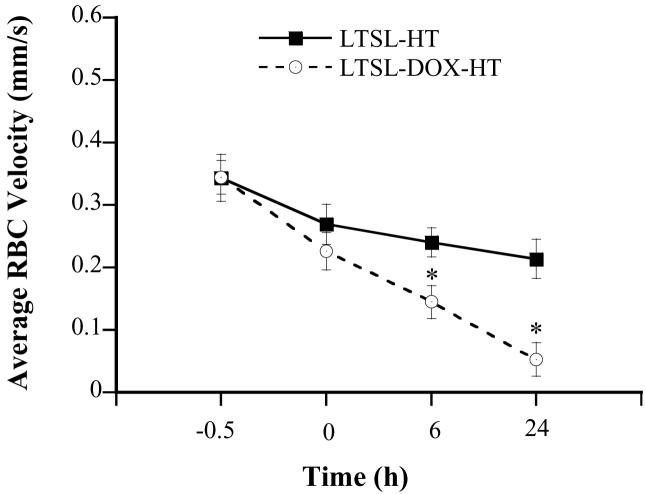
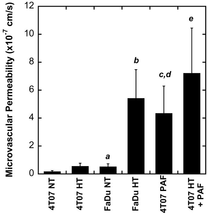
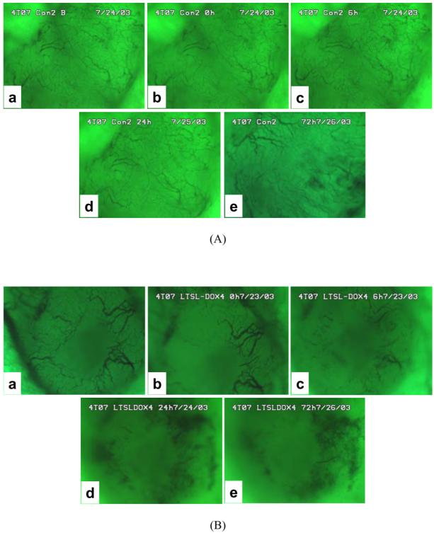
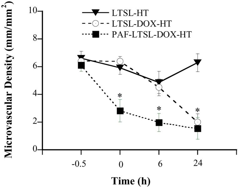
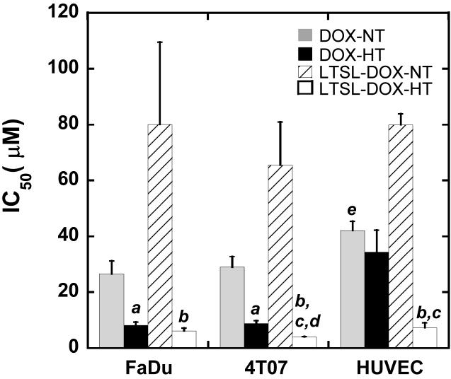
Previous data have demonstrated that doxorubicin (DOX) released from a lysolecithin-containing thermosensitive liposome (LTSL) can shut down blood flow in a human tumor xenograft (FaDu) in mice when the treatment is combined with hyperthermia (HT), suggesting that LTSL-DOX is a potential antivascular agent. To further understand mechanisms of the treatment, we investigated effects of LTSL-DOX (5 mg/kg body weight) plus HT (42 degrees C, 1 h) on microcirculation in another tumor (a murine mammary carcinoma, 4T07) implanted in mouse dorsal skin-fold chambers and dose responses of tumor (FaDu and 4T07) and endothelial cells to LTSL-DOX or free DOX with or without HT. We observed that LTSL-DOXHT could significantly reduce blood flow and microvascular density in 4T07 tumors. The antivascular efficacy of LTSLDOX- HT could be enhanced through increasing tumor microvascular permeability of liposomes by using platelet activating factor (PAF). We also observed that the dose responses of FaDu and 4T07 to DOX in vitro were similar to each other and could be enhanced by HT. Taken together, these data suggested that tumor microvascular permeability was more critical than the sensitivity of tumor cells to DOX in determining the antivascular efficacy of LTSL-DOX-HT treatment.
Figures
Similar articles
-
Efficacy of liposomes and hyperthermia in a human tumor xenograft model: importance of triggered drug release.Cancer Res. 2000 Dec 15;60(24):6950-7. Cancer Res. 2000. PMID: 11156395
-
Comparative effects of thermosensitive doxorubicin-containing liposomes and hyperthermia in human and murine tumours.Int J Hyperthermia. 2010;26(5):485-98. doi: 10.3109/02656731003789284. Int J Hyperthermia. 2010. PMID: 20597627 Free PMC article.
-
Targeting tumor microvessels using doxorubicin encapsulated in a novel thermosensitive liposome.Mol Cancer Ther. 2004 Oct;3(10):1311-7. Mol Cancer Ther. 2004. PMID: 15486198
-
Mild hyperthermia triggered doxorubicin release from optimized stealth thermosensitive liposomes improves intratumoral drug delivery and efficacy.J Control Release. 2013 Jun 10;168(2):142-50. doi: 10.1016/j.jconrel.2013.03.011. Epub 2013 Mar 21. J Control Release. 2013. PMID: 23524188
-
Focused Ultrasound Hyperthermia Mediated Drug Delivery Using Thermosensitive Liposomes and Visualized With in vivo Two-Photon Microscopy.Theranostics. 2017 Jul 7;7(10):2718-2731. doi: 10.7150/thno.19662. eCollection 2017. Theranostics. 2017. PMID: 28819458 Free PMC article.
Cited by
-
Mathematical spatio-temporal model of drug delivery from low temperature sensitive liposomes during radiofrequency tumour ablation.Int J Hyperthermia. 2010;26(5):499-513. doi: 10.3109/02656731003623590. Int J Hyperthermia. 2010. PMID: 20377363 Free PMC article.
-
Novel Nanotechnology-Based Approaches for Targeting HIV Reservoirs.Polymers (Basel). 2022 Jul 29;14(15):3090. doi: 10.3390/polym14153090. Polymers (Basel). 2022. PMID: 35956604 Free PMC article. Review.
-
Phase separation behavior of mixed lipid systems at neutral and low pH: coarse-grained simulations with DMD/LIME.Langmuir. 2015 Jan 27;31(3):1086-94. doi: 10.1021/la504082x. Epub 2015 Jan 15. Langmuir. 2015. PMID: 25549801 Free PMC article.
-
Guided Delivery of Polymer Therapeutics Using Plasmonic Photothermal Therapy.Nano Today. 2012 Jun 1;7(3):158-167. doi: 10.1016/j.nantod.2012.04.002. Epub 2012 May 24. Nano Today. 2012. PMID: 22737178 Free PMC article.
-
Hyperthermia and Temperature-Sensitive Nanomaterials for Spatiotemporal Drug Delivery to Solid Tumors.Pharmaceutics. 2020 Oct 22;12(11):1007. doi: 10.3390/pharmaceutics12111007. Pharmaceutics. 2020. PMID: 33105816 Free PMC article. Review.
References
-
- Ranson M, Howell A, Cheeseman S, Margison J. Liposomal drug delivery. Cancer Treat Rev. 1996;22(5):365–79. - PubMed
-
- Tardi PG, Boman NL, Cullis PR. Liposomal doxorubicin. J Drug Target. 1996;4(3):129–40. - PubMed
-
- Gregoriadis G. Liposome Technology. CRC press; Boca Raton: 1993.
-
- Lasic DD, Papahadjopoulos D. Liposomes revisited. Science. 1995;267(5202):1275–1276. - PubMed
Publication types
MeSH terms
Substances
Grants and funding
LinkOut - more resources
Full Text Sources