

doi: 10.1042/BJ20081237.
Arf family GTP loading is activated by, and generates, positive membrane curvature
Affiliations
- PMID: 18597672
- PMCID: PMC2518064
- DOI: 10.1042/BJ20081237
Item in Clipboard
Arf family GTP loading is activated by, and generates, positive membrane curvature
Biochem J.
.
Abstract
Small G-proteins belonging to the Arf (ADP-ribosylation factor) family serve as regulatory proteins for numerous cellular processes through GTP-dependent recruitment of effector molecules. In the present study we demonstrate that proteins in this family regulate, and are regulated by, membrane curvature. Arf1 and Arf6 were shown to load GTP in a membrane-curvature-dependent manner and stabilize, or further facilitate, changes in membrane curvature through the insertion of an amphipathic helix.
Figures
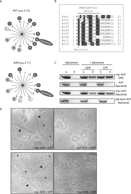
(A) Helical-wheel representation of amino acids (a.a.) 2–13 in Arf1 (top wheel) and 2–11 in Arf6 (bottom wheel). Hydrophobic and charged amino acids are highlighted by dark grey circles or light grey circles respectively. Myristoylation of the second residue (glycine) is depicted by the long oval containing the word ‘myristoyl’. (B) Sequence alignment of the N-terminal amino acids of Arf/Arl family members. Hydrophobic amino acids are highlighted in dark grey. (C) Liposome co-sedimentation assays. Liposomes generated from brain-derived lipids (Folch fraction I) were incubated with the indicated proteins and GTP or GDP before centrifugation and analysis of supernatants (S) and pellets (P) by SDS/PAGE and Coomassie Blue staining. A control experiment where protein was incubated in the absence of liposomes is shown. The proteins used were myristoylated Arf1 purified from Sf9 insect cells [myr-Arf1 (Sf9)], Arf1 purified from bacteria [Arf1 (bacteria)], myristoylated Arf1 purified from bacteria co-expressing N-myristoyltransferase [myr-Arf1 (bacteria)] and N-terminally truncated Arf1 purified from bacteria [ΔN-term Arf1 (bacteria)]. (D) Electron micrographs of negatively stained 800 nm liposomes incubated in the presence of equal concentrations of proteins (10 μM) and nucleotides (100 μM) as indicated. Arrowheads indicate tubules that are approx. 45 nm in diameter and asterisks indicate tubules that are 13–17 nm wide. Scale bars represent 100 nm. Abbreviation: myr, myristoylated.
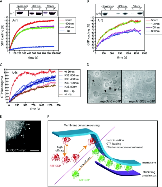
(A)–(C) Indicated proteins were incubated together with or without liposomes (−lip) of the indicated diameters and GTP. GTP loading was analysed in real time by measuring tryptophan fluorescence (see the Experimental section). Fluorescence is presented as a percentage of the value at 1000 s for 50 nm liposomes and Arf1 and Arf6 respectively. Insets in (A) and (B) shows liposome binding assays of Arf1 (A) and Arf6 (B) in the absence or presence of liposomes of different diameters. S, supernatant; P, pellet. Results shown in (A)–(C) are representative of three independent experiments with the same set of proteins and lipids. (D) Electron micrographs of negatively stained liposomes incubated together with GTP-loaded Arf6 (left) and Arf6 K3E (right). The scale bar represents 100 nm. The asterisks and arrowheads have the same meaning as in Figure 1. myr, myristoylated. (E) Epifluorescent micrograph of a HeLa cell expressing a myc-tagged constitutively active Arf6 mutant (Arf6 Q67L-myc). Arrowheads indicate tubular localization. The scale bar represents 5 μm. (F) Schematic model of the GTP-dependent role of Arfs as curvature sensors at the rim of a budding vesicle. Arfs loaded with GDP (ARF–GDP) display a transient membrane binding while Arfs bound to GTP (ARF–GTP) stay membrane-associated within the diffusion barrier generated by positive membrane curvature.
Comment in
-
Arfs and membrane lipids: sensing, generating and responding to membrane curvature.Biochem J. 2008 Sep 1;414(2):e1-2. doi: 10.1042/BJ20081438. Biochem J. 2008. PMID: 18687059
Similar articles
-
Arfs and membrane lipids: sensing, generating and responding to membrane curvature.Biochem J. 2008 Sep 1;414(2):e1-2. doi: 10.1042/BJ20081438. Biochem J. 2008. PMID: 18687059
-
EFA6 controls Arf1 and Arf6 activation through a negative feedback loop.Proc Natl Acad Sci U S A. 2014 Aug 26;111(34):12378-83. doi: 10.1073/pnas.1409832111. Epub 2014 Aug 11. Proc Natl Acad Sci U S A. 2014. PMID: 25114232 Free PMC article.
-
The structural basis of Arfaptin-mediated cross-talk between Rac and Arf signalling pathways.Nature. 2001 May 10;411(6834):215-9. doi: 10.1038/35075620. Nature. 2001. PMID: 11346801
-
Membrane curvature and the control of GTP hydrolysis in Arf1 during COPI vesicle formation.Biochem Soc Trans. 2005 Aug;33(Pt 4):619-22. doi: 10.1042/BST0330619. Biochem Soc Trans. 2005. PMID: 16042557 Review.
-
Localization and function of Arf family GTPases.Biochem Soc Trans. 2005 Aug;33(Pt 4):639-42. doi: 10.1042/BST0330639. Biochem Soc Trans. 2005. PMID: 16042562 Review.
Cited by
-
The late stage of COPI vesicle fission requires shorter forms of phosphatidic acid and diacylglycerol.Nat Commun. 2019 Jul 30;10(1):3409. doi: 10.1038/s41467-019-11324-4. Nat Commun. 2019. PMID: 31363100 Free PMC article.
-
Biophysical mechanism for ras-nanocluster formation and signaling in plasma membrane.PLoS One. 2009 Jul 9;4(7):e6148. doi: 10.1371/journal.pone.0006148. PLoS One. 2009. PMID: 19587789 Free PMC article.
-
Structural basis for membrane binding and remodeling by the exomer secretory vesicle cargo adaptor.Dev Cell. 2014 Sep 8;30(5):610-24. doi: 10.1016/j.devcel.2014.07.014. Dev Cell. 2014. PMID: 25203211 Free PMC article.
-
Membrane Curvature Sensing by Amphipathic Helices Is Modulated by the Surrounding Protein Backbone.PLoS One. 2015 Sep 14;10(9):e0137965. doi: 10.1371/journal.pone.0137965. eCollection 2015. PLoS One. 2015. PMID: 26366573 Free PMC article.
-
Structural elucidation of how ARF small GTPases induce membrane tubulation for vesicle fission.bioRxiv [Preprint]. 2023 Dec 20:2023.12.19.572083. doi: 10.1101/2023.12.19.572083. bioRxiv. 2023. PMID: 38187566 Free PMC article. Preprint.
References
-
- Gillingham A. K., Munro S. The small G proteins of the Arf family and their regulators. Annu. Rev. Cell Dev. Biol. 2007;23:579–611. - PubMed
-
- D'souza-Schorey C., Chavrier P. ARF proteins: roles in membrane traffic and beyond. Nat. Rev. Mol. Cell Biol. 2006;7:347–358. - PubMed
-
- Amor J. C., Harrison D. H., Kahn R. A., Ringe D. Structure of the human ADP-ribosylation factor 1 complexed with GDP. Nature. 1994;372:704–708. - PubMed
-
- Menetrey J., Macia E., Pasqualato S., Franco M., Cherfils J. Structure of Arf6–GDP suggests a basis for guanine nucleotide exchange factors specificity. Nat. Struct. Biol. 2000;7:466–469. - PubMed
Publication types
MeSH terms
Substances
Grants and funding
LinkOut - more resources
Full Text Sources