. 2008 Jul 29;47(30):7937-46.
doi: 10.1021/bi800723n. Epub 2008 Jul 3.

Distinct flippases translocate glycerophospholipids and oligosaccharide diphosphate dolichols across the endoplasmic reticulum

Affiliations

Distinct flippases translocate glycerophospholipids and oligosaccharide diphosphate dolichols across the endoplasmic reticulum

Sumana Sanyal et al. Biochemistry. .

Abstract

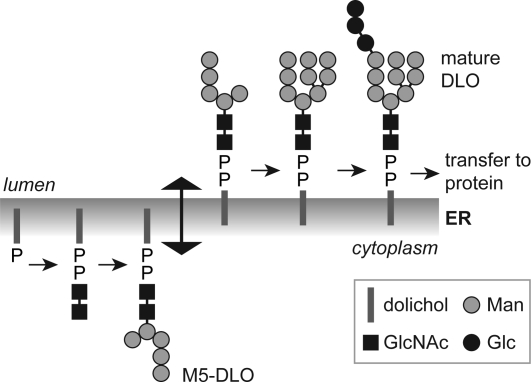
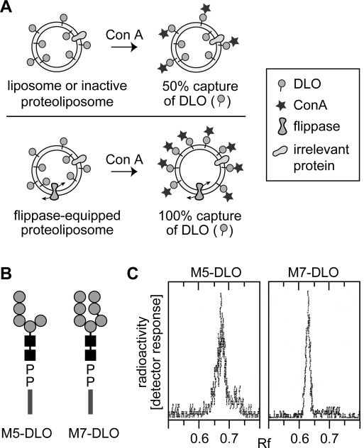
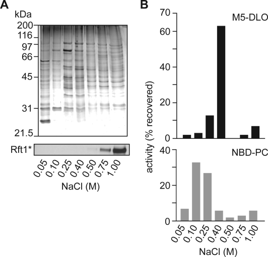
Transbilayer movement, or flip-flop, of lipids across the endoplasmic reticulum (ER) is required for membrane biogenesis, protein glycosylation, and GPI anchoring. Specific ER membrane proteins, flippases, are proposed to facilitate lipid flip-flop, but no ER flippase has been biochemically identified. The glycolipid Glc 3Man 9GlcNAc 2-PP-dolichol is the oligosaccharide donor for protein N-glycosylation reactions in the ER lumen. Synthesis of Glc 3Man 9GlcNAc 2-PP-dolichol is initiated on the cytoplasmic side of the ER and completed on the lumenal side, requiring flipping of the intermediate Man 5GlcNAc 2-PP-dolichol (M5-DLO) across the ER. Here we report the reconstitution of M5-DLO flipping in proteoliposomes generated from Triton X-100-extracted Saccharomyces cerevisiae microsomal proteins. Flipping was assayed by using the lectin Concanavalin A to capture M5-DLOs that had been translocated from the inner to the outer leaflet of the vesicles. M5-DLO flipping in the reconstituted system was ATP-independent and trypsin-sensitive and required a membrane protein(s) that sedimented at approximately 4 S. Man 7GlcNAc 2-PP-dolichol, a higher-order lipid intermediate, was flipped >10-fold more slowly than M5-DLO at 25 degrees C. Chromatography on Cibacron Blue dye resin enriched M5-DLO flippase activity approximately 5-fold and resolved it from both the ER glycerophospholipid flippase activity and the genetically identified flippase candidate Rft1 [Helenius, J., et al. (2002) Nature 415, 447-450]. The latter result indicates that Rft1 is not the M5-DLO flippase. Our data (i) demonstrate that the ER has at least two distinct flippase proteins, each specifically capable of translocating a class of phospholipid, and (ii) provide, for the first time, a biochemical means of identifying the M5-DLO flippase.


Figures

Figure 1
Figure 1
Topology model for the dolichol pathway. The assembly of the dolichol-linked oligosaccharide (DLO) precursor of protein N-glycans is initiated on the cytoplasmic face of the ER when GlcNAc-P is transferred from UDP-GlcNAc to dolichol phosphate. Synthesis continues with the addition of another GlcNAc residue (from UDP-GlcNAc) to generate GlcNAc2-PP-dolichol and five mannose residues (from GDP-mannose) to generate the branched structure Man5GlcNAc2-PP-dolichol (M5-DLO). M5-DLO is flipped to the lumenal face of the ER where it is extended by four mannose residues (from dolichol-P-mannose) and three glucose residues (from dolichol-P-glucose) to yield mature DLO (Glc3Man9GlcNAc2-PP-dolichol). This lipid is the oligosaccharide donor for the protein N-glycosylation reaction catalyzed by oligosaccharyltransferase (OST) in the ER lumen. Flipping of M5-DLO across the ER is depicted as bidirectional, consistent with data presented in this paper.
Figure 2
Figure 2
Strategy for assaying DLO translocation in reconstituted vesicles. (A) Assay for assessing DLO flipping in reconstituted vesicles. Unilamellar vesicles with [3H]DLO symmetrically distributed across the membrane are incubated with Con A. Con A captures DLO species that are initially located in the outer leaflet, as well as those that gain access to the outer leaflet after being translocated from the inner leaflet through the action of a flippase. After Con A binding is allowed to go to completion, the sample is extracted with organic solvent. DLOs that are bound to Con A precipitate with the protein, while free DLOs are extracted. In liposomes or inactive proteoliposomes (vesicles containing membrane proteins but not a flippase), 50% of the [3H]DLOs are expected to be captured by Con A, corresponding to the pool of DLO in the outer leaflet. [3H]DLOs in the inner leaflet of these vesicles cannot access Con A and are extracted by the solvent (top panel). For proteoliposomes containing a flippase (bottom panel), Con A binds all [3H]DLOs since those originally in the inner leaflet are flipped to the outer leaflet and captured by the lectin; thus, ∼100% of the DLOs are expected to be precipitated with Con A in this situation. In mixtures of vesicles in which some possess a flippase while others do not, the percent of [3H]DLO captured by Con A is predicted to be intermediate between 50 and 100%, reflecting the proportion of flippase-containing vesicles in the population. (B) Structures of M5-DLO and M7-DLO. The symbols used for dolichol, mannose, and GlcNAc are as described in the legend of Figure 1. The chemical structure of the oligosaccharide moiety of M5-DLO is Manα1−2Manα1−2Manα1−3(Manα1−6)Manβ1−4GlcNAcβ1−4GlcNAc; the oligosaccharide moiety of M7-DLO is Manα1−2Manα1−2Manα1−3(Manα1−2Manα1−3Manα1−6)Manβ1−4GlcNAcβ1−4GlcNAc. (C) Thin layer chromatography of [3H]M5-DLO and [3H]M7-DLO. Radiolabeled M5-DLO and M7-DLO were prepared from [3H]mannose-labeled yeast as described in . An aliquot of each preparation was analyzed by thin layer chromatography; chromatograms were visualized using a Berthold LB2842 radioactivity scanner. Each chromatogram contained a single peak of radioactivity; the relevant section of the chromatograms is shown.
Figure 3
Figure 3
M5-DLO flipping in proteoliposomes reconstituted from TE. (A) Characterization of TE. Microsomes from strain YCF40 were extracted with Triton X-100 as described in . Soluble (TE) and insoluble (Insol) fractions were separated by ultracentrifugation; sample equivalents were analyzed by SDS−PAGE and immunoblotting using antibodies against organelle marker proteins: ER, Sec61, Dpm1, and Rft1*; vacuole, Vph1; plasma membrane, Gas1; and Golgi/endosomes, Pep12. The immunoblotting profile indicates that ER membrane proteins are selectively extracted into the TE. (B) Protein reconstitution verified by flotation analysis. Proteoliposomes (prepared using TE from YCF40 cells, as well as trace amounts of [3H]phosphatidylcholine) were permeabilized, made up to 30% (w/w) sucrose, and layered under a sucrose step gradient. The gradient was centrifuged (SW41 rotor, 28000 rpm, 1.5 h), and fractions were collected as indicated and analyzed for protein and phospholipid content, as well as for Rft1*; the amount of phospholipid was determined by scintillation counting to detect [3H]phosphatidylcholine. The data show that the vesicles float to the 20%−10% and 10%−5% (w/w) sucrose interfaces, with the denser vesicles containing a higher protein:phospholipid ratio as well as the majority of Rft1*. (C) Cryo-electron micrographs of reconstituted vesicles. The scale bar is 100 nm. (D) M5-DLO flipping in proteoliposomes reconstituted from TE. Different amounts of TE were used during reconstitution to generate different proteoliposome samples with protein:phospholipid ratios ranging from 0 to 50 mg/mmol. The samples were incubated on ice with Con A for 30 min and processed as described in to determine the fraction of [3H]M5-DLO captured by Con A. The graph (representative of more than five experiments) shows a monoexponential increase in the percent of [3H]M5-DLO captured, from a value of ∼50% for protein-free vesicles to ∼80% for vesicles with a PPR of ∼50 mg/mmol. Identical results were obtained when the assay was carried out at 25 °C. The predicted maximum of 100% [3H]M5-DLO bound is not experimentally observed. The initial slope of the graph suggests that M5-DLO flippase represents ∼1% (w/w) of proteins in the TE (see the text for details of the calculation). (E) M5-DLO flipping is trypsin-sensitive. TE was treated with trypsin (30 μg/mL) for different amounts of time (from 0 to 90 min, as indicated) at 30 °C. Reactions were stopped with 30 μg/mL soybean trypsin inhibitor, and mixtures were reconstituted and assayed for [3H]M5-DLO flipping (◼). Samples incubated with trypsin in the presence of trypsin inhibitor were processed in parallel (◻). The percent of [3H]M5-DLO captured in the control sample was ∼67%, corresponding to a PPR of ∼15 mg/mmol. The data (mean and range) are derived from two independent experiments.
Figure 4
Figure 4
M7-DLO is flipped more slowly than M5-DLO. (A) Comparison of M7-DLO and M5-DLO flipping at 25 °C. Proteoliposomes with a PPR of ∼20 mg/mmol were reconstituted with [3H]M5-DLO (◻) or [3H]M7-DLO (◼) and assayed for DLO flipping at 25 °C. The kinetics of flipping are shown. The earliest time point analyzed was 5 min, corresponding to the time taken for completion of Con A binding to detergent-solubilized DLOs at 25 °C. The data (mean and range) are derived from two independent experiments. (B) Identical protein dependence of M7-DLO and M5-DLO flipping. M7-DLO and M5-DLO flipping were analyzed in vesicles prepared with different PPRs. Assays were carried out for 40 min at 25 °C.
Figure 5
Figure 5
Glycerophospholipid flippase is more abundant than M5-DLO flippase. Resolution of the two flippase activities by velocity gradient sedimentation. (A) Dose−response plots indicate that glycerophospholipid flippase is ∼3-fold more abundant than M5-DLO flippase. Glycerophospholipid flippase activity was assayed using NBD-PC as a reporter (○; the line represents a monoexponential fit); M5-DLO flippase activity was assayed in the same reconstituted samples (only the fit to the data is shown; data points are the same as those shown in Figure 3D). (B) TE was analyzed by velocity gradient sedimentation as described in . Fractions were collected from the top. The figure shows a silver-stained SDS−PAGE gel of fraction equivalents. (C) Resolution of NBD-PC and M5-DLO flippase activities. Gradient fractions were reconstituted and assayed for NBD-PC and M5-DLO flippase activities. The percent of activity (relative to the load) that was recovered in each fraction is indicated. Sedimentation standards analyzed in a parallel gradient are indicated at the top. Protein recovery in the fractions relative to the load was ∼80%; the enrichment of activity in the peak fractions was ∼4-fold in each case. The data are representative of five independent experiments.
Figure 6
Figure 6
Resolution of glycerophospholipid flippase activity, M5-DLO flippase activity, and the flippase candidate Rft1* by Cibacron dye resin chromatography. (A) Fractionation of TE on Cibacron Blue dye resin. TE from Rft1*-containing YCF40 cells was loaded onto Cibacron blue dye resin, and the bound material was eluted in six sequential steps with salt. The 50 mM step corresponds to unbound (flow-through) material. The top panel shows a Coomassie-stained SDS−PAGE gel of the eluted fractions. The bottom panel shows the same fractions taken for immunoblotting to locate the elution behavior of Rft1*. (B) Resolution of NBD-PC and M5-DLO flippase activities. Fractions eluted from the Cibacron blue dye resin were reconstituted and assayed for M5-DLO (top) and NBD-PC (bottom) flippase activities. The percent of activity recovered (relative to the load) is indicated. Less than 5% of activity was recovered in the flow-through fraction (50 mM NaCl). Protein recovery in the fractions relative to the load was ∼85%; the enrichment of M5-DLO and NBD-PC flippase activities was 4.6- and 6.6-fold, respectively. The data are representative of three independent experiments.

Similar articles

Cited by

References

    1. Hanover J. A.; Lennarz W. J. (1980) N-Linked glycoprotein assembly. Evidence that oligosaccharide attachment occurs within the lumen of the endoplasmic reticulum. J. Biol. Chem. 255, 3600–3604. - PubMed
    1. Snider M. D.; Robbins P. W. (1982) Transmembrane organization of protein glycosylation: Mature oligosaccharide-lipid is located on the luminal side of microsomes from Chinese hamster ovary cells. J. Biol. Chem. 257, 6796–6801. - PubMed
    1. Hirschberg C. B.; Snider M. D. (1987) Topography of glycosylation in the rough endoplasmic reticulum and Golgi apparatus. Annu. Rev. Biochem. 56, 63–87. - PubMed
    1. Lennarz W. J. (1987) Protein glycosylation in the endoplasmic reticulum: Current topological issues. Biochemistry 26, 7205–7210. - PubMed
    1. Helenius J.; Aebi M. (2002) Transmembrane movement of dolichol-linked carbohydrates during N-glycoprotein biosynthesis in the endoplasmic reticulum. Semin. Cell Dev. Biol. 13, 171–178. - PubMed

Publication types

MeSH terms