. 2008 Sep 5;283(36):25014-26.
doi: 10.1074/jbc.M802140200. Epub 2008 Jul 2.

Palmitoylation and membrane interactions of the neuroprotective chaperone cysteine-string protein

Affiliations

Palmitoylation and membrane interactions of the neuroprotective chaperone cysteine-string protein

Jennifer Greaves et al. J Biol Chem. .

Abstract

Cysteine-string protein (CSP) is an extensively palmitoylated DnaJ-family chaperone, which exerts an important neuroprotective function. Palmitoylation is required for the intracellular sorting and function of CSP, and thus it is important to understand how this essential modification of CSP is regulated. Recent work identified 23 putative palmitoyl transferases containing a conserved DHHC domain in mammalian cells, and here we show that palmitoylation of CSP is enhanced specifically by co-expression of the Golgi-localized palmitoyl transferases DHHC3, DHHC7, DHHC15, or DHHC17. Indeed, these DHHC proteins promote stable membrane attachment of CSP, which is otherwise cytosolic. An inverse correlation was identified between membrane affinity of unpalmitoylated CSP mutants and subsequent palmitoylation: mutants with an increased membrane affinity localize to the endoplasmic reticulum (ER) and are physically separated from the Golgi-localized DHHC proteins. Palmitoylation of an ER-localized mutant could be rescued by brefeldin A treatment, which promotes the mixing of ER and Golgi membranes. Interestingly though, the palmitoylated mutant remained at the ER following brefeldin A washout and did not traffic to more distal membrane compartments. We propose that CSP has a weak membrane affinity that allows the protein to locate its partner Golgi-localized DHHC proteins directly by membrane "sampling." Mutations that enhance membrane association prevent sampling and lead to accumulation of CSP on cellular membranes such as the ER. The coupling of CSP palmitoylation to Golgi membranes may thus be an important requirement for subsequent sorting.


Figures

FIGURE 1.
FIGURE 1.
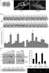
DHHC proteins mediate CSP palmitoylation. A, left panel, PC12 cells transfected with EGFP-CSP were incubated in [3H]palmitate for 4 h. Cells were lysed and EGFP-CSP recovered by immunoprecipitation. Precipitated samples were resolved by SDS-PAGE and transferred to nitrocellulose for immunoblotting analysis using monoclonal anti-GFP (blot) or processed for fluorographic detection of incorporated radiolabel (3H). Right panel, a lysate from HEK293 cells transfected with EGFP-CSP probed by immunoblotting with anti-GFP. B, HEK293 cells were transfected with EGFP-CSP together with empty pEFBOS-HA (-) or with each of the 23 DHHC constructs (numbered 1-23). Lysates were prepared from transfected cells ∼20 h post-transfection and resolved by SDS-PAGE and transferred to nitrocellulose for immunoblotting analysis using anti-GFP. C, EGFP-CSP plasmid together with the indicated amounts of DHHC3 vector were transfected into HEK293 cells. Note that the total amount of plasmid in each sample was maintained constant by including empty pEFBOS-HA vector in appropriate amounts. Lysates were prepared from the transfected cells, resolved by SDS-PAGE, and transferred to nitrocellulose for immunoblotting analysis using anti-GFP or anti-HA. D, lysates were prepared from HEK293 cells transfected with each of the 23 DHHC proteins and probed by immunoblotting with anti-HA. The position of molecular weight standards is shown on the left side of all panels; asterisks denote palmitoylated CSP and arrowheads indicate unpalmitoylated CSP.
FIGURE 2.
FIGURE 2.
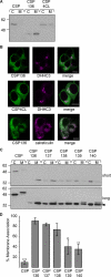
Analysis of HA-DHHCs and giantin localization in HEK293 cells. HEK cells plated on coverslips were transfected with HA-tagged DHHC constructs and ∼20 h later were fixed, permeabilized, and stained with Alexa Fluor 488-conjuagted anti-giantin (1:200) and rhodamine-conjugated anti-HA (1:100). The coverslips were then mounted in Mowiol and imaged using a Zeiss LSM510 Axiovert laser scanning confocal microscope. For clarity, a rough outline of the cell membranes (solid line) and the nuclei (dashed line, n) is shown for the merged images. Scale bars represent 10 μm.
FIGURE 3.
FIGURE 3.
DHHC proteins regulate CSP membrane binding. A, PC12 and HEK293 cells were transfected with EGFP-CSP and fractionated into cytosol (C) and membrane (M) fractions. Equal volumes of the recovered samples were resolved by SDS-PAGE and transferred to nitrocellulose for immunoblotting analysis using anti-GFP. B, confocal images of EGFP-CSP distribution in PC12 cells and HEK293 cells. Scale bars represent 10 μm. C, HEK293 cells were transfected with EGFP-CSP in the absence (-) or presence of each of the 23 HA-tagged DHHC constructs. The cells were processed as described for A. D, the percent membrane association ± S.E. of EGFP-CSP transfected together with each of the DHHC proteins (indicated by 1-23) was determined by densitometry, and the averaged data are presented (n = 5). *, p value of <0.03; **, p value of <0.004 compared with CSP in the absence of DHHC co-expression. E, HEK293 cells were co-transfected with EGFP-CSP and wild-type HA-tagged DHHC3/DHHC7 or with the inactive DHHC mutants, DHHC3 (C157S)/DHHC7 (C160S). Cells were fractionated into cytosol (C) and membrane (M) fractions, and equal volumes were resolved by SDS-PAGE, transferred to nitrocellulose, and probed with antibodies against GFP and HA. Representative blots are shown in the left panel. The right panel shows the percent membrane association ± S.E. of EGFP-CSP under the different transfection conditions determined by quantification of immunoblots (n = 6). *, p value of <0.03, **, p value of <0.003, and ***, p value of <0.0006 compared with EGFP-CSP in the absence of DHHC expression. F, HEK293 cells were co-transfected with EGFP-CSP and DHHC3, DHHC7, DHHC15, or DHHC17 and fractionated into cytosol (C) and membrane (M) fractions. The remaining insoluble (I) cell material was solubilized in SDS-dissociation buffer. Equal volumes of the recovered fractions were resolved by SDS-PAGE and transferred to nitrocellulose for immunoblotting analysis using anti-GFP. G, primers recognizing DHHC3, DHHC7, DHHC15, and DHHC17 were used in PCR reactions that contained purified plasmid DNA (3, DHHC3 template; 7, DHHC7 template; 15, DHHC15 template; 17, DHHC17 template), no DNA (-) or cDNA (C). Position of selected standards (in kilobase pairs) is shown on the left. The predicted sizes of amplification products were: DHHC3, 903 bp; DHHC7, 928 bp; DHHC15, 716 bp; and DHHC17, 1164 bp. The position of molecular weight standards is shown on the left side of all panels; asterisks denote palmitoylated CSP, and arrowheads indicate unpalmitoylated CSP.
FIGURE 4.
FIGURE 4.
Membrane binding and palmitoylation of CSP mutants. A, HEK293 cells transfected with EGFP-CSP, CSP136, or CSP4CL were fractionated into cytosol (C) and membrane (M) fractions ∼20 h post-transfection. Equal volumes of the recovered fractions were resolved by SDS-PAGE and analyzed by immunoblotting using anti-GFP. B, HEK293 cells were transfected with EGFP-CSP136 and HA-DHHC3 (top panel), EGFP-CSP4CL and HA-DHHC3 (middle panel), or with EGFP-CSP136 alone (bottom panel). Cells were fixed, permeabilized, and incubated with rhodamine-conjugated anti-HA (1:100) (top and middle panels) or with anti-calreticulin antibody (1:50) followed by Alexa Fluor 543-conjugated rabbit secondary antibody (1:200, bottom panel). Coverslips were mounted in Mowiol and examined using a Zeiss LSM510 Axiovert laser scanning confocal microscope. Scale bars represent 10 μm. C, HEK293 cells were transfected with wild-type EGFP-CSP and the indicated C-terminal truncation mutants. Cells were then fractionated into cytosol (C) and membrane (M) fractions, which were examined by immunoblotting with anti-GFP. Shown is a short and long exposure of the same blot. D, averaged data ± S.E. for percent membrane binding of the mutants analyzed in panel C (n = 5). *, p value of <0.02; **, p value of <0.002; and ***, p value of <0.000008 compared with CSP136. For all immunoblots shown, the positions of molecular weight standards are shown on the left; arrowheads indicate unpalmitoylated CSP, whereas asterisks highlight palmitoylated CSP.
FIGURE 5.
FIGURE 5.
Effect of BFA on CSP palmitoylation and membrane association in HEK293 cells. A, HEK293 cells were transfected with EGFP-CSP in the presence/absence of DHHC3 or DHHC7. 4 h after transfection, fresh medium was added either with or without 30 μg/ml BFA and, after an additional 4 h, the cells were fractionated into cytosol (C) and membrane (M) fractions. Equal volumes of the fractions were probed with an antibody against GFP. Positions of molecular weight standards are shown on the left. Arrowheads indicate unpalmitoylated CSP, whereas asterisks highlight palmitoylated CSP. B, averaged data ± S.E. for percent membrane binding of EGFP-CSP in the absence or presence of DHHC3/7 co-expression and with or without BFA treatment (n = 3). *, p value of <0.05. C, cells were transfected with HA-DHHC3 and ∼20 h later were incubated in the absence (left panel) or presence (right panel) of 30 μg/ml BFA/10 μg/ml cycloheximide for 90 min. The cells were then washed, fixed, permeabilized, stained with Alexa Fluor 488-conjugated anti-HA, and examined usinga Zeiss LSM510 Axiovert laser scanning confocal microscope. For clarity, a rough outline of the cell membranes (solid line) is shown for untreated cells (-BFA). Scale bars represent 10 μm.
FIGURE 6.
FIGURE 6.
BFA does not affect membrane binding or palmitoylation of EGFP-CSP in PC12 cells. A, PC12 cells transfected with EGFP-CSP or empty vector (EGFP) were incubated with 35S-labeled cysteine/methionine for 15 min, washed, and chased for various times as indicated ranging from 0 to 60 min. The labeled cells were fractionated into cytosol (C) and membrane (M) fractions, from which GFP-tagged proteins were recovered by immunoprecipitation and subsequently analyzed by immunoblotting with anti-GFP (GFP). Duplicate gels were developed using autoradiography ([35S]). Note that the 35S-labeled band detected in the EGFP cytosol fraction is a nonspecific band that migrates more slowly than cytosolic EGFP-CSP. B, cells were transfected with EGFP-SNAP25B. Approximately 40 h post-transfection, the cells were incubated in the absence or presence of 30 μg/ml BFA for 15 min, and then incubated in 35S-labeled cysteine/methionine for 15 min, washed, and chased for a further 60 min (for BFA samples, BFA was present throughout the pulse-chase period). The labeled cells were fractionated into cytosol (C) and membrane (M) fractions, from which EGFP-SNAP25B was recovered by immunoprecipitation and subsequently analyzed by immunoblotting with anti-GFP (GFP). Duplicate gels were developed using autoradiography ([35S]). The percent membrane binding ± S.E. of EGFP-SNAP25 in the presence and absence of BFA was determined by densitometry of autoradiographs. *, p value of <0.005 compared with EGFP-SNAP25 in the absence of BFA. C, cells transfected with EGFP-CSP were treated as described for SNAP25. D, the percent membrane binding ± S.E. of EGFP-CSP in the presence and absence of BFA was determined by densitometry of autoradiographs (left panel). The percent palmitoylation of EGFP-CSP was determined as a percentage of the total membrane-bound pool (right panel). Arrowheads indicate unpalmitoylated CSP, whereas asterisks denote palmitoylated CSP. The positions of molecular weight standards are shown on the left of all panels.
FIGURE 7.
FIGURE 7.
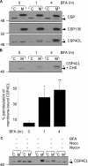
Effect of BFA on palmitoylation of CSP proteins in PC12 cells. A, PC12 cells transfected with EGFP-CSP, CSP136, or CSP4CL were treated with 30 μg/ml BFA for 1 or 4 h or left untreated (0). The cells were then fractionated into cytosol (C) and membrane (M) fractions, and equal volumes of each fraction were separated by SDS-PAGE and transferred to nitrocellulose for immunoblotting analysis using anti-GFP. B, as in panel A except shown only for EGFP-CSP4CL, which was treated with or without BFA in the presence of 10 μg/ml cycloheximide (CHX). The top panel shows a representative immunoblot, whereas the bottom panel shows averaged data ± S.E. for percent palmitoylation of membrane-bound CSP4CL (n = 5). *, p value of <0.02 and **, p < 0.003 compared with percent palmitoylation in the absence of BFA treatment. C, cells were incubated for 4 h with or without 30 μg/ml BFA in the presence or absence of 10 μg/ml nocodazole as indicated. Noco+ indicates where nocodazole was added 2 h before the addition of BFA and maintained throughout BFA treatment. Cycloheximide (10 μg/ml) was present in all samples. For all immunoblots shown, the positions of molecular weight standards are shown on the left, and unpalmitoylated and palmitoylated CSP bands are indicated by arrowheads and asterisks, respectively.
FIGURE 8.
FIGURE 8.
Effect of BFA on palmitoylation of CSP proteins in HEK293 cells. Cells were transfected with EGFP-CSP136 or EGFP-CSP4CL in the presence or absence of HA-DHHC3/7. 4 h post-transfections, the cells were incubated in the absence or presence of 30 μg/ml BFA for a further 4 h. The cells were then fractionated into cytosol (C) and membrane (M) fractions, equal volumes of which were resolved by SDS-PAGE and probed by immunoblotting with anti-GFP. Panel A shows a representative immunoblot, whereas panel B shows averaged data for percent palmitoylation ± S.E. of membrane-bound CSP4CL (without co-expression of DHHCs) in the absence or presence of BFA treatment (n = 7). *, p value of <0.0000005. Positions of molecular weight standards are shown on the left; the arrowhead denotes unpalmitoylated CSP, whereas the asterisk highlights palmitoylated CSP.
FIGURE 9.
FIGURE 9.
Intracellular localization of EGFP-CSP4CL following BFA treatment and washout. PC12 cells transfected with EGFP-CSP4CL or DHHC17-EGFP were either untreated (control), incubated in 30 μg/ml BFA and 10 μg/ml cycloheximide for 2 h(+BFA), or BFA/cycloheximide-treated and then washed and incubated in the presence of cycloheximide for 4 h (BFA washout). Cells were examined using a Zeiss LSM510 Axiovert laser scanning confocal microscope. For clarity, a rough outline of the cell membrane (solid line) and nuclei (dashed line, n) is shown for DHHC17-EGFP-expressing cells that were untreated or subjected to BFA washout. Scale bars represent 10 μm. B, DHHC17-EGFP-expressing cells under all treatments were scored for a Golgi localization or an ER (dispersed) localization. The total number of cells counted was 61 for the control condition, 50 for BFA treatment, and 74 for BFA washout.
FIGURE 10.
FIGURE 10.
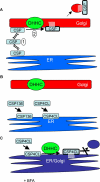
Membrane binding and palmitoylation of CSP. A, CSP utilizes a weak membrane affinity to sample intracellular membranes (1). Upon binding to Golgi membranes, CSP is recognized and palmitoylated by its partner DHHC proteins (2). Palmitoylation leads to stable membrane binding of CSP and may facilitate forward transport. B, the enhanced membrane affinity of CSP136 and CSP4CL leads to tight binding to the most abundant cellular membranes, such as the ER, and physical separation from Golgi-localized DHHC proteins. C, BFA treatment induces the fusion of ER and Golgi membranes and puts CSP4CL and DHHC proteins on the same membrane compartment. This membrane mixing allows palmitoylation of CSP4CL but is not sufficient to allow transport out of the ER.

Similar articles

Cited by

References

    1. Resh, M. D. (2006) Nat. Chem. Biol. 2 584-590 - PubMed
    1. Linder, M. E., and Deschenes, R. J. (2007) Nat. Rev. Mol. Cell Biol. 8 74-84 - PubMed
    1. Greaves, J., and Chamberlain, L. H. (2007) J. Cell Biol. 176 249-254 - PMC - PubMed
    1. Nadolski, M. J., and Linder, M. E. (2007) FEBS J. 274 5202-5210 - PubMed
    1. Hayashi, T., Rumbaugh, G., and Huganir, R. L. (2005) Neuron 47 709-723 - PubMed

Publication types

MeSH terms