. 2008 Jun 25;28(26):6691-702.
doi: 10.1523/JNEUROSCI.1701-08.2008.

Endocytosis of prion protein is required for ERK1/2 signaling induced by stress-inducible protein 1

Affiliations

Endocytosis of prion protein is required for ERK1/2 signaling induced by stress-inducible protein 1

Fabiana A Caetano et al. J Neurosci. .

Abstract

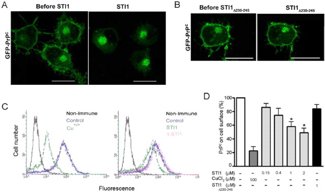
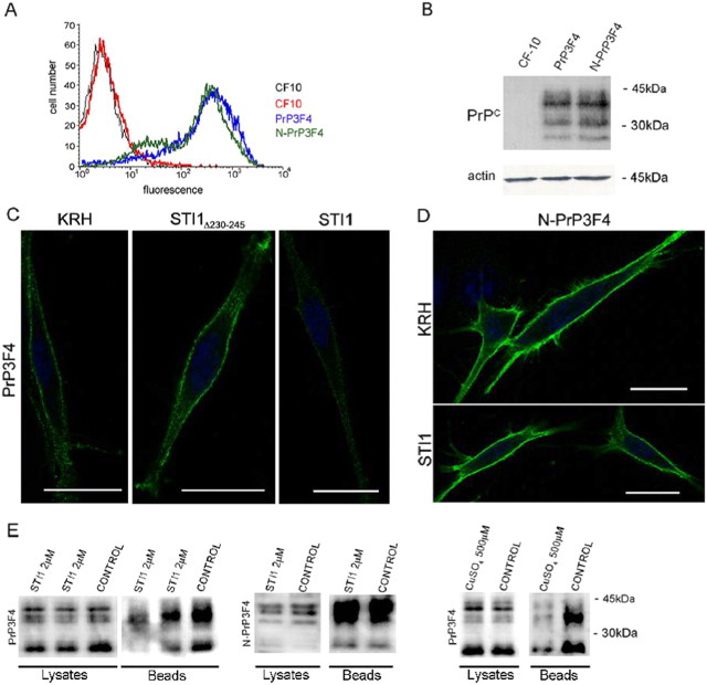
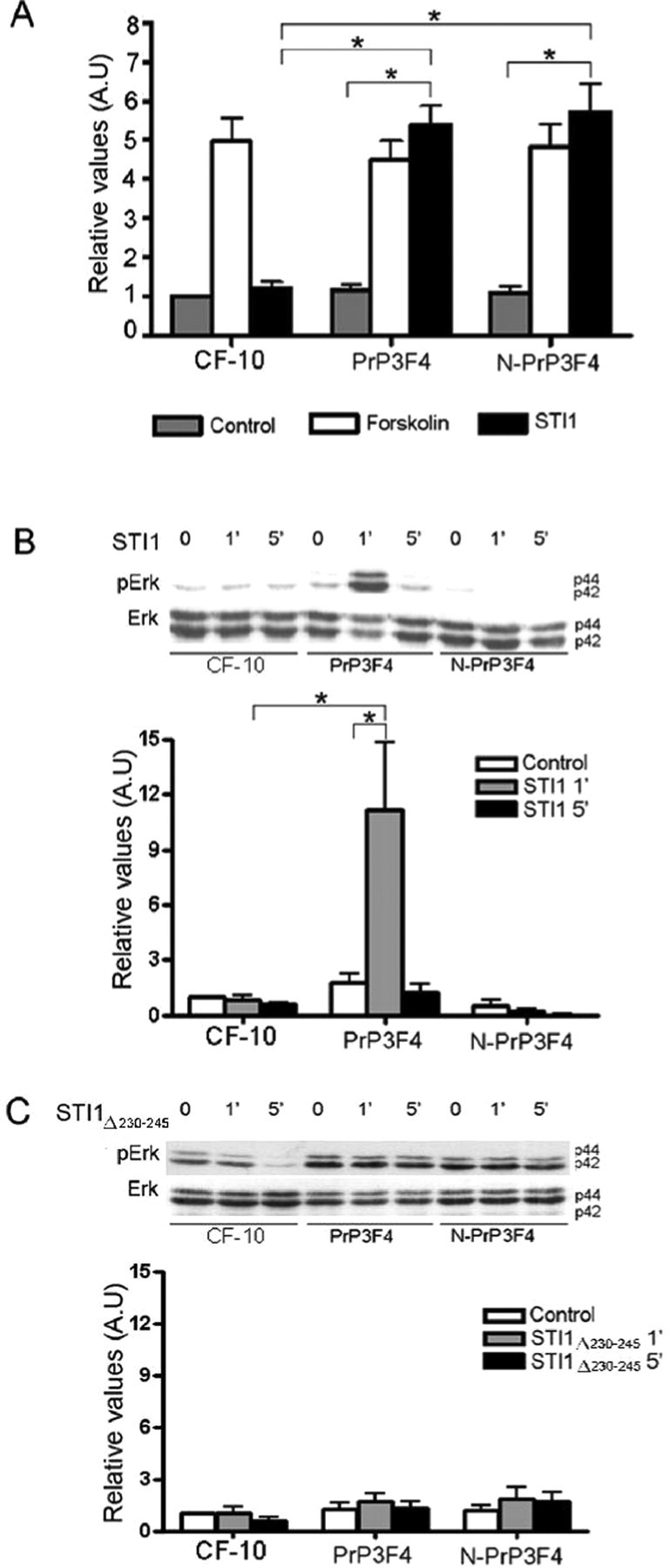
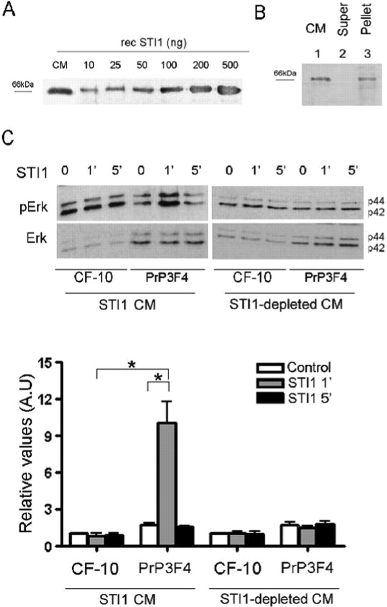
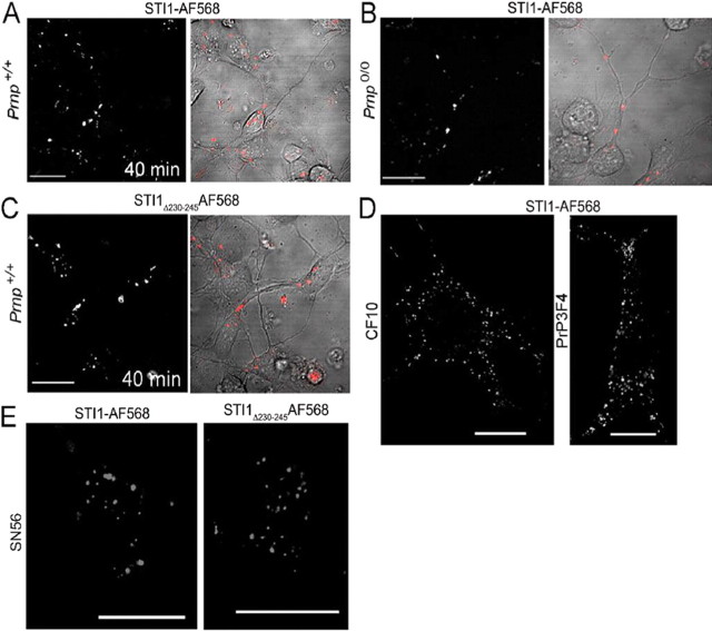
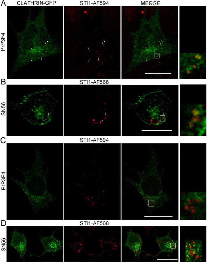
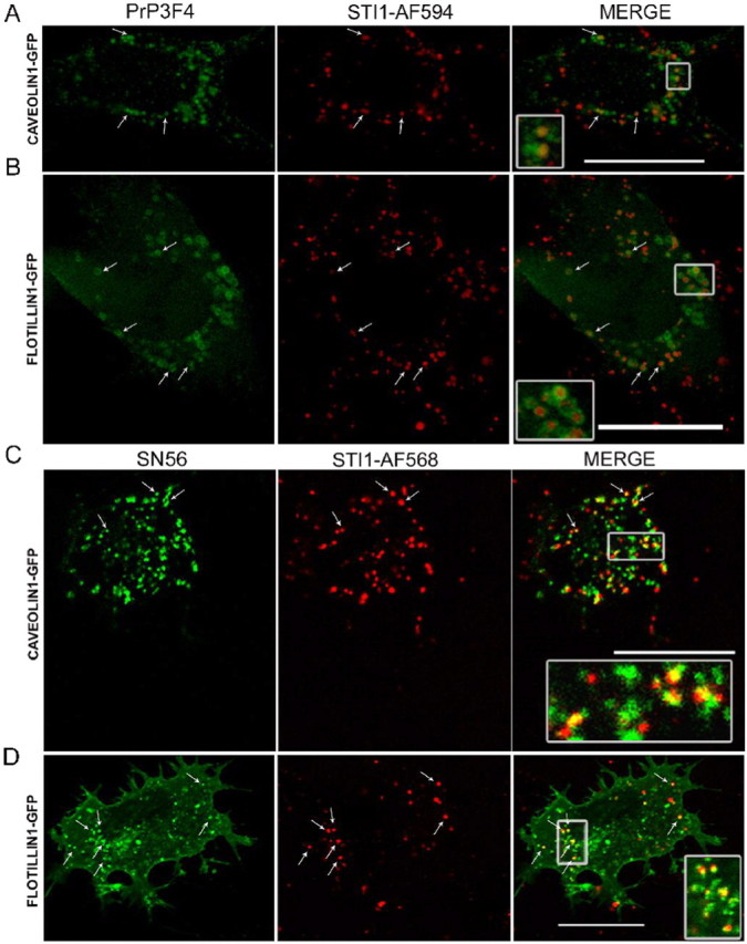
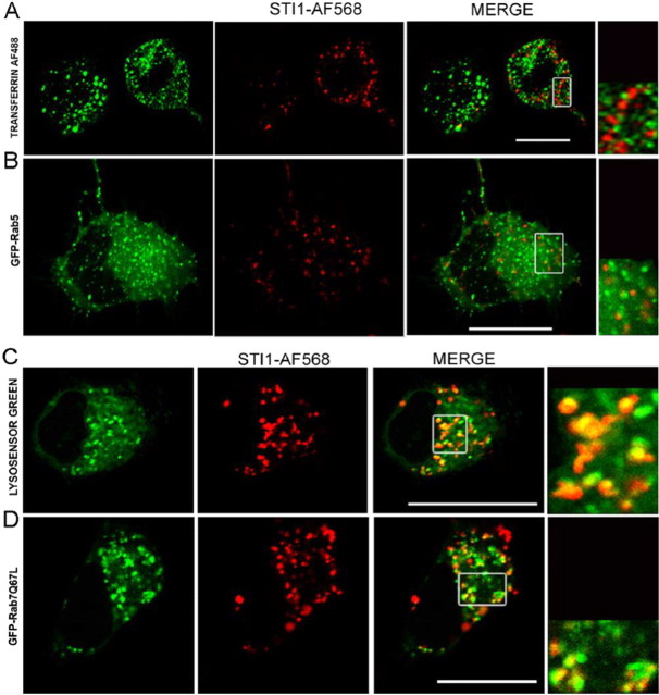
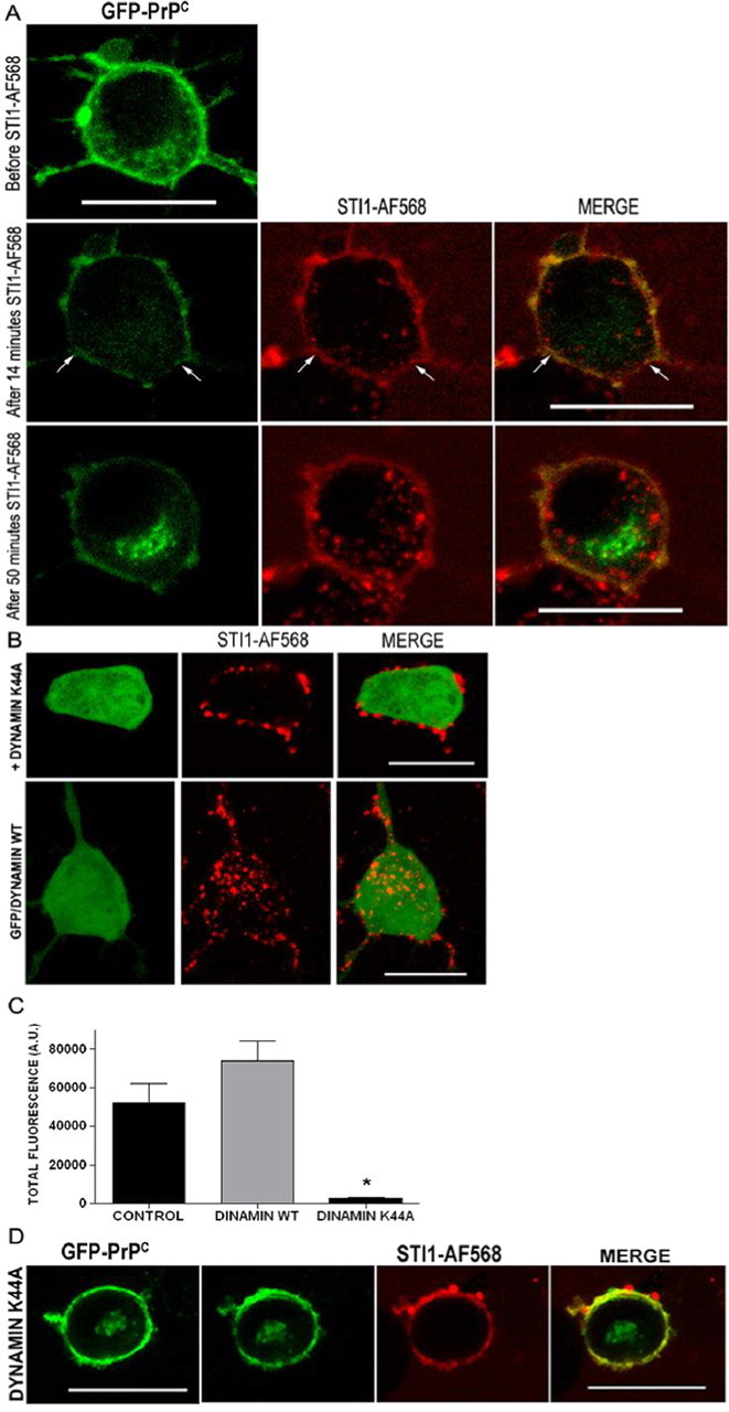
The secreted cochaperone STI1 triggers activation of protein kinase A (PKA) and ERK1/2 signaling by interacting with the cellular prion (PrP(C)) at the cell surface, resulting in neuroprotection and increased neuritogenesis. Here, we investigated whether STI1 triggers PrP(C) trafficking and tested whether this process controls PrP(C)-dependent signaling. We found that STI1, but not a STI1 mutant unable to bind PrP(C), induced PrP(C) endocytosis. STI1-induced signaling did not occur in cells devoid of endogenous PrP(C); however, heterologous expression of PrP(C) reconstituted both PKA and ERK1/2 activation. In contrast, a PrP(C) mutant lacking endocytic activity was unable to promote ERK1/2 activation induced by STI1, whereas it reconstituted PKA activity in the same condition, suggesting a key role of endocytosis in the former process. The activation of ERK1/2 by STI1 was transient and appeared to depend on the interaction of the two proteins at the cell surface or shortly after internalization. Moreover, inhibition of dynamin activity by expression of a dominant-negative mutant caused the accumulation and colocalization of these proteins at the plasma membrane, suggesting that both proteins use a dynamin-dependent internalization pathway. These results show that PrP(C) endocytosis is a necessary step to modulate STI1-dependent ERK1/2 signaling involved in neuritogenesis.


Figures

Figure 1.
Figure 1.
A, B, STI1 induces PrPC internalization. SN56 cells expressing GFP-PrPC were treated with 1 μm STI1 (A) or STI1Δ230–245 (B) for 45 min at 37°C. The left and right panels show the green fluorescence from GFP-PrPC before and after the incubation with STI1 or STI1Δ 230–245, respectively. A and B represent Z projections acquired before and after the perfusion. Images are representative of nine and five independent experiments with multiple culture plates in which 40 and 12 cells were analyzed. Scale bars, 20 μm. C, Flow cytometry assay from SN56 cells treated with 500 μm Cu2+, 1 μm wild-type STI1, or 1 μm STI1Δ230–245 for 40 min. Cells were incubated with nonimmune or anti-PrPC mouse serum followed by R-phycoerythrin-labeled anti-mouse IgG. D, The fluorescence for cell-surface PrPC in untreated cells as described in C was set up to 100%, and the levels of PrPC cell-surface expression after treatment with the indicated concentrations of Cu2+, STI1, or STI1Δ230–245 were normalized to untreated cells. The results shown are the mean values of six independent experiments. Error bars represent SEM. ANOVA followed by Tukey's HSD test was used for comparisons. *p < 0.05.
Figure 2.
Figure 2.
Internalization of PrPC is dependent of the N-terminal basic motif. The PrPC-null cell line CF-10 was transfected with an expression vector encoding wild-type PrPC (PrP3F4) or a mutated PrPC (N-PrP3F4) protein, the internalization of which is impaired, and stably transfected cells were sorted. A, Flow cytometry of nonpermeabilized cells detected using anti-PrPC antibodies (except in CF-10 cells, black lines that were incubated only with secondary antibodies). B, Western blot assays using anti-PrPC antibodies show similar expression of ectopic proteins. C, PrP3F4 cells were kept in KRH or treated with 2 μm STI1Δ230–245 (second column) or STI1 (third column) for 20 min at 37°C. After treatment, cells were fixed and immunostained for cell-surface PrPC using the 3F4 antibody. D, As in C, the N-PrP3F4 mutant cells were treated with 2 μm STI1 for 20 min at 37°C. Scale bars, 20 μm. E, PrP3F4- or N-PrP3F4-expressing cells were treated with STI1 or KRH for 5 min and iced. Cell-surface PrPC after these treatments was detected by biotinylation of cell-surface proteins. Biotinylated proteins were isolated using Neutravidin beads, subjected to SDS-PAGE, and immunobloted using a mouse anti-PrPC antibody. The lysates represent the expression of PrP3F4 or N-PrP3F4 proteins, and beads represent biotinylated cell-surface PrPC after treatment with STI1 or KRH (control). Note that STI1 decreased the amount of PrP3F4 in the membrane in the two lanes (duplicates) labeled STI1 compared with control, whereas N-PrP3F4 was not decreased. Treatment with 500 μm CuSO4 for 10 min was used to test for efficient detection PrPC cell-surface sequestration. The blots are representative of six or seven experiments, respectively.
Figure 3.
Figure 3.
PrPC endocytic trafficking is necessary for STI-PrPC-dependent ERK1/2 but not for PKA activation. A, Cells were treated with forskolin or 1 μm of STI1, and the PKA activity was evaluated. B, Cells were treated with 0.5 μm STI1 for 1 or 5 min, and ERK1/2 activity (pErk) was analyzed. C, Cells were treated with 0.5 μm STI1Δ230–245 for 1 or 5 min, and ERK1/2 activity was analyzed. The basal activity of CF-10 cells without treatment was normalized to 1, and the other values are relative to it. The results show the mean values of five (A) or four (B, C) independent datasets. Error bars represent SEM. ANOVA followed by Tukey's HSD test was used for comparisons. *p < 0.05. A.U., Arbitrary units.
Figure 4.
Figure 4.
STI1 secreted by astrocytes induces ERK1/2 activation. A, Increasing amounts of recombinant STI1 (from 10 to 500 ng) and 30 μl of 200× concentrated CM was loaded onto SDS-PAGE. Immunoblotting was developed with anti-STI1 antibodies. B, STI1 was immunoprecipitated from 150 μl of concentrated CM. The supernatant and the pellet were resolved by SDS-PAGE and detected with anti-STI1 antibodies: lane 1, 30 μl of concentrated CM; lane 2, 30 μl of immunodepleted concentrated CM; lane 3, 30 μl pellet from immunoprecipitation. C, CF-10 or CF-10-expressing PrP3F4 were treated with 50 μl of concentrated CM (STI1 final concentration of 5 nm) or immunodepleted concentrated CM for 1 or 5 min, and ERK1/2 activity was analyzed. The basal activity of CF-10 cells without treatment was normalized to 1, and the other values are relative to it. The results show the mean values of three independent datasets. Error bars represent SEM. ANOVA followed by Tukey's HSD test was used for comparisons. *p < 0.05. A.U., Arbitrary units.
Figure 5.
Figure 5.
STI1 is internalized independently of PrPC. A–E, Wild-type Prnp+/+ (A, C); PrPC-null, Prnp0/0 primary hippocampal neurons (B), CF-10 and CF-10 PrP3F4 cells (D); or SN56 cells (E) were incubated with 1 μm STI1–AF568 or STI1Δ230–245–AF568, as indicated, for 40 min at 37°C. Optical sections of representative cells from at least three independent experiments done in multiple cultures are shown. Scale bars, 20 μm.
Figure 6.
Figure 6.
A fraction of internalized STI1 colocalizes with clathrin. PrP3F4 or SN56 cells, as indicated, were transfected with clathrin–GFP and incubated with 1 μm STI1–AF594 or STI1–AF568 for various periods of time. A, B, Representative images for the first minutes after incubation with STI1. Arrows point to some vesicles where clathrin–GFP and fluorescent STI1 are found. C, D, Representative images of transfected cells 20 min after incubation with STI1. Right panels present a magnified view of the region indicated in the merged images. Arrows indicate some of the colocalization spots. Images are representative of at least 30 cells from several culture dishes on 3 different days. Scale bars, 20 μm.
Figure 7.
Figure 7.
STI1 is also found in raft-derived vesicles. A, B, PrP3F4 cells were transfected with caveolin-1–GFP (A) or flotillin1–GFP (B), and living cells were perfused with STI1–AF594 for 30 min at 37°C. These images are representative examples of colocalization patterns in the first 15 min of incubation with STI1. C, D, SN56 cells were transfected with caveolin-1–GFP (C) or flotillin1–GFP (D) and incubated with STI1–AF568 for 40 min at 37°C. Insets show a magnified view of the designated box. Colocalization between STI1 and the two markers is seen in yellow in the superimposed images, and arrows show some of the colocalization spots. Images are representative of at least 40 cells from several dishes analyzed on 5 different days for each condition. Scale bars, 20 μm.
Figure 8.
Figure 8.
Subcellular localization of STI1. A, SN56 cells were double labeled with STI1–AF568 and transferrin–AF488 for 40 min at 37°C. B, Cells expressing GFP-Rab5 were incubated with STI1–AF568 for 40 min at 37°C. Right panels present a magnified view of the region indicated in the merged images. C, SN56 cells were double labeled with STI1–AF568 and the aciditrophic probe Lysosensor Green (first panel) for 60 min. D, Cells expressing constitutive active mutant GFP-Rab7Q67L were incubated with STI1–AF568 for 40 min at 37°C (second panel). The merged image shows colocalizations in yellow. The images are representative of three experiments with multiple dishes. At least 30 cells were analyzed in each condition. Scale bars, 20 μm.
Figure 9.
Figure 9.
STI1 interacts with PrPC predominantly at the cell surface. A, SN56 cells expressing GFP-PrPC (green) were treated with 1 μm STI1–AF568 (red) for distinct periods of time at 37°C. Images are representative five independent experiments with multiple dishes in which 40 and 12 cells were analyzed. Arrows indicate colocalization spots between GFP-PrPCc (green) and STI1 (red) in forming vesicles. B, SN56 cells were cotransfected with the constructs GFP and dynamin I K44A (top row) or wild-type (WT) dynamin I (bottom row), and living cells were incubated with 1 μm STI1–AF568 for 40 min at 37°C. C, Quantitative analysis of the STI1–AF568 internalization in the presence of dynamin I K44A. Quantification was done using ImageJ software (control, n = 27 cells; dynamin I, n = 67 cells; dynamin I K44A, n = 85 cells) by scoring every cell for the total values of fluorescence internalized. The results are the mean values of fluorescence per cell from multiple dishes examined on 3 different days. Error bars represent SEM. For these experiments, a Kruskal–Wallis one-way ANOVA was used (H = 120; p < 0.001) followed by a Dunn's post hoc test (*p < 0.05). A.U., Arbitrary units. D, SN56 cells coexpressing GFP-PrPC and dynamin I K44A were treated with 1 μm STI1–AF568 at 37°C. Z-series were acquired before (GFP-PrPC) and 40 min after (second to fourth panels) treatment with STI1. Images are representative of four independent experiments in which 15 cells were analyzed. Scale bars, 20 μm.

Similar articles

Cited by

References

    1. Americo TA, Chiarini LB, Linden R. Signaling induced by hop/STI-1 depends on endocytosis. Biochem Biophys Res Commun. 2007;358:620–625. - PubMed
    1. Barbosa J, Jr, Ferreira LT, Martins-Silva C, Santos MS, Torres GE, Caron MG, Gomez MV, Ferguson SS, Prado MA, Prado VF. Trafficking of the vesicular acetylcholine transporter in SN56 cells: a dynamin-sensitive step and interaction with the AP-2 adaptor complex. J Neurochem. 2002;82:1221–1228. - PubMed
    1. Baron GS, Magalhães AC, Prado MAM, Caughey B. Mouse-adapted scrapie infection of SN56 cells: greater efficiency with microsome-associated versus purified PrP-res. J Virol. 2006;80:2106–2117. - PMC - PubMed
    1. Baumann F, Tolnay M, Brabeck C, Pahnke J, Kloz U, Niemann HH, Heikenwalder M, Rulicke T, Burkle A, Aguzzi A. Lethal recessive myelin toxicity of prion protein lacking its central domain. EMBO J. 2007;26:538–547. - PMC - PubMed
    1. Bucci C, Thomsen P, Nicoziani P, McCarthy J, van Deurs B. Rab7: a key to lysosome biogenesis. Mol Biol Cell. 2000;11:467–480. - PMC - PubMed

Publication types

MeSH terms

Substances

LinkOut - more resources