. 2008 Sep;26(9):2349-60.
doi: 10.1634/stemcells.2008-0080. Epub 2008 Jun 12.

Fate mapping and lineage analyses demonstrate the production of a large number of striatal neuroblasts after transforming growth factor alpha and noggin striatal infusions into the dopamine-depleted striatum

Affiliations

Fate mapping and lineage analyses demonstrate the production of a large number of striatal neuroblasts after transforming growth factor alpha and noggin striatal infusions into the dopamine-depleted striatum

Antoine de Chevigny et al. Stem Cells. 2008 Sep.

Abstract

Infusion of transforming growth factor alpha (TGFalpha) into the adult dopamine (DA)-depleted striatum generates a local population of nestin(+)/proliferating cell nuclear antigen (PCNA)(+) newborn cells. The precise origin and fate of these new striatal cells are unknown, making it difficult to direct them for neural repair in Parkinson's disease. Experiments in rats using 5-bromo-2'-deoxyuridine (BrdU) to label neural progenitor cells showed that during TGFalpha infusion in the DA-depleted striatum, newborn striatal cells formed a homogeneous population of precursors, with the majority coexpressing nestin, Mash1, Olig2, and epidermal growth factor receptor, consistent with the phenotype of multipotent C cells. Upon TGFalpha pump withdrawal, the subventricular zone (SVZ) was repopulated by neuroblasts. Strikingly, during this period, numerous clusters of doublecortin(+)/polysialylated neuronal cell adhesion molecule(+) neuroblasts were also produced in the ipsilateral medial striatum. In parallel, striatal BrdU(+)/glial fibrillary acidic protein(+) astrocytes were generated, but no BrdU(+)/O4(+)/CNPase(+) oligodendrocytes were generated. Infusion of the neuralizing bone morphogenetic protein antagonist noggin after TGFalpha pump withdrawal increased the neuroblast-to-astrocyte ratio among new striatal cells by blocking glial differentiation but did not alter striatal neurogenesis. At no time or treatment condition were differentiated neurons generated, including DA neurons. Using 6-hydroxydopamine-lesioned nestin-CreER(T2)/R26R-YFP mice that allow genetic fate-mapping of SVZ nestin(+) cells, we show that TGFalpha-generated striatal cells originate from SVZ nestin(+) precursors that confirmed data from the rats on the phenotype and fate of striatal nestin(+)/PCNA(+) cells upon TGFalpha withdrawal. This work demonstrates that a large population of multipotent striatal C-like cells can be generated in the DA-depleted striatum that do not spontaneously differentiate into DA neurons.


Figures
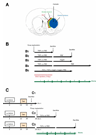
Figure 1
Figure 1. Schematic of experimental paradigms
(A) Schematic of the rat brain at the level of the striatum, showing the infusion site and tissue regions examined. (B) BrdU was administered daily during the 2 first weeks of infusion (shown in red). Adult 6-OHDA lesioned rats were sacrificed either on day 14 during TGFα or PBS infusion (B1), on day 28 after infusion of the second agent (PBS or noggin) for a further 14 days (B2, B3), on day 41 after TGFα then PBS infusion (B4), or on day 28 during infusion of PBS, TGFα, TGFα+noggin or noggin (B5). (C) Tamoxifen was administered to young adult nestin-CreERT2/R26R-YFP mice daily between days 14 and 19 after 6-OHDA injection. (C1) A first group of mice was sacrificed 2 days after the end of tamoxifen regimen. For the 2 other groups, pumps were implanted 1 week after the last tamoxifen dose. Nestin-CreERT2/R26R-YFP mice were sacrificed either on day 14 during TGFα or PBS infusion (C2), or on day 28 after infusion of PBS for a further 14 days period (C3).
Figure 2
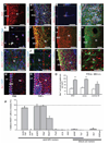
Figure 2. Distribution and phenotypes of ectopic striatal cells during intrastriatal TGFα infusion in adult 6-OHDA lesioned rats
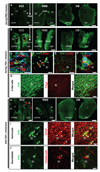
(A) Distribution of BrdU+ cells in the ipsilateral hemisphere (coronal section at the level of the striatum). (B) Nestin (blue) and Olig2 (green) were co-expressed in the majority of BrdU+ cells (red) in the ipsilateral striatum. (C) Orthogonal projection showing a BrdU+/nestin+/Olig2+ cell. EGFR (green, D) and Mash1 (green, E) were also expressed by striatal BrdU+/Olig2+ cells. (F) Some, but not all, BrdU+ cells in the ipsilateral striatal wave and SVZ expressed the endogenous proliferation marker PCNA. (G) BrdU+ (red) / DCX+ (green) cells were virtually absent in TGFα infused rats, and DCX expression was downregulated in SVZ (control not shown). (H) β-catenin (green) was upregulated in the ipsilateral SVZ and striatal wave. (I, J) No BrdU+ cells (red) expressed either Tuj1 (green) or GFAP (blue) in the striatal wave (orthogonal projection in J). (K, L) Few striatal BrdU+ cells (red) were Iba1+ microglial cells (green) and none were CNPase+ oligodendrocytes (blue). Only one BrdU+ cell expresses Iba1 in (L). (M, N) Representative images of Iba1+ microglia in the SVZ and ipsilateral striatum during PBS or TGFα infusion. (O) Upon quantification, a higher Iba1+ microglial density was found in the ipsilateral SVZ and striatum during TGFα administration (grey) than during PBS infusion (white). (P) Histogram showing the percentage of marker-positive cells among striatal BrdU+ cells. Scale bars: A, B, F, G, K, M, N, 100 µm; C, E, 20 µm; D, H, 50 µm; I, J, L, 40 µm.
Figure 3
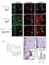
Figure 3. Distribution of immature dividing cells during PBS or during and after TGF-alpha withdrawal
(A, C) Distribution of immature dividing cells in the ipsilateral striatum. (A) During PBS infusion in the DA-depleted striatum, PCNA+ (green) dividing cells were present in the ipsilateral SVZ, but rarely detected in the adjacent striatum. (B) TGFα striatal infusion for 2 weeks induced a large population of PCNA+ cells in the ipsilateral DA-depleted striatum. (C) Two weeks after withdrawal of the TGFα infusing pump, the distribution of PCNA+ cells resembled that of PBS infused rats. (D–F) Distribution of BrdU+ cells. (D) During PBS infusion, BrdU+ cells (red) were confined to the ipsilateral and contralateral SVZs. (E) During TGFα infusion, BrdU+ cells formed a striatal wave in the ipsilateral striatum (right panel), while in the contralateral hemisphere they formed hyper-proliferative nodules protruding from the SVZ towards the source of TGFα (left panel). (F) Two weeks after TGFα withdrawal, contralateral ventricular BrdU+ hyper-proliferative nodules were not found (left panel). BrdU+ cells were observed in the ipsilateral striatum (right), although their distribution pattern (C, right) and number had changed. Note the ~ 35% reduction in total BrdU+ cell number after TGFα withdrawal (G). (H–K) Programmed cell death is induced by TGFα treatment. (H, I) TUNEL+ cells in ipsilateral SVZ and striatum of rats infused with PBS (H) or TGFα (I), 2 weeks after its withdrawal. Arrowheads show TUNEL+ cells. The number of TUNEL+ cells was significantly higher in rats sacrificed after TGFα infusion than in their control counterparts (K). (J) In one rat that died 5 days after TGFα withdrawal, ventricular nodules were found and contained a high proportion of TUNEL+ cells. Scale bars: A–B, 50 µm; D–F, 100 µm; H, I, 200 µm; J, 50 µm.
Figure 4
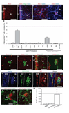
Figure 4. Phenotypes of newborn striatal cells in the rat striatum 2 weeks after TGFα withdrawal
(A) Numerous BrdU+ cells remained in the ipsilateral striatum 2 weeks after TGFα osmotic pump withdrawal and its replacement by a PBS containing pump. (B) These striatal BrdU+ cells (red) did not express Olig2 (green) at this time, but most of them expressed nestin (blue). (C) Higher magnification of the ipsilateral DA-depleted striatum showing BrdU and nestin expression. (D) High magnification confocal scan of a BrdU+/GFAP+ cell in the ipsilateral striatum. (E) Histogram comparing the percentage of marker-positive cells among striatal BrdU+ cells. (F–M) Large number of ectopic striatal neuroblasts were found in the ipsilateral striatum 2 weeks after TGFα withdrawal. (F–H) The ipsilateral SVZ and striatum stained for BrdU and DCX. Some of the neuroblast clusters contained BrdU+ cells (F), but mostly the neuroblast clusters were BrdU (G). (H) Clusters of DCX+ neuroblasts were numerous and distributed throughout the striatum. (I) Most DCX+ cells inside striatal neuroblast clusters expressed PCNA, demonstrating a mitotic state. (J) Neuroblasts in striatal clusters coexpressed PSANCAM (green) and the neuronal marker Tuj1 (red). Arrowheads show striatal PSANCAM+/Tuj1+ cells. (K, L) Most PSANCAM+ neuroblast clusters (red) were directly apposed to blood vessels and astrocyte processes (green). (M) Histogram showing an increased number of striatal neuroblasts 2 weeks after TGFα withdrawal as compared to control, PBS infused rats. Scale bars: A, 100 µm; B, 50 µm; C, 25 µm; D, 20 µm, F, G, I–K, 25 µm; H, 50 µm.
Figure 5
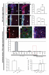
Figure 5. Phenotypes of striatal newborn cells in the rat striatum after TGFα and noggin sequential infusions
(A–D) Increased SVZ neurogenesis after noggin infusion in the DA-depleted rat striatum. (A) PSANCAM+ cells in the SVZ of PBS (left panel) and noggin infused rats (right panel). (B) Cell counts revealed significantly more SVZ neuroblasts in rats treated with noggin than in rats treated with PBS (unpaired t test, p < 0.01). (C) PCNA+ and nestin+ profiles in PBS (left) and noggin infused rats (right). (D) Cell counts revealed similar numbers of nestin+/PCNA+ cells in the SVZ after PBS or noggin infusion. (E–J) Striatal fate of newborn cells after TGFα/noggin consecutive infusions. (E) Numerous BrdU+ cells (red) were detected in the ipsilateral striatum after TGFα and noggin sequential infusions. (F) Orthogonal projection of a striatal BrdU+ cell (red) that neither expressed Tuj1 (green), nor GFAP (blue). (G) Higher magnification image of the region in (E) showed that most striatal BrdU+ cells expressed nestin (blue). (H) Histogram comparing the percentage of marker-positive cells among striatal BrdU+ cells. (I) Histogram comparing the proportion of BrdU+ cells colabeled with GFAP, in rats sequentially infused with TGFα and then PBS, or TGFα and then noggin. (J) Mean striatal number of DCX+ neuroblasts per ipsilateral striatum in the 4 conditions. Scale bars: A, G, 50 µm; C, E, 100 µm; F, 25 µm.
Figure 6
Figure 6. 6-OHDA lesioned nestin-CreERT2/R26R-YFP mice show that TGFα-induced C-like cells and their progeny arise from SVZ nestin+ cells
(A) Mice that received an intrastriatal infusion of PBS for 2 weeks showed YFP+ cells (green) that were restricted to SVZs (left panel), RMS (middle panel) and olfactory bulbs (right panel). (B) Strikingly, mice that received an intrastriatal infusion of TGFα for 14 days exhibited an increased number of YFP+ cells (green) in SVZs (left), RMS (middle) and olfactory bulbs (right). Such cells contributed to YFP+ cellular waves in the ipsilateral striatum, septum as well as corpus callosum and deep cortical layers (see left and middle panels). (C) YFP+ cells (green) in the ipsilateral striatum coexpressed PCNA (red) and nestin (blue). (D) Most ipsilateral striatal YFP+ cells (green) expressed BrdU (red), thus confirming a proliferative state. (E) Single plan confocal scan showing that no YFP+ cells (green) were GFAP+ astrocytes (red) in mice sacrificed during TGFα infusion. (F) YFP+ cells (green) coexpressing EGFR (red) and Olig2 (blue) in the DA-depleted striatum. (G) At a lower magnification, most ipsilateral striatal YFP+ cells (green) coexpressed Olig2 (red). (G–I) Distribution and phenotypes of SVZ-derived NPCs 2 weeks after TGFα withdrawal. (G) Numerous YFP+ cells (green) were observed in the SVZs, ipsilateral striatum, septum (left), RMS (middle) and olfactory bulbs (right). (H) Numerous ipsilateral striatal YFP+ cells (green, left) coexpressed GFAP (red, middle). Right panel depicts a merged image. (I) Other striatal YFP+ cells (green, left) coexpressed PSANCAM (red, middle), indicating a neuroblast phenotype (see right panel for a merged image). Scale bars: A, B, H, 500 µm; C, D, 10 µm, E, 400 µm; F, 5 µm; G, 50 µm, I, J, 20 µm.
Figure 7
Figure 7. Phenotypes of SVZ-derived cells in the olfactory bulb of 6-OHDA lesioned nestin-CreERT2/R26R-YFP mice during and after TGFα infusion
(A–C) During TGFα infusion, olfactory bulb YFP+ cells were morphologically similar to striatal C-like cells (A) and did not express NeuN (B). (C) Orthogonal projection of a YFP+ cell that coexpressed Olig2 in the olfactory bulb. (D–E) Two weeks after TGFα withdrawal, most olfactory bulb YFP+ cells had the typical morphology of granular neurons (D) and expressed NeuN (E). EPL: external plexiform layer, GCL, granule cell layer. Scale bars: A, D, 50 µm; B, C, E, 10 µm.

Similar articles

Cited by

References

    1. Cooper O, Isacson O. Intrastriatal transforming growth factor alpha delivery to a model of Parkinson's disease induces proliferation and migration of endogenous adult neural progenitor cells without differentiation into dopaminergic neurons. J Neurosci. 2004;24:8924–8931. - PMC - PubMed
    1. Hoglinger GU, Rizk P, Muriel MP, et al. Dopamine depletion impairs precursor cell proliferation in Parkinson disease. Nat Neurosci. 2004;7:726–735. - PubMed
    1. Isacson O. The production and use of cells as therapeutic agents in neurodegenerative diseases. Lancet Neurol. 2003;2:417–424. - PubMed
    1. Isacson O, Bjorklund LM, Schumacher JM. Toward full restoration of synaptic and terminal function of the dopaminergic system in Parkinson's disease by stem cells. Ann Neurol. 2003;53 Suppl 3:S135–S146. discussion S146-138. - PubMed
    1. Alvarez-Buylla A, Garcia-Verdugo JM. Neurogenesis in adult subventricular zone. J Neurosci. 2002;22:629–634. - PMC - PubMed

Publication types

MeSH terms