Centrosome amplification can initiate tumorigenesis in flies
- PMID: 18555779
- PMCID: PMC2653712
- DOI: 10.1016/j.cell.2008.05.039
Centrosome amplification can initiate tumorigenesis in flies
Abstract
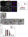
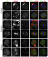
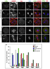
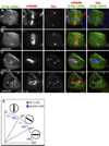
Centrosome amplification is a common feature of many cancer cells, and it has been previously proposed that centrosome amplification can drive genetic instability and so tumorigenesis. To test this hypothesis, we generated Drosophila lines that have extra centrosomes in approximately 60% of their somatic cells. Many cells with extra centrosomes initially form multipolar spindles, but these spindles ultimately become bipolar. This requires a delay in mitosis that is mediated by the spindle assembly checkpoint (SAC). As a result of this delay, there is no dramatic increase in genetic instability in flies with extra centrosomes, and these flies maintain a stable diploid genome over many generations. The asymmetric division of the larval neural stem cells, however, is compromised in the presence of extra centrosomes, and larval brain cells with extra centrosomes can generate metastatic tumors when transplanted into the abdomens of wild-type hosts. Thus, centrosome amplification can initiate tumorigenesis in flies.
Figures


Comment in
-
A tale of too many centrosomes.Cell. 2008 Aug 22;134(4):572-5. doi: 10.1016/j.cell.2008.08.007. Cell. 2008. PMID: 18724931 Review.
Similar articles
-
Mechanisms to suppress multipolar divisions in cancer cells with extra centrosomes.Genes Dev. 2008 Aug 15;22(16):2189-203. doi: 10.1101/gad.1700908. Epub 2008 Jul 28. Genes Dev. 2008. PMID: 18662975 Free PMC article.
-
PLP inhibits the activity of interphase centrosomes to ensure their proper segregation in stem cells.J Cell Biol. 2013 Sep 30;202(7):1013-22. doi: 10.1083/jcb.201303141. J Cell Biol. 2013. PMID: 24081489 Free PMC article.
-
Dynamic centriolar localization of Polo and Centrobin in early mitosis primes centrosome asymmetry.PLoS Biol. 2020 Aug 6;18(8):e3000762. doi: 10.1371/journal.pbio.3000762. eCollection 2020 Aug. PLoS Biol. 2020. PMID: 32760088 Free PMC article.
-
Deregulation of the centrosome cycle and the origin of chromosomal instability in cancer.Adv Exp Med Biol. 2005;570:393-421. doi: 10.1007/1-4020-3764-3_14. Adv Exp Med Biol. 2005. PMID: 18727509 Review.
-
The centrosome in normal and transformed cells.DNA Cell Biol. 2004 Aug;23(8):475-89. doi: 10.1089/1044549041562276. DNA Cell Biol. 2004. PMID: 15307950 Review.
Cited by
-
Expanding roles of centrosome abnormalities in cancers.BMB Rep. 2023 Apr;56(4):216-224. doi: 10.5483/BMBRep.2023-0025. BMB Rep. 2023. PMID: 36945828 Free PMC article. Review.
-
Galectin-3, a novel centrosome-associated protein, required for epithelial morphogenesis.Mol Biol Cell. 2010 Jan 15;21(2):219-31. doi: 10.1091/mbc.e09-03-0193. Epub 2009 Nov 18. Mol Biol Cell. 2010. PMID: 19923323 Free PMC article.
-
The Protein Phosphatase 2A regulatory subunit Twins stabilizes Plk4 to induce centriole amplification.J Cell Biol. 2011 Oct 17;195(2):231-43. doi: 10.1083/jcb.201107086. Epub 2011 Oct 10. J Cell Biol. 2011. PMID: 21987638 Free PMC article.
-
Protein phosphatase 2A-SUR-6/B55 regulates centriole duplication in C. elegans by controlling the levels of centriole assembly factors.Dev Cell. 2011 Apr 19;20(4):563-71. doi: 10.1016/j.devcel.2011.03.007. Dev Cell. 2011. PMID: 21497766 Free PMC article.
-
Identification of Saccharomyces cerevisiae spindle pole body remodeling factors.PLoS One. 2010 Nov 12;5(11):e15426. doi: 10.1371/journal.pone.0015426. PLoS One. 2010. PMID: 21103054 Free PMC article.
References
-
- Al-Hajj M, Clarke MF. Self-renewal and solid tumor stem cells. Oncogene. 2004;23:7274–7282. - PubMed
-
- Baker JD, Adhikarakunnathu S, Kernan MJ. Mechanosensory-defective, male-sterile unc mutants identify a novel basal body protein required for ciliogenesis in Drosophila. Development. 2004;131:3411–3422. - PubMed
-
- Basto R, Lau J, Vinogradova T, Gardiol A, Woods CG, Khodjakov A, Raff JW. Flies without centrioles. Cell. 2006;125:1375–1386. - PubMed
-
- Bello B, Reichert H, Hirth F. The brain tumor gene negatively regulates neural progenitor cell proliferation in the larval central brain of Drosophila. Development. 2006;133:2639–2648. - PubMed
-
- Betschinger J, Mechtler K, Knoblich JA. Asymmetric segregation of the tumor suppressor brat regulates self-renewal in Drosophila neural stem cells. Cell. 2006;124:1241–1253. - PubMed
Publication types
MeSH terms
Substances
Grants and funding
LinkOut - more resources
Full Text Sources
Other Literature Sources
Molecular Biology Databases
Research Materials

