

The TLR2-MyD88-NOD2-RIPK2 signalling axis regulates a balanced pro-inflammatory and IL-10-mediated anti-inflammatory cytokine response to Gram-positive cell walls
- PMID: 18549453
- PMCID: PMC4966886
- DOI: 10.1111/j.1462-5822.2008.01189.x
The TLR2-MyD88-NOD2-RIPK2 signalling axis regulates a balanced pro-inflammatory and IL-10-mediated anti-inflammatory cytokine response to Gram-positive cell walls
Abstract
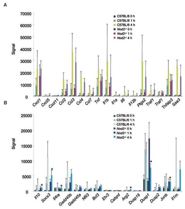
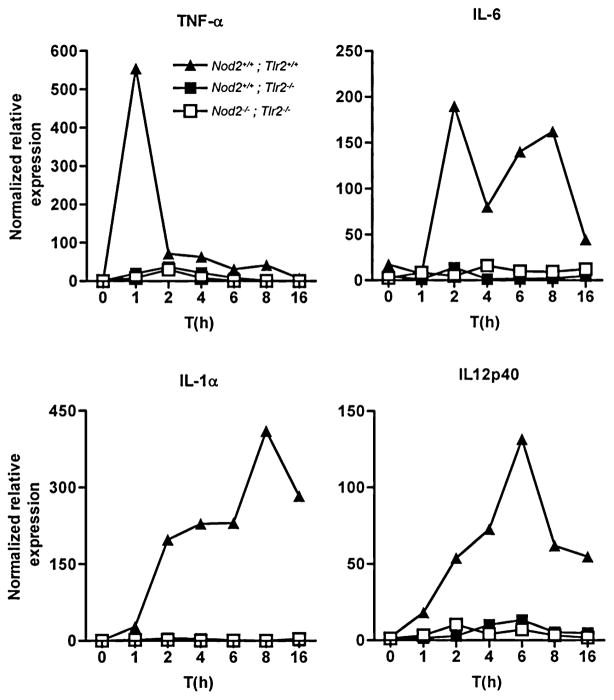
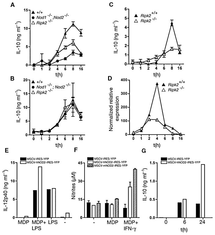
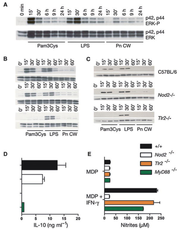
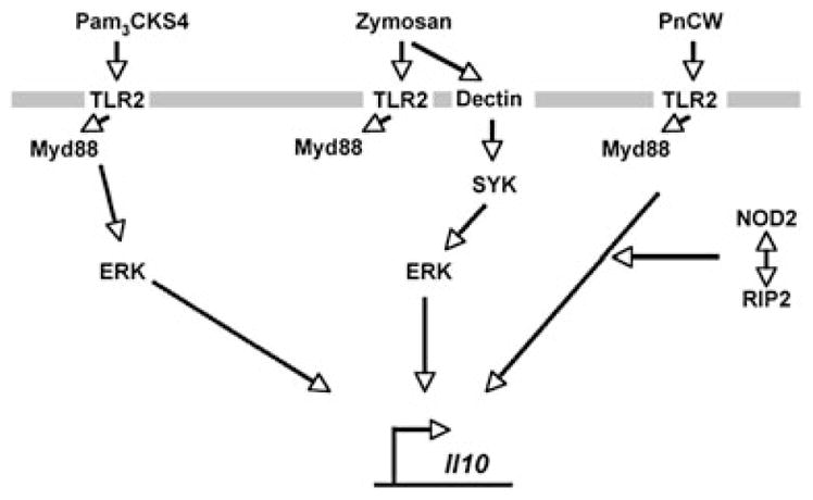
Systemic infection with Streptococcus pneumoniae is associated with a vigorous pro-inflammatory response to structurally complex cell wall fragments (PnCW) that are shed during cell growth and antibiotic-induced autolysis. Consistent with previous studies, inflammatory cytokine production induced by PnCW was dependent on TLR2 but independent of NOD2, a cytoplasmic NLR protein. However, in parallel with the pro-inflammatory response, we found that PnCW also induced prodigious secretion of anti-inflammatory IL-10 from macrophages. This response was dependent on TLR2, but also involved NOD2 as absence of NOD2-reduced IL-10 secretion in response to cell wall and translated into diminished downstream effects on IL-10-regulated target gene expression. PnCW-mediated production of IL-10 via TLR2 required RIPK2 a kinase required for NOD2 function, and MyD88 but differed from that known for zymosan in that ERK pathway activation was not detected. As mutations in NOD2 are linked to aberrant immune responses, the temporal and quantitative effects of activation of the TLR2-NOD2-RIPK2 pathway on IL-10 secretion may affect the balance between pro- and anti-inflammatory responses to Gram-positive bacteria.
Figures

Similar articles
-
Morphine inhibits murine dendritic cell IL-23 production by modulating Toll-like receptor 2 and Nod2 signaling.J Biol Chem. 2011 Mar 25;286(12):10225-32. doi: 10.1074/jbc.M110.188680. Epub 2011 Jan 18. J Biol Chem. 2011. PMID: 21245149 Free PMC article.
-
NOD2 pathway activation by MDP or Mycobacterium tuberculosis infection involves the stable polyubiquitination of Rip2.J Biol Chem. 2007 Dec 14;282(50):36223-9. doi: 10.1074/jbc.M703079200. Epub 2007 Oct 18. J Biol Chem. 2007. PMID: 17947236
-
The crosstalk between TLR2 and NOD2 in Aspergillus fumigatus keratitis.Mol Immunol. 2015 Apr;64(2):235-43. doi: 10.1016/j.molimm.2014.11.021. Epub 2014 Dec 27. Mol Immunol. 2015. PMID: 25549945
-
Recent advances in the development of RIPK2 modulators for the treatment of inflammatory diseases.Front Pharmacol. 2023 Mar 7;14:1127722. doi: 10.3389/fphar.2023.1127722. eCollection 2023. Front Pharmacol. 2023. PMID: 36959850 Free PMC article. Review.
-
RIPK2: a promising target for cancer treatment.Front Pharmacol. 2023 May 30;14:1192970. doi: 10.3389/fphar.2023.1192970. eCollection 2023. Front Pharmacol. 2023. PMID: 37324457 Free PMC article. Review.
Cited by
-
Integrin CD11b attenuates colitis by strengthening Src-Akt pathway to polarize anti-inflammatory IL-10 expression.Sci Rep. 2016 May 18;6:26252. doi: 10.1038/srep26252. Sci Rep. 2016. PMID: 27188220 Free PMC article.
-
LRRK2 and RIPK2 variants in the NOD 2-mediated signaling pathway are associated with susceptibility to Mycobacterium leprae in Indian populations.PLoS One. 2013 Aug 28;8(8):e73103. doi: 10.1371/journal.pone.0073103. eCollection 2013. PLoS One. 2013. PMID: 24015287 Free PMC article. Clinical Trial.
-
Beyond peptidoglycan for Nod2.Nat Immunol. 2009 Oct;10(10):1053-4. doi: 10.1038/ni1009-1053. Nat Immunol. 2009. PMID: 19767725 No abstract available.
-
Transcriptomic Profiling of the Adaptive and Innate Immune Responses of Atlantic Salmon to Renibacterium salmoninarum Infection.Front Immunol. 2020 Oct 28;11:567838. doi: 10.3389/fimmu.2020.567838. eCollection 2020. Front Immunol. 2020. PMID: 33193341 Free PMC article.
-
Collaborative action of Toll-like and NOD-like receptors as modulators of the inflammatory response to pathogenic bacteria.Mediators Inflamm. 2014;2014:432785. doi: 10.1155/2014/432785. Epub 2014 Dec 1. Mediators Inflamm. 2014. PMID: 25525300 Free PMC article. Review.
References
-
- Benjamini Y, Drai D, Elmer G, Kafkafi N, Golani I. Controlling the false discovery rate in behavior genetics research. Behav Brain Res. 2001;125:279–284. - PubMed
-
- Borm ME, van Bodegraven AA, Mulder CJ, Kraal G, Bouma G. The effect of NOD2 activation on TLR2-mediated cytokine responses is dependent on activation dose and NOD2 genotype. Genes Immun. 2008;9:274–278. - PubMed
-
- Chin AI, Dempsey PW, Bruhn K, Miller JF, Xu Y, Cheng G. Involvement of receptor-interacting protein 2 in innate and adaptive immune responses. Nature. 2002;416:190–194. - PubMed
-
- Dillon S, Agrawal A, Van Dyke T, Landreth G, McCauley L, Koh A, et al. A Toll-like receptor 2 ligand stimulates Th2 responses in vivo, via induction of extracellular signal-regulated kinase mitogen-activated protein kinase and c-Fos in dendritic cells. J Immunol. 2004;172:4733–4743. - PubMed
Publication types
MeSH terms
Substances
Grants and funding
LinkOut - more resources
Full Text Sources
Molecular Biology Databases
Research Materials
Miscellaneous
