. 2008 Jun;20(6):1586-602.
doi: 10.1105/tpc.108.060020. Epub 2008 Jun 6.

Light-induced phosphorylation and degradation of the negative regulator PHYTOCHROME-INTERACTING FACTOR1 from Arabidopsis depend upon its direct physical interactions with photoactivated phytochromes

Affiliations

Light-induced phosphorylation and degradation of the negative regulator PHYTOCHROME-INTERACTING FACTOR1 from Arabidopsis depend upon its direct physical interactions with photoactivated phytochromes

Hui Shen et al. Plant Cell. 2008 Jun.

Abstract

The phytochrome (phy) family of photoreceptors regulates changes in gene expression in response to red/far-red light signals in part by physically interacting with constitutively nucleus-localized phy-interacting basic helix-loop-helix transcription factors (PIFs). Here, we show that PIF1, the member with the highest affinity for phys, is strongly sensitive to the quality and quantity of light. phyA plays a dominant role in regulating the degradation of PIF1 following initial light exposure, while phyB and phyD and possibly other phys also influence PIF1 degradation after prolonged illumination. PIF1 is rapidly phosphorylated and ubiquitinated under red and far-red light before being degraded with a half-life of approximately 1 to 2 min under red light. Although PIF1 interacts with phyB through a conserved active phyB binding motif, it interacts with phyA through a novel active phyA binding motif. phy interaction is necessary but not sufficient for the light-induced phosphorylation and degradation of PIF1. Domain-mapping studies reveal that the phy interaction, light-induced degradation, and transcriptional activation domains are located at the N-terminal 150-amino acid region of PIF1. Unlike PIF3, PIF1 does not interact with the two halves of either phyA or phyB separately. Moreover, overexpression of a light-stable truncated form of PIF1 causes constitutively photomorphogenic phenotypes in the dark. Taken together, these data suggest that removal of the negative regulators (e.g., PIFs) by light-induced proteolytic degradation might be sufficient to promote photomorphogenesis.


Figures

Figure 1.
Figure 1.
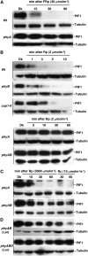
PIF1 Stability Is Highly Sensitive to the Quality and Quantity of Light. Native PIF1 is rapidly degraded under Rp (1 μmol·m−2) (A) or FRp (10 or 30 μmol·m−2) (B) light conditions. Four-day-old dark-grown seedlings were exposed to Rp or FRp light and then incubated in the dark for the durations indicated before being harvested for protein extraction. Protein extracts from dark-grown wild-type and pif1 null mutant seedlings are also included in the first and second lanes, respectively. Approximately 30 μg of total protein in each lane was separated on an 8% polyacrylamide gel, transferred to PVDF membranes, and probed with anti-PIF1 antibody. A similar blot was probed with anti-tubulin antibody. The bands corresponding to PIF1 and tubulin are labeled.
Figure 2.
Figure 2.
phyA Plays a Dominant Role during the Initial Light Exposure, While phyB, phyD, and Other phys Regulate PIF1 Stability under Prolonged Light Exposure. Protein gel blots showing native PIF1 levels in wild-type, phyA, phyB, and phyAB backgrounds. Four-day-old dark-grown seedlings were exposed to FRp (A), Rp (B), or continuous red light (Rc) ([C] and [D]) at the indicated fluences and then incubated in the dark for the times indicated (except Rc) before harvesting for protein extraction. phyAB and phyABD shown in (D) are in the Landsberg erecta (Ler) ecotype; all other mutants are in the Columbia ecotype.
Figure 3.
Figure 3.
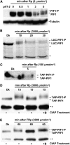
Light Induces Rapid Phosphorylation prior to Degradation of PIF1. (A) Native PIF1 migrates as two bands (PIF1 and PIF1-P) following Rp (2 μmol·m−2). A blot probed with anti-PIF1 antibody is shown. (B) LUC-PIF1 also exhibits a slower migrating band (LUC-PIF1-P) after Rp (3000 μmol·m−2). Proteins from plants expressing LUC-PIF1 were probed with anti-LUC antibody. (C) TAP-PIF1 shows a slower migrating band (TAP-PIF1-P) and is also degraded after Rp (100 μmol·m−2). Proteins from plants expressing TAP-PIF1 were probed with anti-MYC antibody that recognizes the TAP tag. Dotted lines separate the two forms of PIF1 in (A) to (C). (D) and (E) The Rp- and FRp-induced slow-migrating band is a phosphorylated form of PIF1. TAP-PIF1 was immunoprecipitated from protein extracts prepared using 4-d-old dark-grown 35S:TAP-PIF1 seedlings kept in the dark or exposed to either Rp (3000 μmol·m−2; [D]) or FRp (3000 μmol·m−2; [E]) followed by dark incubation. The immunoprecipitated pellets from the Rp- or FRp-exposed samples were dissolved in buffer and incubated without (−) or with (+) native CIAP or with boiled CIAP (+B). Samples were then separated on 6.5% SDS-PAGE gels and probed with anti-MYC antibody. Asterisks denote cross-reacting bands.
Figure 4.
Figure 4.
Light Induces Rapid Phosphorylation and Ubiquitination prior to Degradation of PIF1. (A) LUC-PIF1 shows high molecular weight bands (LUC-PIF1-ubi) after Rp (3000 μmol·m−2). A blot probed with anti-LUC antibody is shown. (B) TAP-PIF1 shows high molecular weight bands (TAP-PIF1-ubi) and is also degraded following Rp (100 μmol·m−2), while TAP-GFP is stable under these conditions. A blot probed with anti-MYC antibody that recognizes the TAP tag is shown. Asterisks denote cross-reacting bands. (C) and (D) The Rp- and FRp-induced slow-migrating bands are ubiquitinated forms of PIF1. TAP-PIF1 was immunoprecipitated from protein extracts prepared using 4-d-old dark-grown seedlings either kept in the dark or exposed briefly to Rp light (3000 μmol·m−2; [C]) or FRp light (3000 μmol·m−2; [D]). The immunoprecipitated samples were then separated on 6.5% SDS-PAGE gels and probed with anti-Ubi or anti-MYC antibody.
Figure 5.
Figure 5.
The APB and APA Motifs Present in the N-Terminal 150–Amino Acid Region of PIF1 Are Necessary for Its Pfr-Specific Interaction with phyA and phyB Both in Vitro and in Vivo. (A) Schematic representation of the GAD-PIF1 baits (left) and full-length phy (phy) preys (right) used in coimmunoprecipitation assays. Mutations made in GAD-PIF1 for testing phyB binding are shown above the diagram, and those for testing phyA binding are shown below the diagram. (B) and (C) Autoradiographs show in vitro interactions of wild-type PIF1 or each of four PIF1 mutants with the Pr or Pfr form of phyB (B) or with single, double (2M), or triple (3M) mutants of PIF1 with the Pr or Pfr form of phyA (C). The left lane in each panel shows the input, and the other lanes show the pellet fractions from coimmunoprecipitation assays performed with in vitro synthesized bait and prey proteins. The phyA and phyB holoproteins were reconstituted by adding the chromophore. The baits were immunoprecipitated using anti-GAD antibody. (D) LUC-PIF1-3M shows much less affinity for the Pfr form of phyA and phyB compared with LUC-PIF1 in in vivo coimmunoprecipitation assays. The input and pellet fractions from in vivo coimmunoprecipitation assays are indicated. Total protein was extracted from 4-d-old dark-grown seedlings either exposed to Rp light (R; 3000 μmol·m−2) or kept in the dark (D). Coimmunoprecipitations were performed using the anti-PIF1 antibody or with an unrelated IgG as a control. The immunoprecipitated samples were then probed with anti-phyA, anti-phyB, or anti-LUC antibody.
Figure 6.
Figure 6.
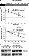
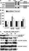
Interactions with the Pfr Form of phyA and phyB Are Necessary for the Light-Induced Phosphorylation and Degradation of PIF1. (A) Design of the cycloheximide chase assays. Relative lucerifase activity for phy interaction–deficient mutants was measured in 4-d-old dark-grown seedlings pretreated with cycloheximide (CHX) in the dark for 3 h, exposed to R (3000 μmol·m−2) light, and then incubated in the dark for the indicated times (min). (B) and (C) Assays show the kinetics of degradation of LUC-PIF1-G47A (B) and LUC-PIF1-2M and LUC-PIF1-3M (C) compared with wild-type LUC-PIF1. LUC-PIF1G47A is deficient in phyB interaction, LUC-PIF1-2M is deficient in phyA interaction, and LUC-PIF1-3M is deficient in both phyA and phyB interaction, as shown in Figure 5. Means ± se of five biological replicates are shown. (D) The abundance and phosphorylation status of LUC-PIF1 and LUC-PIF1-3M fusion proteins prior to and after exposure to Rp determined on protein gel blots using anti-LUC antibody. The dotted line separates the two forms of PIF1. The asterisk denotes a cross-reacting band.
Figure 7.
Figure 7.
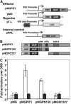
Transcriptional Activation Domains Are Located at the N Terminus of PIF1. (A) Constructs used for the experiment. The effector constructs were designed to express a GAL4 DNA binding domain (DBD)–PIF1 fusion (pMGPIF1) or the GAL4 DNA binding domain alone (pMG). The reporter construct (pT-L) expresses a firefly luciferase (LUC) from the 35S minimal promoter fused to the gal4 DNA binding site (DBS). The internal control (pRNL) expresses a renilla luciferase (RNL LUC) from the 35S promoter. (B) PIF1 deletion constructs used to map the transcriptional activation domains. Each effector construct in (A) and (B) is fused to β-glucuronidase (GUS) to permit the determination of the expression level of the fusion proteins. (C) Three-day-old etiolated Arabidopsis seedlings were cobombarded with the reporter and effector constructs. Seedlings were treated for 15 min with FR light and then incubated in darkness for 16 h. Means ± se from four biological replicates are shown. Transcriptional activity was measured in seedling extracts by a dual-luciferase assay system (Promega). Fold activation is expressed as transcriptional activation activity of DBD-GUS-PIF1 over transcriptional activity of DBD-GUS (white bars) and normalized with GUS activity for the amount of protein expressed by each construct (blue bars).
Figure 8.
Figure 8.
Both the N and C Termini of PIF1 Are Necessary for the Light-Induced Degradation of PIF1. (A) Design of the PIF1 deletion constructs fused to LUC. The white boxes represents a nuclear localization signal (NLS). (B) LUC activity was measured from 4-d-old dark-grown seedlings transferred to R (10 μmol·m−2·s−1) or FR (10 μmol·m−2·s−1) light for 1 h as described (Shen et al., 2005). Means ± se of five biological replicates are shown. Some constructs showed greater stability of the fusion protein in light relative to darkness for unknown reasons. (C) Protein gel blots showing truncated PIF1 fusion proteins are neither phosphorylated nor degraded under light, but the wild-type LUC-PIF1 is both phosphorylated and degraded under light. The dotted line separates the two forms of PIF1. Asterisks denote a cross-reacting band.
Figure 9.
Figure 9.
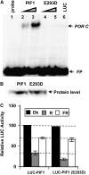
DNA Binding Is Not Necessary for the Light-Induced Degradation of PIF1. (A) The PIF1E293D mutant does not bind to a G-box DNA sequence element (POR C; Su et al., 2001; Moon et al., 2008). In vitro translated PIF1 or PIF1E293D was incubated with a radiolabeled fragment of POR C in a DNA gel shift assay. Lane 1, free probe; lanes 2 and 3, increasing amounts of wild-type PIF1; lanes 4 and 5, increasing amounts of PIF1E293D mutant protein; lane 6, unrelated luciferase protein as a negative control. FP, free probes. (B) Comparison of the levels of wild-type and mutant PIF1 proteins produced by in vitro transcription and translation. (C) Relative LUC assays were performed under the conditions described for Figure 8. Means ± se of five biological replicates are shown.
Figure 10.
Figure 10.
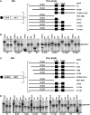
Both the N and C Termini of phyA and phyB Are Necessary for Interaction with PIF1. (A) and (C) Schematic representations of the GAD-PIF1 bait (left) and various phy preys (right) used for coimmunoprecipitation assays. (B) and (D) Autoradiographs showing interactions of full-length PIF1 with the Pr and Pfr forms of full-length and truncated versions of phyA (B) and phyB (D). Input and pellet fractions are shown from the in vitro coimmunoprecipitation assays performed as described (see Figure 5) (Huq et al., 2004). The CT samples are from the C-terminal halves of either phyA or phyB that do not bind to chromophore and are not labeled as Pr or Pfr.
Figure 11.
Figure 11.
Overexpression of the Light-Stable, Truncated Form of PIF1 (C327) Induces a Constitutive Photomorphogenic Phenotype in the Dark. (A) Visible cotyledon-opening phenotypes of various lines grown in the dark for 4 d. (B) to (D) Measurement of cotyledon angles (B), cotyledon areas (C), and hypocotyl lengths (D) of various lines grown in the dark for 4 d (means ± se; n ≥ 30). (E) Photosynthetic gene expression is higher in the C327 lines compared with the wild type in the dark. RNA was extracted from 4-d-old dark-grown seedlings and probed for the indicated photosynthetic (RBCS and CAB3) or nonphotosynthetic control (18S) transcripts. (F) Luciferase activity of various LUC fusion proteins as an indicator of C327 protein amounts in the independent transgenic lines. Relative LUC assays were performed from 4-d-old dark-grown seedlings as described (Shen et al., 2005).
Figure 12.
Figure 12.
Simplified Model of PIF Function in phy Signaling Pathways. Left, in the dark, phys are localized to the cytosol, while PIFs are constitutively localized to the nucleus and negatively regulate photomorphogenesis. Right, light signals promote nuclear migration of phys by inducing the photoconversion of the Pr form to the active Pfr form. In the nucleus, the photoactivated phys interact with PIFs, resulting in the phosphorylation of PIF1 and other PIFs either directly or indirectly. The phosphorylated forms of PIFs are then polyubiquitinated by a ubi ligase and subsequently degraded by the 26S proteasome. The light-induced proteolytic removal of PIFs relieves the negative regulation, thus promoting photomorphogenesis. X indicates an unknown factor that might be involved in the light-induced phosphorylation of PIFs. P, phosphorylated form. This figure is adapted and modified from Castillon et al. (2007).

Similar articles

Cited by

References

    1. Al-Sady, B., Kikis, E.A., Monte, E., and Quail, P.H. (2008). Mechanistic duality of transcription factor function in phytochrome signaling. Proc. Natl. Acad. Sci. USA 105 2232–2237. - PMC - PubMed
    1. Al-Sady, B., Ni, W., Kircher, S., Schafer, E., and Quail, P.H. (2006). Photoactivated phytochrome induces rapid PIF3 phosphorylation prior to proteasome-mediated degradation. Mol. Cell 23 439–446. - PubMed
    1. Bauer, D., Viczian, A., Kircher, S., Nobis, T., Nitschke, R., Kunkel, T., Panigrahi, K.C., Adam, E., Fejes, E., Schafer, E., and Nagy, F. (2004). Constitutive photomorphogenesis 1 and multiple photoreceptors control degradation of phytochrome interacting factor 3, a transcription factor required for light signaling in Arabidopsis. Plant Cell 16 1433–1445. - PMC - PubMed
    1. Castillon, A., Shen, H., and Huq, E. (2007). Phytochrome interacting factors: central players in phytochrome-mediated light signaling networks. Trends Plant Sci. 12 514–521. - PubMed
    1. Chen, M., Chory, J., and Fankhauser, C. (2004). Light signal transduction in higher plants. Annu. Rev. Genet. 38 87–117. - PubMed

Publication types

MeSH terms

LinkOut - more resources