P-selectin glycoprotein ligand-1 is highly expressed on Ly-6Chi monocytes and a major determinant for Ly-6Chi monocyte recruitment to sites of atherosclerosis in mice
- PMID: 18519846
- PMCID: PMC2596619
- DOI: 10.1161/CIRCULATIONAHA.108.771048
P-selectin glycoprotein ligand-1 is highly expressed on Ly-6Chi monocytes and a major determinant for Ly-6Chi monocyte recruitment to sites of atherosclerosis in mice
Abstract
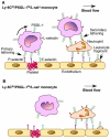
Background: Ly-6C(hi) monocytes are key contributors to atherosclerosis in mice. However, the manner in which Ly-6C(hi) monocytes selectively accumulate in atherosclerotic lesions is largely unknown. Monocyte homing to sites of atherosclerosis is primarily initiated by rolling on P- and E-selectin expressed on endothelium. We hypothesize that P-selectin glycoprotein ligand-1 (PSGL-1), the common ligand of P- and E-selectin on leukocytes, contributes to the preferential homing of Ly-6C(hi) monocytes to atherosclerotic lesions.
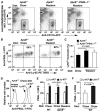
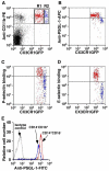
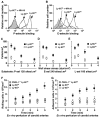
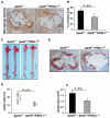
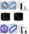
Methods and results: To test this hypothesis, we examined the expression and function of PSGL-1 on Ly-6C(hi) and Ly-6C(lo) monocytes from wild-type mice, ApoE(-/-) mice, and mice lacking both ApoE and PSGL-1 genes (ApoE(-/-)/PSGL-1(-/-)). We found that Ly-6C(hi) monocytes expressed a higher level of PSGL-1 and had enhanced binding to fluid-phase P- and E-selectin compared with Ly-6C(lo) monocytes. Under in vitro flow conditions, more Ly-6C(hi) monocytes rolled on P-, E-, and L-selectin at slower velocities than Ly-6C(lo) cells. In an ex vivo perfused carotid artery model, Ly-6C(hi) monocytes interacted preferentially with atherosclerotic endothelium compared with Ly-6C(lo) monocytes in a PSGL-1-dependent manner. In vivo, ApoE(-/-) mice lacking PSGL-1 had impaired Ly-6C(hi) monocyte recruitment to atherosclerotic lesions. Moreover, ApoE(-/-)/PSGL-1(-/-) mice exhibited significantly reduced monocyte infiltration in wire injury-induced neointima and in atherosclerotic lesions. ApoE(-/-)/PSGL-1(-/-) mice also developed smaller neointima and atherosclerotic plaques.
Conclusions: These data indicate that PSGL-1 is a new marker for Ly-6C(hi) monocytes and a major determinant for Ly-6C(hi) cell recruitment to sites of atherosclerosis in mice.
Figures

Comment in
-
Diversity of denizens of the atherosclerotic plaque: not all monocytes are created equal.Circulation. 2008 Jun 24;117(25):3168-70. doi: 10.1161/CIRCULATIONAHA.108.783068. Circulation. 2008. PMID: 18574058 Review. No abstract available.
Similar articles
-
Core2 1-6-N-glucosaminyltransferase-I is crucial for the formation of atherosclerotic lesions in apolipoprotein E-deficient mice.Arterioscler Thromb Vasc Biol. 2009 Feb;29(2):180-7. doi: 10.1161/ATVBAHA.108.170969. Epub 2008 Dec 4. Arterioscler Thromb Vasc Biol. 2009. PMID: 19057022 Free PMC article.
-
Impaired infarct healing in atherosclerotic mice with Ly-6C(hi) monocytosis.J Am Coll Cardiol. 2010 Apr 13;55(15):1629-38. doi: 10.1016/j.jacc.2009.08.089. J Am Coll Cardiol. 2010. PMID: 20378083 Free PMC article.
-
P-selectin glycoprotein ligand-1 deficiency leads to cytokine resistance and protection against atherosclerosis in apolipoprotein E deficient mice.Atherosclerosis. 2012 Jan;220(1):110-7. doi: 10.1016/j.atherosclerosis.2011.10.012. Epub 2011 Oct 17. Atherosclerosis. 2012. PMID: 22041028 Free PMC article.
-
Adhesion molecules and atherogenesis.Acta Physiol Scand. 2001 Sep;173(1):35-43. doi: 10.1046/j.1365-201X.2001.00882.x. Acta Physiol Scand. 2001. PMID: 11678724 Review.
-
P-selectin glycoprotein ligand-1 plays a crucial role in the selective recruitment of leukocytes into the atherosclerotic arterial wall.Trends Cardiovasc Med. 2009 May;19(4):140-5. doi: 10.1016/j.tcm.2009.07.006. Trends Cardiovasc Med. 2009. PMID: 19818951 Free PMC article. Review.
Cited by
-
Platelet Membrane Nanocarriers Cascade Targeting Delivery System to Improve Myocardial Remodeling Post Myocardial Ischemia-Reperfusion Injury.Adv Sci (Weinh). 2024 Apr;11(16):e2308727. doi: 10.1002/advs.202308727. Epub 2024 Feb 12. Adv Sci (Weinh). 2024. PMID: 38345237 Free PMC article.
-
Inflammatory Cell Recruitment in Cardiovascular Disease.Front Cell Dev Biol. 2021 Feb 18;9:635527. doi: 10.3389/fcell.2021.635527. eCollection 2021. Front Cell Dev Biol. 2021. PMID: 33681219 Free PMC article. Review.
-
Nanoparticle Based Cardiac Specific Drug Delivery.Biology (Basel). 2023 Jan 4;12(1):82. doi: 10.3390/biology12010082. Biology (Basel). 2023. PMID: 36671774 Free PMC article. Review.
-
Glycosyltransferases, glycosylation and atherosclerosis.Glycoconj J. 2014 Dec;31(9):605-11. doi: 10.1007/s10719-014-9560-8. Epub 2014 Oct 8. Glycoconj J. 2014. PMID: 25294497 Review.
-
Inflammatory Markers Related to Innate and Adaptive Immunity in Atherosclerosis: Implications for Disease Prediction and Prospective Therapeutics.J Inflamm Res. 2021 Feb 16;14:379-392. doi: 10.2147/JIR.S294809. eCollection 2021. J Inflamm Res. 2021. PMID: 33628042 Free PMC article. Review.
References
-
- Ross R. Atherosclerosis--an inflammatory disease. N Engl J Med. 1999;340:115–126. - PubMed
-
- Libby P. Inflammation in atherosclerosis. Nature. 2002;420:868–874. - PubMed
-
- Geissmann F, Jung S, Littman DR. Blood monocytes consist of two principal subsets with distinct migratory properties. Immunity. 2003;19:71–82. - PubMed
Publication types
MeSH terms
Substances
Grants and funding
- HL78679/HL/NHLBI NIH HHS/United States
- R01 HL078679/HL/NHLBI NIH HHS/United States
- P20 RR018758-019002/RR/NCRR NIH HHS/United States
- HL080569/HL/NHLBI NIH HHS/United States
- P20 RR018758-057590/RR/NCRR NIH HHS/United States
- P20 RR018758-020004/RR/NCRR NIH HHS/United States
- P20 RR018758-047441/RR/NCRR NIH HHS/United States
- P20 RR018758-057593/RR/NCRR NIH HHS/United States
- P20 RR018758/RR/NCRR NIH HHS/United States
- RR018758/RR/NCRR NIH HHS/United States
- P01 HL085607-020003/HL/NHLBI NIH HHS/United States
- HL085607/HL/NHLBI NIH HHS/United States
- P20 RR018758-029002/RR/NCRR NIH HHS/United States
- P20 RR018758-030004/RR/NCRR NIH HHS/United States
- R01 HL080569/HL/NHLBI NIH HHS/United States
- P01 HL085607/HL/NHLBI NIH HHS/United States
- P20 RR018758-039002/RR/NCRR NIH HHS/United States
- P01 HL085607-010003/HL/NHLBI NIH HHS/United States
- P20 RR018758-047438/RR/NCRR NIH HHS/United States
LinkOut - more resources
Full Text Sources
Medical
Molecular Biology Databases
Miscellaneous

