Review
. 2008 Sep;10(9):1527-48.
doi: 10.1089/ars.2008.2046.

Evolution of catalases from bacteria to humans

Affiliations
Review

Evolution of catalases from bacteria to humans

Marcel Zamocky et al. Antioxid Redox Signal. 2008 Sep.

Abstract

Excessive hydrogen peroxide is harmful for almost all cell components, so its rapid and efficient removal is of essential importance for aerobically living organisms. Conversely, hydrogen peroxide acts as a second messenger in signal-transduction pathways. H(2)O(2) is degraded by peroxidases and catalases, the latter being able both to reduce H(2)O(2) to water and to oxidize it to molecular oxygen. Nature has evolved three protein families that are able to catalyze this dismutation at reasonable rates. Two of the protein families are heme enzymes: typical catalases and catalase-peroxidases. Typical catalases comprise the most abundant group found in Eubacteria, Archaeabacteria, Protista, Fungi, Plantae, and Animalia, whereas catalase-peroxidases are not found in plants and animals and exhibit both catalatic and peroxidatic activities. The third group is a minor bacterial protein family with a dimanganese active site called manganese catalases. Although catalyzing the same reaction (2 H(2)O(2)--> 2 H(2)O+ O(2)), the three groups differ significantly in their overall and active-site architecture and the mechanism of reaction. Here, we present an overview of the distribution, phylogeny, structure, and function of these enzymes. Additionally, we report about their physiologic role, response to oxidative stress, and about diseases related to catalase deficiency in humans.


Figures
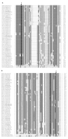
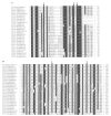
FIG. 1
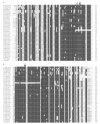
FIG. 1. Multiple sequence alignment of selected typical catalases
ClustalX, version 1.81, was used with Gonnet 250 protein weight matrix. Gap-opening penalty 9.00 and gap-extension penalty 0.20 was used in slow-accurate alignment algorithm. Residue-specific penalties and hydrophilic penalties were activated, and the gap-separation distance was optimised to 8. (A) Distal side of the prosthetic heme group. (B) Proximal side of heme with essential tyrosine. Black, Similarity scheme with highest similarity; dark grey, high similarity; light grey, low similarity; arrow, catalytically important residues. Abbreviations for Linnaean names and ID numbers correspond to PeroxiBase nomenclature (; see http://peroxibase.isb-sib.ch/ for all details).
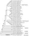
FIG. 2
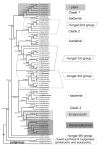
FIG. 2. Reconstructed phylogenetic tree of 70 typical catalases from all main living kingdoms
Presented is the tree obtained with the neighbor-joining method and 1,000 bootstrap cycles. Very similar trees were reconstructed also with maximum parsimony (1,000 bootstraps) and maximum likelihood (100 bootstraps) methods. Rooting of the tree was performed by using the fused synthase and oxygenase sequence as an outgroup. Numbers on the branches represent bootstrap values for NJ/MP/ML methods, respectively. The phylogenetic relations were reconstructed by using MEGA4 (91) and Phylip packages (http://evolution.gs.washington.edu/phylip.html). For NJ and ML methods, the Jones-Taylor-Thornton protein matrix was used with an optimized γ parameter = 1.90. For the MP method, the CNI level was set to 1 with initial tree search by random addition with 10 replicates. The outgroup (for rooting purposes) is labeled with a rhombus. Sequences with known 3D structures are underlined. The groupings in major subfamilies are marked by different boxes with self-explanation on the right side.
FIG. 3
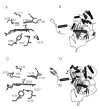
FIG. 3. Structural comparison of catalase A from Saccharomyces cerevisiae and catalase-1 from Neurospora crassa
(A) Heme-cavity architecture of catalase A from Saccharomyces cerevisiae. The conserved distal residues His70, Ser104, Val111, Asn143, Phe148, and Phe156 are shown; the proximal heme ligand is Tyr355, hydrogen-bonded to Arg375. The figure was constructed by using the coordinates deposited in the Protein Data Bank (accession code 1a4e). (B) Monomeric structure of catalase A from Saccharomyces cerevisiae, showing the assignment of secondary structure elements and the prosthetic group. (C) View of the active site of catalase-1 from Neurospora crassa, showing the conserved distal residues His92, Ser131, Val133, Asp165, Phe170, and Phe178, as well as the proximal residues Tyr379, Arg375, and Gly237. The figure was constructed by using the coordinates deposited in the Protein Data Bank (accession code 1sy7). (D) Monomeric structure of catalase-1 from Neurospora crassa, showing the assignment of secondary structure elements and the prosthetic group.
FIG. 4
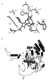
FIG. 4. Structure of NADPH-binding pocket in Homo sapiens catalase
(A) View showing the NADPH-binding pocket with residues that are involved in hydrogen bonding with NADPH. (B) Monomeric structure of catalase from Homo sapiens, showing the assignment of secondary structure elements and the NADPH-binding pocket. The figure was constructed by using the coordinates deposited in the Protein Data Bank (accession code 1dgb).
FIG. 5
FIG. 5. Selected parts of the multiple sequence alignment of 43 catalase–peroxidases (KatGs)
Thirty-two complete fungal sequences are presented together with closely related bacterial counterparts. Similarity scheme is identical with that in Fig. 1. The substitution matrix and the alignment algorithm were the same as used in recent experimental work on this topic (69). (A) Distal side of the heme group with the catalytic triad. (B) Distal side of heme with important hydrogen bond location. (C) Proximal side of the prosthetic heme group with the essential histidine. (D) Proximal side of heme with important hydrogen bond location. Arrow, Catalytically important residues. Abbreviations for Linnaean names and ID numbers correspond to PeroxiBase nomenclature (; see http://peroxibase.isb-sib.ch/ for all details).
FIG. 6
FIG. 6. Reconstructed phylogenetic tree of bacterial and fungal KatGs obtained with the neighbor-joining method and 1,000 bootstrap replications
An almost identical tree was obtained with maximum parsimony method by using 1,000 bootstrap replications. The phylogenetic relations were reconstructed by using the MEGA4 software package (91). The optimized parameters for the NJ and MP methods were the same as those used in recent experimental work on this topic (69). Numbers on the branches represent bootstrap values for NJ/MP methods, respectively. Sequences with known 3D structures are underlined. The outgroup is labeled with a rhombus.
FIG. 7
FIG. 7. Structure of catalase–peroxidase from Burkholderia pseudomallei
(A) Distal active residues are covalent linked Met264, Tyr238, and Try111, the catalytically active His112, Arg108, and Asp141 that contribute to the stabilization of the H-bonding network of the access channel. (B) Monomeric structure of catalase–peroxidase from Burkholderia pseudomallei showing the assignment of secondary structure elements and the prosthetic group. The figure was constructed by using the coordinates deposited in the Protein Data Bank (accession code 1mwv).
FIG. 8
FIG. 8. Inferred phylogenetic tree of 30 manganese catalases rooted with an outgroup
The phylogenetic relations were reconstructed by using MEGA4 (91) and Phylip packages (http://evolution.gs.washington.edu/phylip.html). Presented is the tree obtained with the neighbor-joining method and 1,000 bootstrap replications. Almost identical trees were obtained with maximum-parsimony and maximum-likelihood methods. Applied parameters were the same as those used for Fig. 2 (see earlier), with the only exception that uniform rates of substitutions were applied for NJ and ML methods. Numbers on the branches represent bootstrap values for NJ/MP/ML methods, respectively. Sequences with known 3D structures are underlined. The outgroup is labeled with a rhombus.
FIG. 9
FIG. 9. Structure of manganese catalase of Lactobacillus plantarum
(A) View of the dinuclear manganese complex at the active site in Lactobacillus plantarum. All coordinating ligands are depicted. Dark grey spheres, Manganese ions; light grey spheres, Coordinated solvent. (B) Monomeric structure of Mn catalase from Lactobacillus plantarum showing the assignment of secondary structure elements and the prosthetic group. The figure was constructed by using the coordinates deposited in the Protein Data Bank (accession code 1jku).
FIG. 10
FIG. 10. Selected parts of multiple sequence alignment of 30 bacterial manganese catalases
ClustalX, version 1.81, was used with Gonet 250 protein weight matrix. Gap-opening penalty 8.00 and gap-extension penalty 0.20 were used in a slow-accurate algorithm. Residue-specific penalties and hydrophilic penalties were activated, and the gap-separation distance was optimized to 6. The similarity scheme is identical with that in Fig. 1. Abbreviations for Linnaean names and ID numbers correspond to PeroxiBase nomenclature (; see http://peroxibase.isb-sib.ch/ for all details).

Similar articles

Cited by

References

    1. Alfonso-Prieto M, Borovik A, Carpena X, Murshudov G, Melik-Adamyan W, Fita I, Rovira C, Loewen PC. The structures and electronic configuration of compound I intermediates of Helicobacter pylori and Penicillium vitale catalases determined by X-ray crystallography and WM/MM density functional theory calculations. J Am Chem Soc. 2007;129:4193–4205. - PubMed
    1. Antonyuk SV, Melik-Adamyan VR, Popov AN, Lamzin VS, Hempstead PD, Harrison PM, Artymiuk PJ, Barynin VV. Three-dimensional structure of the enzyme dimanganese catalase from Thermus thermophilus at 1A resolution. Crystallogr Rep. 2000;45:105–116.
    1. Baker RD, Cook CO, Goodwin DC. Properties of catalase-peroxidase lacking its C-terminal domain. Biochem Biophys Res Commun. 2004;320:833–839. - PubMed
    1. Barynin VV, Whittaker MM, Antonyuk SV, Lamzin VS, Harrison PM, Artymiuk PJ, Whittaker JW. Crystal structure of manganese catalase from Lactobacillus plantarum. structure. 2001;9:725–738. - PubMed
    1. Bayne ACV, Mockett RJ, Orr WC, Sohal RS. Enhanced catabolism of mitochondrial superoxide/hydrogen peroxide and aging in transgenic. Drosophila. Biochem J. 2005;391:277–284. - PMC - PubMed

Publication types

LinkOut - more resources