. 2008 May 20;6(5):e128.
doi: 10.1371/journal.pbio.0060128.

Raf activation is regulated by tyrosine 510 phosphorylation in Drosophila

Affiliations

Raf activation is regulated by tyrosine 510 phosphorylation in Drosophila

Fan Xia et al. PLoS Biol. .

Abstract

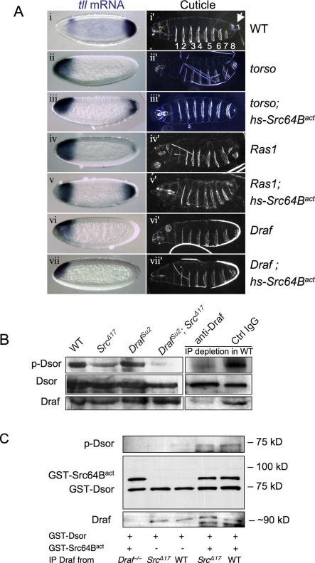
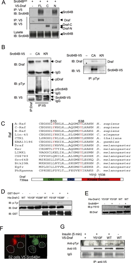
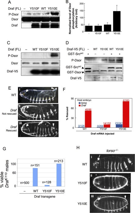
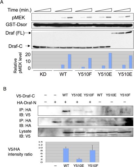
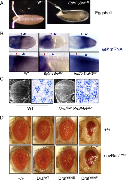
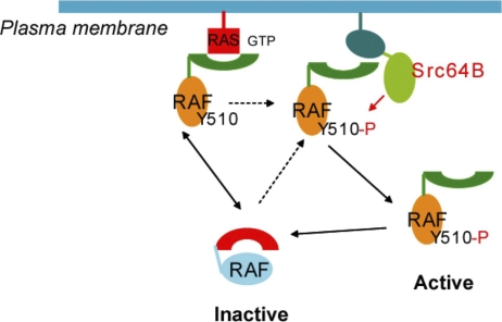
The proto-oncoprotein Raf is pivotal for mitogen-activated protein kinase (MAPK) signaling, and its aberrant activation has been implicated in multiple human cancers. However, the precise molecular mechanism of Raf activation, especially for B-Raf, remains unresolved. By genetic and biochemical studies, we demonstrate that phosphorylation of tyrosine 510 is essential for activation of Drosophila Raf (Draf), which is an ortholog of mammalian B-Raf. Y510 of Draf is phosphorylated by the c-src homolog Src64B. Acidic substitution of Y510 promotes and phenylalanine substitution impairs Draf activation without affecting its enzymatic activity, suggesting that Y510 plays a purely regulatory role. We further show that Y510 regulates Draf activation by affecting the autoinhibitory interaction between the N- and C-terminal fragments of the protein. Finally, we show that Src64B is required for Draf activation in several developmental processes. Together, these results suggest a novel mechanism of Raf activation via Src-mediated tyrosine phosphorylation. Since Y510 is a conserved residue in the kinase domain of all Raf proteins, this mechanism is likely evolutionarily conserved.


Conflict of interest statement

Competing interests. The authors have declared that no competing interests exist.

Figures

Figure 1
Figure 1. Src64B Can Function Downstream or in Parallel to Ras in Activating Draf
(A) Expression of tll mRNA (blue stains) in stage 4 embryos (left column) was detected by in situ hybridization. The right panels show cuticles of mature embryos. All the embryos are shown with anterior to the left. Genotypes are noted to the right. Draf indicates Draf11−29; Ras1, Ras1ΔC40B;and torso, torsoXR1. (i) tll is expressed in anterior and posterior regions of wild-type (WT) embryos. The posterior tll expression is solely dependent on the Torso pathway; the anterior tll expression is repressed by Torso signaling and serves as an internal control. (i′) Wild-type cuticles exhibit eight ventral denticle belts (numbered 1–8) as well as head and tail (terminal) structures. The posterior terminal structures include the eighth ventral denticle belt and the Filzkörper (arrow), which require appropriate posterior tll expression. Embryos maternally null for torso (ii and ii′), Ras1 (iv and iv′), or Draf (vi and vi′) are missing posterior tll expression, and as a consequence, are missing posterior structures and exhibit only seven ventral denticle belts. Transient expression of Src64Bact during early embryogenesis restored the posterior tll expression and the eighth denticle band in torso (iii and iii′) or Ras1 (v and v′) mutant embryos, but not in Draf mutants (vii and vii′). More than 200 embryos were observed for each genotype. Representative pictures are shown. (B) The activity of the Ras-binding–deficient DrafSu2 variant is dependent on Src64B. Cell-free extracts from embryos of indicated genotypes were subjected to in vitro kinase assay using purified GST-Dsor1 (MEK homolog) as substrate. Note DrafSu2 activity is dramatically decreased in Src64BΔ17 background (cf. lanes 3 and 4). Wild-type Draf activity is only mildly decreased in Src64BΔ17 background (cf. lanes 1 and 2). Depletion of Draf by specific antibody abolished the Dsor1 phosphorylation (lane 5). (C) Draf was immunoprecipitated from embryos of indicated genotypes. Draf−/− embryos were maternally null for Draf (negative control). The immunoprecipitates were subjected to kinase assay using GST-Dsor as substrate with or without added Src64Bact. Note the kinase activity of Draf from Src64BΔ17 was undetectable (lane 2). Addition of Src64Bact increased the activity of Draf from both Src64BΔ17 and wild-type embryos to similar levels (lanes 4 and 5).
Figure 2
Figure 2. Src64B Binds to Draf and Phosphorylates Draf on Y510
(A) Src64B binds to Draf mainly through the N-terminal region. Src64Bact was cotransfected with full-length Draf (FL), Draf-N (N), or Draf-C (C) into S2 cells. Transfected Draf was immunoprecipitated (IP) with anti-V5 and blotted (IB) with anti-Src64B. Note Src64Bact was coimmunoprecipitated with Draf or Draf-N, but very little with Draf-C. (B) Src64B binding leads to tyrosine phosphorylation of Draf. S2 cells were transfected with V5-tagged Src64Bact (referred to as CA, constitutive active) or a kinase-dead version (Src64BKR; KR), immunoprecipitated with anti-V5 (left) or anti-pTyr (right), and subjected to SDS-PAGE. Immunoprecipitated Draf from untransfected S2 cells was used as input to mark the position of Draf (left panels, lane 1). Note that the endogenous Draf was co-immunoprecipitated with transfected Src proteins (left panels; lanes 2 and 3). Transfection of Src64Bact (CA), but not Src64BKR, led to tyrosine phosphorylation of coimmunoprecipitated Draf (middle left). Also note that the endogenous Draf was immunoprecipitated by anti-pTyr only when S2 cells were transfected with Src64Bact, but not Src64BKR (right panels). (C) Conservation of Y510 and Y538 in protein kinases. A multisequence alignment of several kinases identifies two conserved tyrosine residues in the kinase domain, which correspond to Y510 and Y538 of Draf (red). Y510 is conserved in all Raf family members as well as in Ksr, whereas Y538 is conserved in all kinases analyzed, including tyrosine kinases such as EGFR and Src64B. The positions of Y510 and Y538 are also indicated in the schematic representation of Draf. (D–F) Src64B phosphorylates Draf-C on Y510. (D) Src64B phosphorylates Draf-C on Y510 in vitro. GST-Src64Bact, His-Draf-CWT, His-Draf-CY510F, and His-Draf-CY538F proteins purified from E. coli were subjected to in vitro kinase assay. The levels of tyrosine phosphorylation on different Draf-C molecules were detected by an anti-pTyr antibody. Increased tyrosine phosphorylation levels in the presence of GST-Src64Bact were detected for His-Draf-CWT (lane 4) and His-Draf-CY538F (lane 6), but not for His-Draf-CY510F (lane 5). WT, wild type. (E) In vitro kinase assay was carried out as in (D), and the proteins were subjected to SDS-PAGE and blotted with an antibody specific for phospho-Y510 of Draf (pY510). Note that anti-pY510 recognized Draf-CWT (lane 4), but not Draf-CY510F (lane 2), following incubation with GST-Src64Bact. (F) Transfection of V5-Src64Bact (red; anti-V5) into S2 cells resulted in increased Y510 phosphorylation of endogenous Draf (green; anti-pY510). Dotted lines circle two adjacent untransfected cells. (G) Y510 phosphorylation correlates with Draf activation in S2 cells. Five minutes following insulin addition, V5-tagged Draf proteins were immunoprecipitated and then subjected to western blots with anti-pTyr. Note that tyrosine phosphorylation was detected in DrafWT (lane 3), but not DrafY510F (lane 2).
Figure 3
Figure 3. Acidic Substitution of Y510 Promotes and Phenylalanine Substitution Impairs Activation of Full-Length Draf In Vitro and In Vivo
(A) DrafY510E (lane 4) exhibited dramatically higher activities by in vitro kinase assay using bacterially expressed full-length Draf variants and GST-Dsor1. Dsor1 phosphorylation was detected by anti-pMEK. WT, wild type. (B) Quantification of results represented in (A). (C) V5-tagged full-length Draf variants were transfected into S2 cells and were immunoprecipitated with anti-V5 and subjected to kinase assay using bacterially expressed Dsor1 as substrate. Note that only DrafY510E exhibited prominent kinase activity toward purified Dsor1, as detected by anti-pMEK (lane 4). (D) V5-tagged full-length DrafWT or DrafY510E were transfected into S2 cells and were immunoprecipitated with anti-V5 and then subjected to kinase assay using bacterially expressed Dsor1 as substrate with or without purified GST-Src64Bact. Note that GST-Src64Bact stimulated the activity of DrafWT to that of DrafY510E (cf. lanes 3 and 4). (E–H) Effects of Y510 substitutions on Draf activity in vivo. (E and F) Injection of mRNA into early stage embryos. (i) Cuticles of a wild-type embryo exhibiting normal head skeleton (arrow), eight ventral denticle belts (numbered), and the Filzkörper (arrowhead). (ii) Cuticles from a buffer-injected (control) Draf −/− embryo exhibit characteristic Draf null phenotypes: collapsed head skeletons (arrow) and loss of all posterior structures (eighth denticle belt and the Filzkörper). (iii) A Draf null embryo rescued by injecting DrafY510E mRNA. Note the restored eighth denticle belt and the Filzkörper (arrowhead). Due to the limited diffusion of injected mRNA or different threshold requirement, the anterior (head skeleton; arrow) defects were not rescued, which serves as an internal control. The phenotypes of rescued torso null embryos are identical to Draf null embryos (unpublished data). (F) DrafY510E expressed from injected mRNA exhibited higher basal and inducible activity than DrafWT (p < 0.001) in vivo. DrafY510F had impaired inducible activity (p < 0.001). Basal Draf activity was measured by the percentage of rescue of posterior structures of torso null embryos by mRNA microinjection (shown in blue). DrafY510E rescued 9.5% of injected torso embryos, whereas no rescue was found for DrafY510F and DrafWT. Inducible Draf activity was measured by the percentage of rescue of posterior structures of Draf null embryos by mRNA microinjection (shown in red). DrafWT, DrafY510F, or DrafY510E rescued 52%, 9.2%, or 81% of Draf null embryos, respectively. The number of injected embryos scored is indicated. Chi-square tests were used to compare the differences, p-values are indicated. (G and H) In vivo activities of Draf transgenes with different Y510 substitutions. (G) Virgin DrafC110/FM7 females were mated with males with a recombinant chromosome carrying hsp70-Gal4 and an indicated UAS-Draf transgene. Surviving F1 DrafC110/Y males were scored and normalized as percent viability relative to control crosses. Note that DrafC110/Y males were not viable in the absence of UAS-Draf transgenes, but were rescued to different degrees by different Draf transgenes. (H) Females of Nanos-Gal4; torsoXR1; UAS-Draf variants were mated to wild-type males, and the F1 progeny were examined for viability and cuticle patterns. Note that expressing DrafWT or DrafY510F did not rescue torsoXR1 phenotypes, whereas expressing DrafY510E fully rescued torsoXR1 female sterility (unpublished data) or restored the posterior cuticle structures.
Figure 4
Figure 4. Y510E Substitution Interferes with Draf Autoinhibitory Interaction
(A) V5-tagged Draf-CKD (K497M; kinase-dead), Draf-CWT, Draf-CY510F, Draf-CY510E, and the full-length DrafY510E (FL) were transfected into S2 cells and immunoprecipitated by anti-V5. The immunoprecipitates were subjected to kinase assay using bacterially expressed Dsor1 as substrate for 0, 5, or 30 min, respectively. The kinase activity was measured by anti-pMEK signals and plotted at the bottom. Note that all Draf-C variants and the full-length DrafY510E exhibited similar kinetics and activities. WT, wild type. (B) V5-tagged Draf-CWT, Draf-CY510F, or Draf-CY510E was cotransfected with or without HA-Draf-N (HA) into S2 cells, and the cells were subjected to immunoprecipitation (IP) by anti-HA and then SDS-PAGE. Quantifications of three independent western blots (IB) are shown in the bottom. Intensity of gel bands of anti-V5 (first row) versus anti-HA (second row) are shown for each Draf-C. Note that compared with Draf-CWT (lane 3), much less Draf-CY510E (lane 4) was coimmunoprecipitated with Draf-N.
Figure 5
Figure 5. Src64B Is Required for Multiple RTK Pathways during Development
(A and B) Src64B is involved in EGFR signaling during oogenesis. (A) Wild-type (WT) eggs have two dorsal appendages arising from the dorsal anterior of the eggshell. The space between these two dorsal appendages represents the dorsal-most cells, which are specified by EGFR signaling. Eggs from Egfr/+; Src64BΔ17 females exhibit a single dorsal appendage (or fusion of two appendages) due to the lack of the dorsal-most cells (“ventralized” phenotype). (B) Expression pattern of the EGFR target gene kek in follicle cells of stage 10 egg chambers were detected by in situ hybridization (blue stains and indicated by arrow). EGFR-independent expression of kek in nurse cells (diffuse blue stain) is the internal control for staining. The oocyte is located to the right and is surrounded by somatic follicle cells. The nurse cells are located to the left of the egg chamber. Arrowheads point to the boundary between nurse cells and the oocyte. (Left) In the wild-type egg chamber, kek is expressed in a gradient in the follicle cells abutting the dorsal-anterior region of the oocyte (arrow). (Middle) The dorsal follicular kek expression in Egfr/+; Src64BΔ17 egg chambers is not detectable (arrow). (Right) Expression of Src64Bact resulted in increased kek expression, such that kek is expressed in expanded domains extending to the ventral region. Higher magnifications of the region of kek expression in dorsal follicle cells as shown in the bottom. (C) Src64B is involved in R7 cell specification during eye differentiation. (i) Scanning electron micrograph and a section of the compound eye are shown for wild type and Drafsu2 Src64B double homozygotes (right). Photoreceptor cells were stained dark blue. Note that the compound eyes of Drafsu2 Src64B double homozygous mutants are slightly smaller and rough. In the wild-type eye (left), each ommatidium contains seven photoreceptors (R1–R7). R7 is located in the center, which is specified by the Sev RTK pathway. In Drafsu2 Src64B double mutants (right), some ommatidia are missing R7 cells (n = 97/438 ommatidia). (D) Effects of expressing Draf variants on eye development. Top row: eyes of GMR-Gal4, UAS-Draf transgenes flies. Note that expressing DrafWT and DrafY510E led to rough eyes, and expressing DrafY510F caused much reduced eye size. Bottom row: eyes of flies carrying GMR-Gal4, UAS-Draf transgenes and sev-RasV12. Note that sev-RasV12 and DrafWT mutually suppressed each other, in agreement with the previous finding that overexpressing DrafWT has dominant-negative effects [61]; DrafY510E enhanced sev-RasV12 phenotypes (eyes blistered); DrafY510F is epistatic to sev-RasV12.
Figure 6
Figure 6. A Proposed Model for Regulation of Draf by Src64B
Inactive Raf assumes a folded or “closed” conformation due to association of the N-terminal regulatory domains with the C-terminal kinase domain. Binding to Ras-GTP transiently dissociates the N-terminus from the C-terminus, forming to an “open” conformation and exposing Y510. Subsequent phosphorylation of Y510 by Src prevents the re-association of the N- and C-termini, stabilizing the “open” conformation of Raf, allowing its further modification or association with other proteins.

Similar articles

Cited by

References

    1. Widmann C, Gibson S, Jarpe MB, Johnson GL. Mitogen-activated protein kinase: conservation of a three-kinase module from yeast to human. Physiol Rev. 1999;79:143–180. - PubMed
    1. Seger R, Krebs EG. The MAPK signaling cascade. FASEB J. 1995;9:726–735. - PubMed
    1. Davies H, Bignell GR, Cox C, Stephens P, Edkins S, et al. Mutations of the BRAF gene in human cancer. Nature. 2002;417:949–954. - PubMed
    1. Rajagopalan H, Bardelli A, Lengauer C, Kinzler KW, Vogelstein B, et al. Tumorigenesis: RAF/RAS oncogenes and mismatch-repair status. Nature. 2002;418:934. - PubMed
    1. Morrison DK, Cutler RE. The complexity of Raf-1 regulation. Curr Opin Cell Biol. 1997;9:174–179. - PubMed

Publication types

MeSH terms