. 2008 Aug;19(8):3180-91.
doi: 10.1091/mbc.e08-02-0225. Epub 2008 May 14.

The PCH family member proline-serine-threonine phosphatase-interacting protein 1 targets to the leukocyte uropod and regulates directed cell migration

Affiliations

The PCH family member proline-serine-threonine phosphatase-interacting protein 1 targets to the leukocyte uropod and regulates directed cell migration

Kate M Cooper et al. Mol Biol Cell. 2008 Aug.

Abstract

Pombe Cdc15 homology (PCH) family members have emerged as important regulators of membrane-cytoskeletal interactions. Here we show that PSTPIP1, a PCH family member expressed in hematopoietic cells, regulates the motility of neutrophil-like cells and is a novel component of the leukocyte uropod where it colocalizes with other uropod components, such as type I PIPKIgamma. Furthermore, we show that PSTPIP1 association with the regulator of endocytosis, dynamin 2, and PSTPIP1 expression impairs transferrin uptake and endocytosis. We also show that PSTPIP1 localizes at the rear of neutrophils with a subpopulation of F-actin that is specifically detected by the binding of an F-actin probe that detects a more stable population of actin. Finally, we show that actin polymerization, but not the microtubule network, is necessary for the polarized distribution of PSTPIP1 toward the rear of the cell. Together, our findings demonstrate that PSTPIP1 is a novel component of the leukocyte uropod that regulates endocytosis and cell migration.


Figures
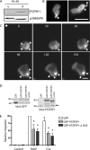
Figure 1.
Figure 1.
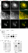
PSTPIP1 localizes to the uropod and regulates cell migration. (A) Immunoblot showing endogenous PSTPIP1 expression in either undifferentiated (u) or differentiated (d) HL-60 cells. Blotting for p38 MAPK shows equal loading. (B) Representative images of human primary neutrophils treated with 100 nM fMLP show localization of endogenous PSTPIP1 toward the rear of the cell. Arrowheads show PSTPIP1 localization at the uropod. Bar, 10 μm. (C) Representative fluorescence time-lapse images of dHL-60 cells that stably express GFP-PSTPIP1. Cells were plated as described in Materials and Methods and exposed to a chemotactic gradient generated by the slow release of C5a from a Femptotip micropipette. Fluorescent time-lapse images were taken at 30-s intervals after exposure to the micropipette. The direction of the micropipette is indicated by a solid white circle. Arrowheads indicate GFP-PSTPIP1 localization. Bar, 10 μm. Shown are representative images from at least three experiments. (D) Immunoblot showing expression of GFP, GFP-PSTPIP1, and GFP-PSTPIP1-ΔSH3 in HL-60 stable cell lines. Differentiated HL-60 cell lines that express GFP, GFP-PSTPIP1 or GFP-PSTPIP1-ΔSH3 were lysed as described in Materials and Methods and probed for GFP or PSTPIP1 as indicated. Arrow indicates endogenous protein expression. (E) Transwell assay was performed in the absence of stimuli (Control) or with C5a or fMLP in the bottom chamber for 3 h using dHL-60 cell lines that stably express control vector (GFP), GFP-PSTPIP1, or GFP-PSTPIP1-ΔSH3. Migration is shown relative to control cells migrating to fMLP (100%). Means ± SEM is shown from four independent experiments. Significant difference compared with control; *p < 0.01, two-way ANOVA, Bonferroni posttests.
Figure 2.
Figure 2.
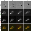
PSTPIP1 and PIPKIγ661 colocalize at the uropod in dHL-60 cells. Representative DIC and fluorescence images of dHL-60 cells that stably express GFP-PSTPIP1 and transiently express mCherry-PIPKIγ661 stimulated with a uniform concentration of 10 nM fMLP. Images were taken at 15-s intervals as indicated. Bar, 10 μm. Arrows show the direction of cell migration.
Figure 3.
Figure 3.
PSTPIP1 colocalizes and interacts with dynamin 2. (A) Representative images of HeLa cells (top panels) and dHL-60 cells (dHL-60; bottom panels) that transiently express mCherry-PSTPIP1 and GFP-dynamin 2. The HeLa cells were fixed as described in Materials and Methods and fluorescence images are shown of the PSTPIP1 (red) and Dynamin 2 (green) and an overlay to show colocalization. Bar, 30 μm. A magnified region of the cell is shown (enlarged region). Bar, 10 μm. Time-lapse images of the dHL-60 cells were taken as described above in the presence of a uniform concentration of fMLP with PSTPIP1 (red), dynamin2 (green), and an overlay. Bar, 10 μm. Arrowheads indicate areas of colocalization. (B) HEK cells transiently transfected with either GFP and PSTPIP1-myc or dynamin 2-GFP and PSTPIP1-myc were lysed; the coimmunoprecipitations with either mouse IgG control or mouse anti-myc were performed as described in Materials and Methods. Immunoblot of the lysates and specific immunoprecipitations are shown. Anti-GFP was used to detect GFP-dynamin 2 and GFP, and anti-PSTPIP1 was used to detect PSTPIP1-myc. All samples were run on the same gel, and the resulting image was adjusted before intervening lanes were removed for clarity. (C) Far Western analysis shows that PSTPIP1 and dynamin 2 directly interact. 35S-labeled PSTPIP was generated from the pcDNA 3.1-PSTPIP1-myc. Exogenously expressed GFP, GFP-dynamin 2, or flag-tagged N-WASP proteins were isolated by immunoprecipitation, resolved by SDS-PAGE, and transferred to nitrocellulose. Membranes were denatured/renatured, blocked, and then incubated with the probe. After an overnight incubation, bound probe was visualized using the STORM PhosphoImager system (left panels). Also shown is the control immunoblot showing amounts of immunoprecipitated proteins (right panels).
Figure 4.
Figure 4.
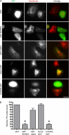
PSTPIP1 expression impairs endocytosis in HeLa cells. (A) HeLa cells were transiently transfected with GFP, GFP-PSTPIP1, GFP-PSTPIP1-ΔSH3 (GFP-ΔSH3), GFP-dynamin 2, or GFP-dynamin 2-K44A (D-K44A-GFP), as indicated. Serum-starved cells were incubated with rhodamine-conjugated transferrin (30 μg/ml) for 10 min and then acid-washed to remove transferrin from the surface. Cells were fixed and imaged. Shown are representative images of GFP expression and rhodamine-transferrin. (B) Quantification of endocytosis. The percent of transfected cells that performed endocytosis of transferrin was determined and expressed relative to GFP control as the mean ± SEM for three independent experiments. Control GFP cells had between 85 and 96% transferrin uptake in each experiment. Significant differences in endocytosis of the raw values between the indicated condition and the GFP-alone cells; *p < 0.01, ANOVA, Tukey posttests.
Figure 5.
Figure 5.
PSTPIP1 does not colocalize with dynamic actin at the leading edge of the cell. Representative DIC and fluorescence images of dHL-60 cells that stably express GFP-PSTPIP1 and transiently express mCherry-actin. Images were taken at 15-s intervals in cells treated with a uniform concentration of 10 nM fMLP. Results are shown for a representative movie from a minimum of three independent experiments. Bar, 10 μm. Arrows show the direction of cell migration.
Figure 6.
Figure 6.
The calponin homology domain from utrophin probes a novel population of actin at the leukocyte uropod. Representative images from dHL-60 cells that transiently express RFP-Utr-CH (red) and GFP-actin (green) and were treated with a uniform concentration of 10 nM fMLP. Images were taken every 15 s as indicated. Results are shown for a representative movie from a minimum of three independent experiments. Bar, 10 μm. Arrows show the direction of cell migration.
Figure 7.
Figure 7.
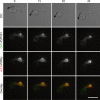
PSTPIP1 colocalizes with the Utr-CH probe at the uropod of dHL-60 cells. Representative DIC and fluorescence images from dHL-60 cells that stably express GFP-PSTPIP1 (green) and transiently express RFP-Utr-CH (red). Cells were treated with a uniform concentration of 10 nM fMLP, and images were taken every 15 s as indicated. Results are shown for a representative movie from a minimum of three independent experiments. Bar, 10 μm. Arrows show the direction of cell migration.
Figure 8.
Figure 8.
Disruption of the actin cytoskeleton perturbs PSTPIP1 localization at the uropod and induces membrane tubulation. (A) Representative DIC and fluorescence images from dHL-60 cells that transiently express RFP-Utr-CH and were treated with a uniform concentration of 10 nM fMLP. Shown is one frame before addition of latrunculin B (1 μM) and one frame 30 s after addition. (B) Representative DIC and fluorescence images from dHL-60 cells that stably express GFP-PSTPIP1 and were treated with a uniform concentration of 10 nM fMLP. Images were taken every 30 s as indicated and latrunculin B (1 μM) was added at 90 s as indicated. Results are shown for a representative movie from a minimum of three independent experiments. Bar, 10 μm.
Figure 9.
Figure 9.
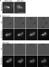
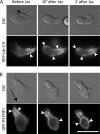
Stabilization of the actin cytoskeleton perturbs PSTPIP1 localization to the uropod. Representative DIC and fluorescence images from dHL-60 cells that transiently express either (A) RFP-Utr-CH or (B) GFP-PSTPIP1 and were treated with a uniform concentration of 10 nM fMLP. Images were taken before addition of jasplakinolide (1 μM) and afterward, with the 30-s and 2-min time points shown. Arrows indicate direction of cell movement. Arrowheads indicate areas of localization. Bar, 10 μm.

Similar articles

Cited by

References

    1. Babior B. M., Matzner Y. The familial Mediterranean fever gene—cloned at last. N. Engl. J. Med. 1997;337:1548–1549. - PubMed
    1. Badour K., Zhang J., Shi F., McGavin M. K., Rampersad V., Hardy L. A., Field D., Siminovitch K. A. The Wiskott-Aldrich syndrome protein acts downstream of CD2 and the CD2AP and PSTPIP1 adaptors to promote formation of the immunological synapse. Immunity. 2003;18:141–154. - PubMed
    1. Bairstow S. F., Ling K., Su X., Firestone A. J., Carbonara C., Anderson R. A. Type Igamma661 phosphatidylinositol phosphate kinase directly interacts with AP2 and regulates endocytosis. J. Biol. Chem. 2006;281:20632–20642. - PubMed
    1. Bennin D. A., Don A. S., Brake T., McKenzie J. L., Rosenbaum H., Ortiz L., DePaoli-Roach A. A., Horne M. C. Cyclin G2 associates with protein phosphatase 2A catalytic and regulatory B′ subunits in active complexes and induces nuclear aberrations and a G1/S phase cell cycle arrest. J. Biol. Chem. 2002;277:27449–27467. - PubMed
    1. Bretscher M. S. Moving membrane up to the front of migrating cells. Cell. 1996;85:465–467. - PubMed

Publication types

LinkOut - more resources