Review
. 2008 Jun;9(6):437-52.
doi: 10.1038/nrn2392. Epub 2008 May 14.

Neuregulin 1 in neural development, synaptic plasticity and schizophrenia

Affiliations
Review

Neuregulin 1 in neural development, synaptic plasticity and schizophrenia

Lin Mei et al. Nat Rev Neurosci. 2008 Jun.

Abstract

Schizophrenia is a highly debilitating mental disorder that affects approximately 1% of the general population, yet it continues to be poorly understood. Recent studies have identified variations in several genes that are associated with this disorder in diverse populations, including those that encode neuregulin 1 (NRG1) and its receptor ErbB4. The past few years have witnessed exciting progress in our knowledge of NRG1 and ErbB4 functions and the biological basis of the increased risk for schizophrenia that is potentially conferred by polymorphisms in the two genes. An improved understanding of the mechanisms by which altered function of NRG1 and ErbB4 contributes to schizophrenia might eventually lead to the development of more effective therapeutics.


Figures

Fig. 1
Fig. 1
a | The six types of neuregulin 1 (NRG1) isoforms are classified according to their distinct amino-terminal sequences. In the type III isoforms, this sequence contains a cysteine-rich domain (CRD) that has a transmembrane domain (TMn). All six types of NRG1 isoforms have an epidermal growth factor (EGF)-like domain. Types I, II, IV and V have an immunoglobulin (Ig)-like domain between the N-terminal sequence and the EGF domain, with or without the spacer region (S), whereas the N-terminal-specific region of types III and VI is connected directly to the EGF domain. Variants are also generated by splicing in the linker regions and the C-terminal regions. Between the two regions is a C-terminal transmembrane domain (TMc). b | Most NRG1 isoforms are synthesized as transmembrane precursor polypeptides (pro-NRG1s) with the EGF domain located in the extracellular region, but in Type III NRG1 both the N- and the C-terminal regions are located inside the cell. Cleavage by tumour necrosis factor-α converting enzyme, β-site of amyloid precursor protein cleaving enzyme or meltrin β(indicated by the lightning arrow) generates mature NRG1s that are soluble, except in the case of Type III NRG1, which is thought to function in a manner that requires cell contact. The processing of Type IV, Type V and Type VI pro-NRG1s is less well-characterized but is thought to resemble that of Type I and Type II.
Fig. 2
Fig. 2
ErbB proteins are type I transmembrane receptor tyrosine kinases. Each has an extracellular region containing two extracellular cysteine-rich domains, a transmembrane domain, a short intracellular juxtamembrane region, a tyrosine kinase domain and a carboxy-terminal tail. In response to neuregulin 1 (NRG1) stimulation, ErbB proteins become dimerized to form homo- and heterodimers, such as ErbB2–ErbB3, ErbB4–ErbB4, ErbB2–ErbB4 and ErbB4–epidermal growth factor receptor (EGFR). ErbB2 does not bind to NRG1 (indicated by the black crosses) but has an active kinase domain; ErbB3 binds to NRG1 but has an impaired tyrosine kinase domain (indicated by the purple cross). Therefore, ErbB2 and ErbB3 need to form heterodimers with each other or with ErbB4 to be functional, whereas ErbB4 homodimers can bind to NRG1 and become activated. Activation of the tyrosine kinase domains leads to auto- and trans-phosphorylation of the intracellular domains, generating docking sites for the adaptor proteins Grb2 and Shc, which activate the Raf–MEK–ERK pathway, and for the p85 subunit of PI3K (indicated in the figure for the ErbB2–ErbB3 heterodimer), which activates the PI3K pathway and subsequently mTOR-dependent protein synthesis. NRG1 activates CDK5, which is also stimulated by neuronal activity. EGFR does not bind to NRG1. Its downstream pathways include ones that involve JNK, Src, Abl, and PKC in addition to ones that involve ERK and PI3K/Akt.
Fig. 3
Fig. 3
a | Inclusion of exon 26 in the intracellular domain of ErbB4 generates CYT-1, whereas its exclusion gives rise to CYT-2 (Refs 188,189). The extracellular juxtamembrane region of ErbB4 is encoded by either exon 16 or exon 15, to generate JMa or JMb, respectively. Different combinations of CYT-1, CYT-2, JMa and JMb thus create 4 isoforms of ErbB4. Exon numbers are shown on the top of corresponding domain structures. b | CYT-1 contains the motif that binds to the p85 subunit of PI3K and thus activates this kinase. Thus, although both isoforms are coupled to the Raf–MEK–ERK pathway, CYT-1 but not CYT-2 activates PI3K and subsequently Akt, , . Both JMa and JMb can be activated by neuregulin 1 (NRG1) to initiate canonical signalling, but only JMa can be cleaved by tumour necrosis factor-α converting enzyme, , (indicated by the lightning arrow) to generate a soluble extracellular polypeptide, ecto-ErbB4 (which contains the NRG1 binding site), and a carboxy-terminal fragment (ErbB4-CTF). CRD, cysteine-rich domain; TK, tyrosine kinase domain; TM, transmembrane domain.
Fig. 4
Fig. 4
In non-canonical forward signalling (bottom cell, right-hand pathway), the carboxy-terminal fragment (ErbB4-CTF) is cleaved by γ-secretase to produce ErbB4-intracellular domain (ErbB4-ICD), which can translocate to the nucleus to regulate gene expression. When it is overexpressed in transfected cells, ErbB4-ICD interacts with several transcriptional regulators, including Eto2, STAT5, Mdm2 and YAP, to mediate the transcriptional activation or repression of heterologous promoters (not shown), , , , , . This interaction might require the phosphorylation of either ErbB4-ICD or the transcriptional regulator and/or the kinase activity of the ICD domain. For example, neuregulin 1 (NRG1) stimulation promotes the association of ErbB4-ICD with TAB2, an adaptor protein, in an ErbB4 kinase-domain-dependent manner. TAB2 also interacts with the nuclear receptor co-repressor, N-CoR, to form a ternary complex that, upon translocation into the nucleus, represses the transcription of genes that are required for the differentiation of neural precursor cells into astrocytes. Backward signalling (top cell) by pro-NRG1 can proceed by two mechanisms. First, the C-terminal fragment of pro-NRG1 (NRG1-CTF), which is generated by extracellular cleavage, can be cleaved again by γ-secretase to generate NRG1-intracellular domain (NRG1-ICD), which can relocate into the nucleus to regulate gene transcription (left-hand pathway). Second, ErbB4 or ecto-ErbB4, which is released by extracellular cleavage, can serve as a ligand for pro-NRG1 or Type III NRG1, which function as receptors (right-hand pathway). It is unknown whether this interaction alters the phosphorylation of pro-NRG1 itself or whether it alters the activation of an intracellular kinase or phosphatase in pro-NRG1-expressing cells. Precisely how the signals are transduced also remains unknown. The cytoplasmic tail of pro-NRG1 interacts with the non-receptor protein kinase LIM kinase 1 (LIMK1), . This kinase has been shown to regulate actin dynamics in many cell types, including neurons. Interestingly, NRG1-ICD is required for NRG1 function in vivo. Treatment of Type III NRG1-expressing neurons with a mixture of ecto-ErbB2 and ecto-ErbB4 promotes neuronal survival in vitro and alters the expression of several apoptotic genes (not shown). Canonical forward signalling (bottom cell, left-hand pathway) is explained in detail in Fig. 2.
Fig. 5
Fig. 5
a | Neuregulin 1 (NRG1) (indicated by blue shading) is released from neurons to promote the formation and maintenance of radial glial cells, which are necessary for the radial migration of neurons from ventricular zones to the pial surface. b | Tangential migration of GABA (γ-aminobutyric acid)-ergic interneurons requires NRG1 in the cortical region; thalamocortical axon navigation through the diencephalon requires corridor cells that express NRG1. c | Myelination and ensheathment of peripheral nerves are controlled by the amounts of NRG1 produced in substrate axons. d | NRG1 from axons might regulate oligodendrocyte development and myelination of axons in the CNS. e | NRG1 is necessary for the formation of neuromuscular junctions (NMJs), probably through effects on terminal Schwann cell differentiation and survival. f | NRG1 stimulates CNS synapse formation. This panel shows typical excitatory synapses, formed between glutamatergic terminals and spines. DT, dorsal thalamus; GP, globus pallidus; LGE, lateral ganglionic eminence; MGE, medial ganglionic eminence. Part b modified, with permission, from Ref. © (2006) Elsevier Science. Part d modified, with permission, from Ref. © (2005) Macmillan Publishers Ltd.
Fig. 6
Fig. 6
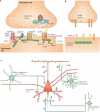
a | ErbB4 interacts specifically with the first and second PDZ domains of postsynaptic density (PSD) protein 95 (PSD95), a scaffold protein, and is localized in the PSD of excitatory synapses. The interaction with PSD95 enhances neuregulin 1 (NRG1) signalling, presumably by increasing ErbB4 homodimerization. NRG1, by activating ErbB4, suppresses long-term potentiation induction and expression, , , , . The mechanisms that underlie this effect remain unclear. Through PSD95, ErbB4 signalling might regulate the properties of NMDA (N-methyl-D-aspartate) receptors (NMDARs), AMPA (α-amino-3-hydroxy-5-methyl-4-isoxazole propionic acid) receptors (AMPARs) and K+ channels (K+ ch). Through the GKAP–Shank–Homer complex, ErbB4 signalling might be involved in regulating the function of metabotropic glutamate receptors (mGluRs). PSD95 might also recruit ErbB4 to the neuroligin–neurexin complex that is essential for synapse formation. ErbB2, on the other hand, interacts with erbin, a protein that contains multiple leucine-rich domains and a PDZ domain, . This interaction has been implicated in regulating NRG1 signalling, . b | ErbB4 is present in the presynaptic terminals of GABA (γ-aminobutyric acid)-ergic interneurons. NRG1 stimulates presynaptic ErbB4 to enhance activity-dependent GABA release through mechanisms that have yet to be identified. c | A working hypothesis for how NRG1 might regulate pyramidal neuron activity. The output of pyramidal neurons in the prefrontal cortex (PFC) is regulated by excitatory glutamatergic neurons (shown in red) and various inhibitory GABAergic interneurons (shown in green). NRG1 regulates glutamatergic transmission and/or plasticity by activating PSD-localized ErbB4. There are at least three types of GABAergic interneurons in the PFC. Wide-arbor basket cells target the somata and proximal dendrites of pyramidal neurons and adjust the integrated synaptic response. Chandelier cells (or axon-targeting interneurons) terminate at or near the axon hillock of pyramidal neurons, forming vertical arrays of terminals termed ‘cartridges’, to regulate the generation and timing of action potentials. Conversely, Martinotti cells terminate on distal dendrites of pyramidal cells to influence the dendritic processing and integration of synaptic inputs, , , , . By controlling activity-dependent GABA release, NRG1 might repress the activity of pyramidal neurons. Part c modified, with permission, from Ref. © (2005) Macmillan Publishers Ltd.
Fig. 7
Fig. 7
a | The neuregulin 1 (NRG1) gene is located in a 1.5 Mb region of DNA, at 8p12−8p21. Roman numerals indicate the type-specific exons. The original deCODE hyplotype including the SNPs SNP8NRG221132, SNP8NRG221533, SNP8NRG241930, SNP8NRG243177 and SNP8NRG433E1006 and two microsatellites (478B14−848 and 420M9−1395) is shown. Exons for individual domains of NRG1 are colour matched with the domain structure in Fig. 1a. b | The 1.15 Mb region of the ERBB4 gene, at 2q33.3−2q34. The SNPs are mainly clustered around exon 3 and in front of exon 13.

Similar articles

Cited by

References

    1. Carlsson A, Lindqvist M. Effect of chlorpromazine or haloperidol on formation of 3methoxytyramine and normetanephrine in mouse brain. Acta Pharmacol. Toxicol. 1963;20:140–144. - PubMed
    1. Laruelle M, Abi-Dargham A. Dopamine as the wind of the psychotic fire: new evidence from brain imaging studies. J. Psychopharmacol. 1999;13:358–371. - PubMed
    1. Harrison PJ, Weinberger DR. Schizophrenia genes, gene expression, and neuropathology: on the matter of their convergence. Mol. Psychiatry. 2005;10:40–68. - PubMed
    1. Tan W, et al. Molecular cloning of a brain-specific, developmentally regulated neuregulin 1 (NRG1) isoform and identification of a functional promoter variant associated with schizophrenia. J. Biol. Chem. 2007;282:24343–24351. - PubMed
    1. Falls DL. Neuregulins: functions, forms, and signalling strategies. Exp. Cell Res. 2003;284:14–30. - PubMed

Publication types