. 2008 May;28(5):698-709.
doi: 10.1016/j.immuni.2008.03.014. Epub 2008 May 1.

Vaccine adjuvants alter TCR-based selection thresholds

Affiliations

Vaccine adjuvants alter TCR-based selection thresholds

Laurent Malherbe et al. Immunity. 2008 May.

Abstract

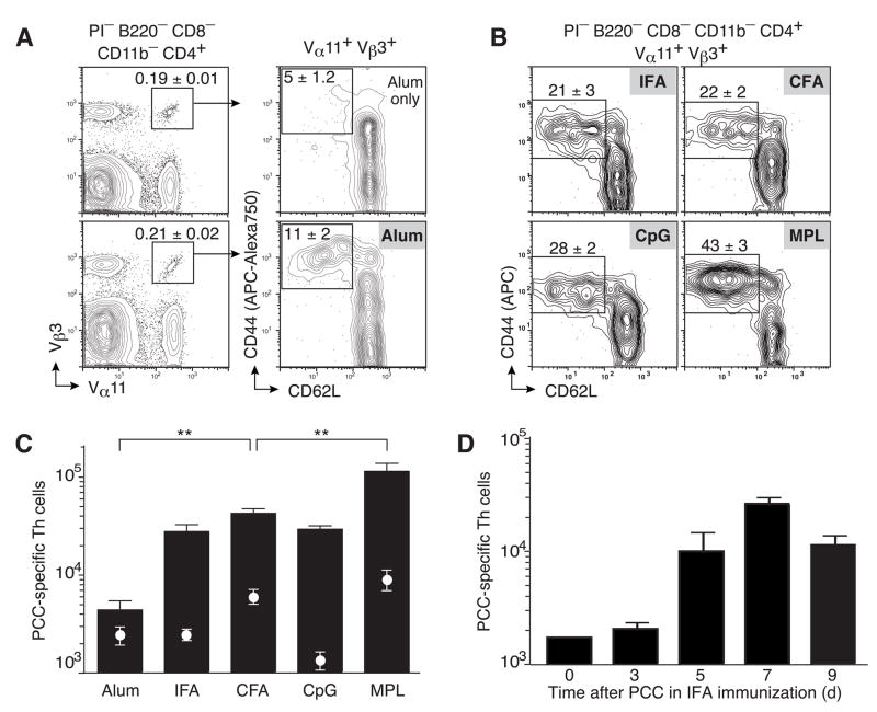
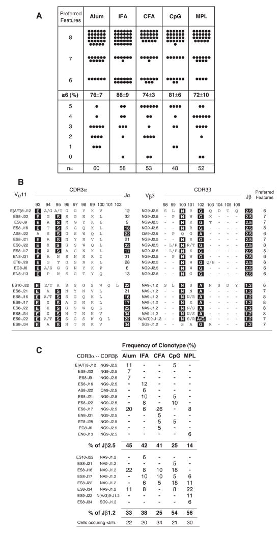
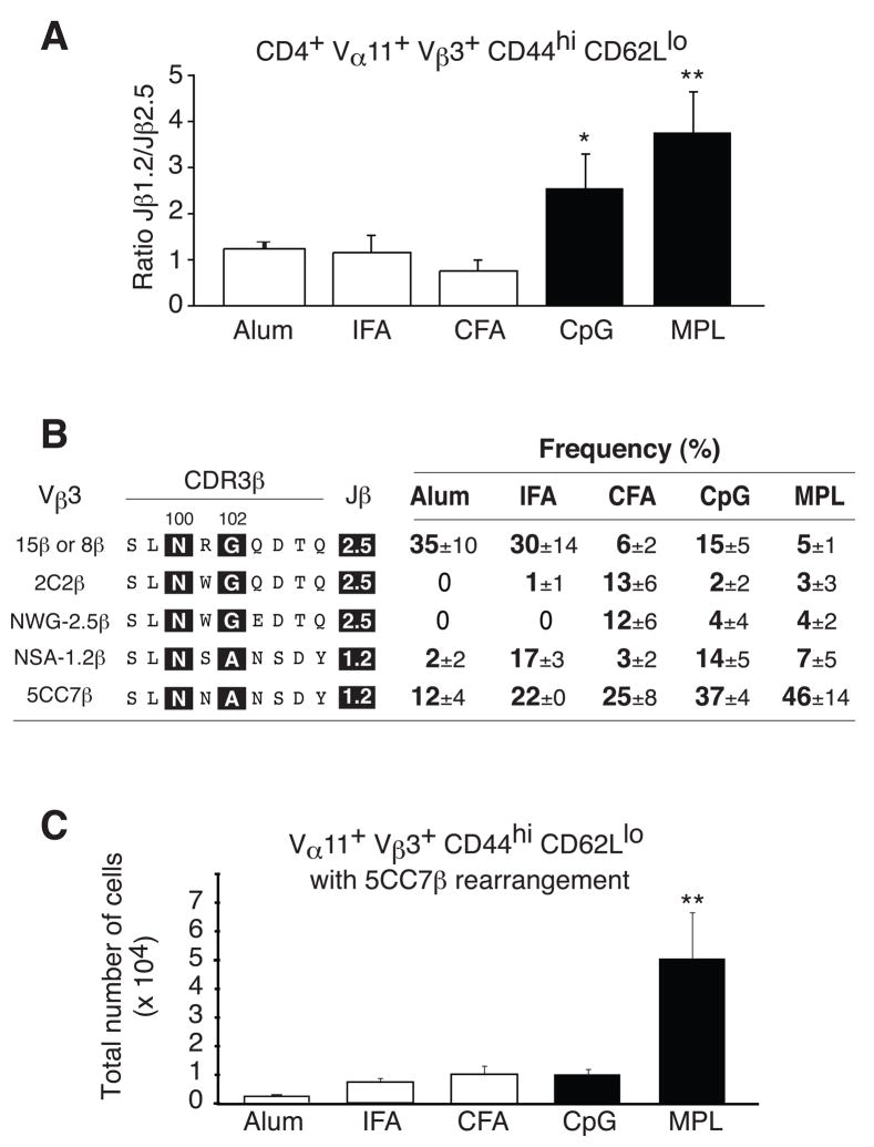
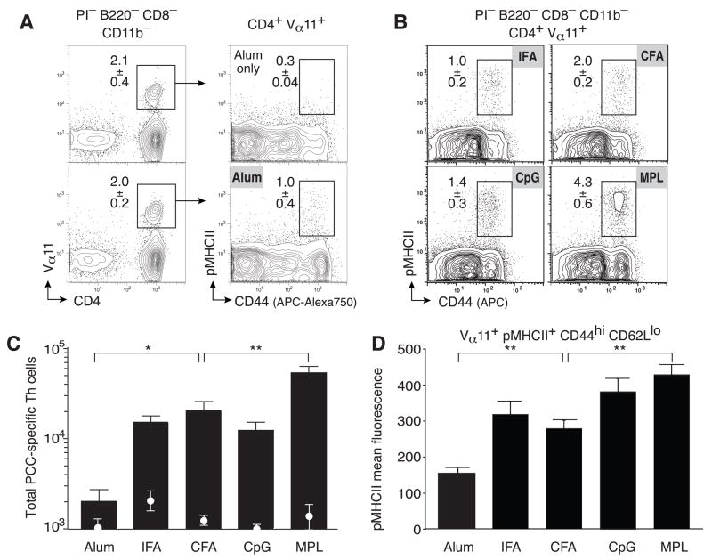
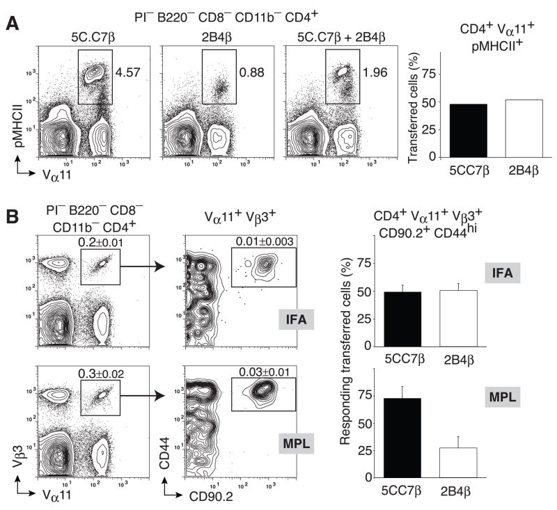
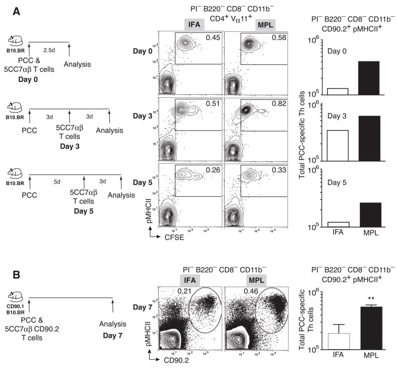
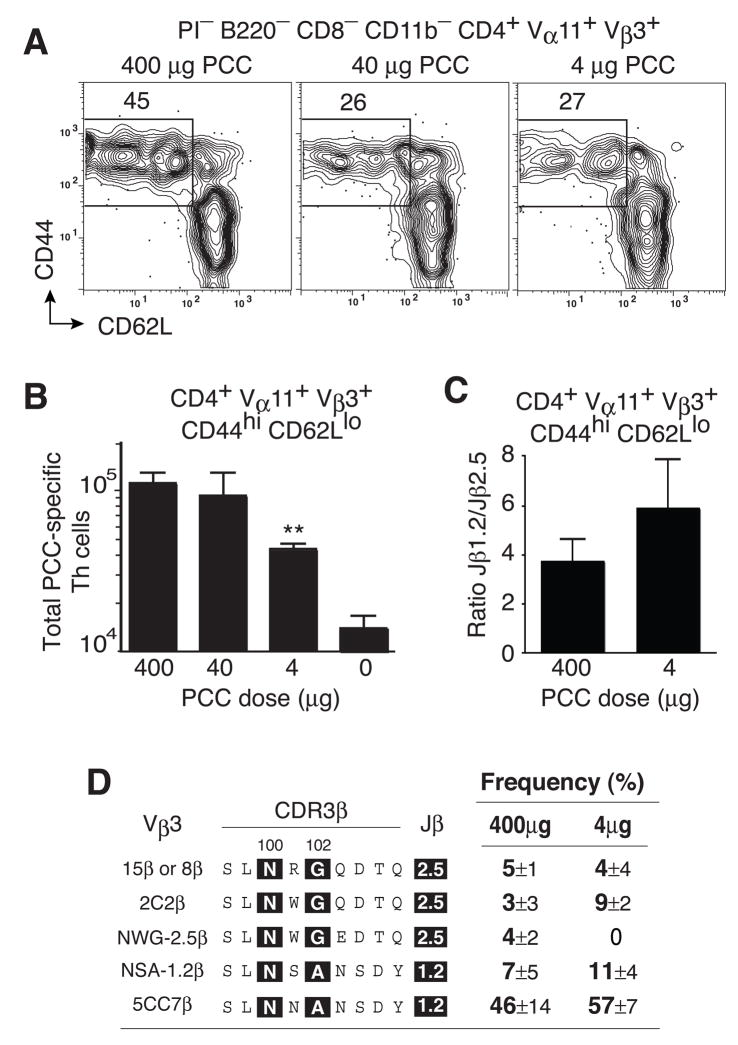
How T cell receptor (TCR) specificity evolves in vivo after protein vaccination is central to the development of helper T (Th) cell function. Most models of clonal selection in the Th cell compartment favor TCR affinity-based thresholds. Here, we demonstrated that depot-forming vaccine adjuvants did not require Toll-like receptor (TLR) agonists to induce clonal dominance in antigen-specific Th cell responses. However, readily dispersible adjuvants using TLR-9 and TLR-4 agonists skewed TCR repertoire usage by increasing TCR selection thresholds and enhancing antigen-specific clonal expansion. In this manner, vaccine adjuvants control the local accumulation of Th cells expressing TCR with the highest peptide MHC class II binding. Clonal composition was altered by mechanisms that blocked the local propagation of clonotypes independently of antigen dose and not as a consequence of interclonal competition. This capacity of adjuvants to modify antigen-specific Th cell clonal composition has fundamental implications for the design of future protein subunit vaccines.


Figures

Figure 1
Figure 1. Local accumulation of antigen-specific Th cells across different adjuvants
(a,b) PCC-specific T helper cells (Vα11+Vβ3+CD44hiCD62Llo) at day 7 in lymph nodes from B10.BR mice immunized with (a) Alum with (lower panels) or without PCC (upper panels) or (b) with PCC and the indicated adjuvant. Profiles gated on propidium iodide (PI) negative cells that are CD4+B220CD8CD11b and Vα11+Vβ3+ as indicated with mean ±SEM (n≥3) percent of cells within insert box (c) Total number of PCC-specific T helper cells 7 days after immunization with Alum (n=7), IFA (n=6), CFA (n=15), CpG (n=6) and MPL (n=25) with (bars) or without PCC (circles); means ±SEM n≥3 p ≤ 0.01 (**) (two-tailed t-test) comparing Alum or MPL to any other adjuvant. (d) Total number of PCC-specific T helper cells 3, 5, 7 or 9 days after immunization with IFA and PCC. Mean ±SEM for at least three animals.
Figure 2
Figure 2. Clonal dominance without TLR agonists or antigen depots
Single-cell repertoire analysis of individual PCC-specific T helper cells (Vα11+Vβ3+CD44hi CD62Llo) sorted from mice immunized with PCC and indicated adjuvant. (a) Each filled circle represents sequence from single cells representing the number of preferred CDR3 features known to be selected in the PCC response (TCR-α: E at α93; S at α 95; CDR3α length of 8aa; and TCRJα 16, 17, 22 and 34. TCR-β: N at β100; A/G at β102; CDR3β length of 9aa; and TCRJβ 1.2 and 2.5). Cells with ≥ 6 preferred features express restricted TCR of the dominant clonotype and the percentage ±SEM; across 3 individual animals with n = number of single cells used in the analysis displayed with individual animals contributing different numbers of sequences, Alum (n=21,23,16); IFA (n=22,18,18); CFA (n=18,17,18); CpG (n=15,16,17) MPL (n=16,19,17) (b) TCR sequences from the dominant clonotypes (≥6 preferred CDR3 features). Columns (left to right): individual CDR3α chain designation (based on amino acids at α93 and α95, CDR3α length and Jα usage); CDR3α, with positions α93E and α95S ‘highlighted’ in black as canonical, motif length depicted; Jα gene usage; individual CDR3β designation (based on amino acids at β100 and β102, CDR3β length and Jβ usage); CDR3β, with positions β100N and β102A or β102G ‘highlighted’ in black as canonical, motif length depicted; Jβ gene segment usage; total number of ‘preferred’ features in both CDR3 regions combined. (c) Penetrance of dominant clonotypes after priming with PCC and the indicated adjuvant as a percent of dominant clonotypes and organized in the same order as (b) with summaries for each adjuvant across Jβ2.5 and Jβ1.2 usage as displayed.
Figure 3
Figure 3. Adjuvants skew TCR β chain usage
(a) Relative abundance of PCC-specific Th cells (Vα11+Vβ3+CD44hiCD62Llo) expressing Jβ1.2 or Jβ2.5 gene segments for the indicated adjuvant (mean ±SEM.; n≥3). (b) Predicted amino acid sequences of CDR3β regions mean ±SEM of five predominant clonotypes (n≥3) for PCC-specific Th cells isolated from mice immunized with indicated adjuvant (c) Total number of PCC-specific Th cells expressing the 5C.C7β (SLNNANSDY) rearrangement mean ±SEM n≥3.
Figure 4
Figure 4. Adjuvants re-set the TCR selection threshold
PCC-specific T helper cells as PICD8B220CD11b cells expressing CD4 and Vα11, binding pMHCII tetramers and expressing high level of CD44 displayed within the inserted box; percent of cells mean±SEM n≥3 at day 7 in lymph nodes from B10.BR mice immunized with (a) Alum with (lower panels) or without PCC (upper panels) or (b) with PCC and the indicated adjuvant. (c) Total numbers of CD4+Vα11+pMHCII+CD44hiCD62Llo T helper cells after immunization with (bars) or without (circles) PCC and the indicated adjuvant; means ±SEM n≥3 p ≤ 0.01 (**) (two-tailed t-test) comparing Alum or MPL to any other adjuvant. (d) Mean fluorescence intensity of pMHCII tetramer staining for PCC-specific T helper cells after immunization with PCC and the indicated adjuvant (mean ±SEM; n≥3).
Figure 5
Figure 5. Blocking the selection of low affinity clonotypes
0.33×10⁵ 5C.C7β and 1.2×10⁵ 2B4β splenocytes were mixed and (a) stained with pMHCII tetramers for pre-immune repertoire analysis or (b) transferred into Thy1.1 syngeneic hosts. Transferred mice were immunized with 400μg PCC in IFA or MPL-based adjuvant. (a) Representative probability contours of pMHCII tetramer staining versus Vα11 for 5C.C7β (left), 2B4β (middle) and the 5C.C7β/2B4β cell mixture as a profile of cells (right) and evaluation of cell origin after cell sorting (Vα11+pMHCII+) and RT-PCR for Jβ1.2/2.5 expression representing 5CC7β and 2B4β respectively (bar graph) (b) Representative probability contours of CD44 and CD90.2 expressions (right panel) by Vα11+ Vβ3+ (left panel) cells from draining lymph nodes of recipient mice 7 days after immunization with IFA (upper panels) or MPL-based adjuvant (lower panels); single antigen-experienced PCC-specific T helper cells (Vα11+Vβ3+CD90.2+CD44hi) were sorted and the relative abundance of 5C.C7β and 2B4β T helper cells was evaluated by single-cell RT-PCR using Jβ1.2 and Jβ2.5 specific primers, respectively. Means ±SEM n=3 for each condition.
Figure 6
Figure 6. TCR-independent enhancement of clonal accumulation
(a) 2×10⁵ CFSE-labeled 5C.C7αβ splenocytes were transferred into syngeneic B10.BR hosts at the time of immunization (upper panels), 3 days (middle panels) or 5 days (lower panels) after immunization with PCC and IFA or MPL-based adjuvant. Representative probability contours of pMHCII tetramer staining versus CFSE for IFA (left) and MPL (middle) immunized recipients. Total number of Vα11+CFSElopMHCIITet+ cells in draining lymph nodes of recipients immunized with IFA or MPL-based adjuvant in bar graphs (far right). (b) 2×10⁵ 5C.C7αβ splenocytes were transferred into CD90.1+ syngeneic hosts and recipients were immunized with PCC and IFA or MPL-based adjuvant. Representative probability contours of pMHCII tetramer staining versus CD90.2 for IFA(left) and MPL (middle) immunized recipients. Total number of Vα11+CD90.2+pMHCII+ cells in draining lymph nodes of recipients immunized with IFA or MPL-based adjuvant (far right), mean ±SEM n=3 p< 0.01 (**)(two-tailed Student’s t-test).
Figure 7
Figure 7. Selection threshold is re-set independent of the antigen dose
(a) PCC-specific T helper cells (Vα11+Vβ3+CD4 4hiCD62Llo) at day 7 in lymph nodes from mice immunized with MPL-based adjuvant containing 400μg (left), 40μg (middle) or 4μg (right) of PCC. (b) Total number of PCC-specific T helper cells after immunization with 400, 40, 4 and 0 μg PCC. mean ±SEM n≥3. p < 0.05 (*) two-tail Student’s t-test, comparing to any other antigen dose. (c) Relative abundance of PCC-specific Th cells expressing Jβ1.2 or Jβ2.5 gene segments for indicated PCC dose, mean ±SEM n≥3 (d) Prevalence of 5 dominant TCRβ chain expressing clonotypes comparing 400ug dose (data from Fig 3c) to 4ug dose, mean ±SEM n=3.

Comment in

Similar articles

Cited by

References

    1. Baldridge JR, Crane RT. Monophosphoryl lipid A (MPL) formulations for the next generation of vaccines. Methods. 1999;19:103–107. - PubMed
    1. Baldridge JR, McGowan P, Evans JT, Cluff C, Mossman S, Johnson D, Persing D. Taking a Toll on human disease: Toll-like receptor 4 agonists as vaccine adjuvants and monotherapeutic agents. Expert Opin Biol Ther. 2004;4:1129–1138. - PubMed
    1. Banchereau J, Steinman RM. Dendritic cells and the control of immunity. Nature. 1998;392:245–252. - PubMed
    1. Bikah G, Pogue-Caley RR, McHeyzer-Williams LJ, McHeyzer-Williams MG. Regulating T helper cell immunity through antigen responsiveness and calcium entry. Nat Immunol. 2000;1:402–412. - PubMed
    1. Billiau A, Matthys P. Modes of action of Freund’s adjuvants in experimental models of autoimmune diseases. J Leukoc Biol. 2001;70:849–860. - PubMed

Publication types

MeSH terms