. 2008 Jun;9(2):225-40.
doi: 10.1007/s10162-008-0119-x. Epub 2008 May 1.

Transplantation of mouse embryonic stem cells into the cochlea of an auditory-neuropathy animal model: effects of timing after injury

Affiliations

Transplantation of mouse embryonic stem cells into the cochlea of an auditory-neuropathy animal model: effects of timing after injury

Hainan Lang et al. J Assoc Res Otolaryngol. 2008 Jun.

Abstract

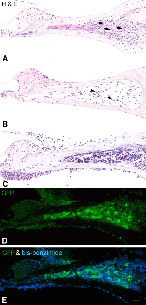
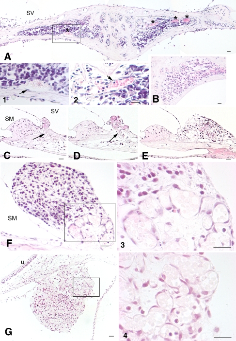
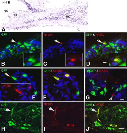
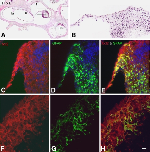
Application of ouabain to the round window membrane of the gerbil selectively induces the death of most spiral ganglion neurons and thus provides an excellent model for investigating the survival and differentiation of embryonic stem cells (ESCs) introduced into the inner ear. In this study, mouse ESCs were pretreated with a neural-induction protocol and transplanted into Rosenthal's canal (RC), perilymph, or endolymph of Mongolian gerbils either 1-3 days (early post-injury transplant group) or 7 days or longer (late post-injury transplant group) after ouabain injury. Overall, ESC survival in RC and perilymphatic spaces was significantly greater in the early post-injury microenvironment as compared to the later post-injury condition. Viable clusters of ESCs within RC and perilymphatic spaces appeared to be associated with neovascularization in the early post-injury group. A small number of ESCs transplanted within RC stained for mature neuronal or glial cell markers. ESCs introduced into perilymph survived in several locations, but most differentiated into glia-like cells. ESCs transplanted into endolymph survived poorly if at all. These experiments demonstrate that there is an optimal time window for engraftment and survival of ESCs that occurs in the early post-injury period.


Figures

FIG. 1
FIG. 1
Approaches used for ESC transplantation and the state of differentiation of ESCs after RA neural induction. A Surgical approach to expose the round window (RW) niche of the gerbil cochlea for ESC transplantation. The basilar membrane comprises the translucent zone and is visible through the RW membrane. The bony osseous spiral lamina, the wall of Rosenthal’s canal (RC), and the central modiolus are located in the opaque zone. Scale bar = 500 μm. B Schematic diagram illustrating three delivery routes of ESCs into: (1) Rosenthal’s canal, (2) perilymph of scala tympani (ST), (3) and endolymph of scala media (SM). The photograph was modified from a radial paraffin section of the basal turn from a normal young gerbil. Sa stapedial arteria, Sp.L spiral ligament, SV scala vestibuli. Scale bar = 100 μm. C The majority of the cultured ESCs stained for nestin (green). Nuclei were countstained with bis-benzimide (blue). Scale bar = 20 μm.
FIG. 2
FIG. 2
Survival of transplanted ESCs in RC of ouabain-exposed cochleas. A An H&E-stained radial section through the basal turn of the gerbil cochlea shows the normal profiles of SGNs (arrows) within RC. B Only a few neurons (arrowheads) remain in RC in a ouabain-exposed cochlea 1 month after recovery. C A large number of ESCs survive in RC of an “early post-injury” (see text) cochlea 3 weeks after transplantation. ESCs are easily identified by their irregular shape and large, dark blue staining nuclei. D, E Immunostaining for GFP (green) in a section adjacent to the one shown in C identifies the grafted cells as ESCs. Nuclei were counterstained with bis-benzimide (blue). Scale bar = 50 μm.
FIG. 3
FIG. 3
Transplanted ESCs survived at several other locations in ouabain-exposed cochleas. All sections except the one in E were obtained from early post-injury cochleas 3–4 weeks after ESC transplantation. A A radial section through the basal organ of Corti shows an ESC mass (arrow) in the perilymphatic space of the scala vestibuli (SV) adjacent to the suprastrial (SS) area. B A mass of grafted ESCs (arrow) survived in the perilymphatic space above RC within the SV. Bony defects (arrowheads) caused by the injection pipette are evident on both the SV and scala tympani (ST) sides. There was a large amount of cell debris with some surviving ESCs (white arrows) outside the RW membrane. C ESCs were seen within the SV in the apical turn. D ESCs (arrow) were present within the perilymphatic space outside the vestibular duct. E Radial section through the basal turn showing the lateral wall of a normal gerbil cochlea. F, G Surviving ESCs (asterisk) are present between Reissner’s membrane (RM) and the SS region in the basal turn. SM scala media, St.V stria vascularis, la lateral ampulla, u utricle. Scale bar = 50 μm.
FIG. 4
FIG. 4
ESCs survived poorly within scala media (SM) after endolymphatic transplantation. A Radial section through upper basal turn of an early post-injury cochlea 11 days after ESCs were injected into endolymph. B Higher magnification view of the boxed area in A indicates that most ESCs within the scala media were dead or dying (arrows). C Another radial section through the organ of Corti in the hook 3 weeks after ESCs were injected into the endolymph. D: Higher magnification view of boxed area in C indicates that most ESCs within the scala media were undergoing apoptosis (arrows). Scale bar (for A and C) = 50 μm. Scale bar (for B and D) = 10 μm.
FIG. 5
FIG. 5
EP values and DPOAEs were reduced after introducing ESCs into the scala media. A The same animal shown in Fig. 4a. Left panel shows that CAP responses were absent across all frequencies in the treated ear. EP values were reduced about 20–30 mV at turn one, turn two, and turn three compared to the mean EPs from a group of control gerbils (n = 12). Right panel demonstrates that DPOAEs were reduced across all frequencies. Dotted line indicates the noise floor of the system. B An early post-injury cochlea 3 weeks after ESCs were injected into the perilymphatic space. Left panel demonstrates that CAP responses were absent across all frequencies in the treated ear, whereas EP values were relatively normal. Right panel showed that DPOAEs remained similar to control levels in this ear.
FIG. 6
FIG. 6
Remodeling of blood vessels in early post-injury cochleas. A H&E-stained section through RC taken 3 weeks after ESCs were introduced into RC. Increased blood vessel areas (asterisks) are evident within RC. 1, 2 Higher magnification views of the boxed areas in A showed the morphological characteristics of endothelial cells (arrows) and red blood cells. B Radial section through RC taken from a normal gerbil ear. CF Sections were taken from another ouabain-exposed cochlea 3 weeks after transplantation. Microvessels (arrows) were found arising from RC across the broken bone and forming a cluster underneath an ESC mass in the supralimbal region. CE are about 20 μm apart from each other. F is about 40 μm distance from E. 3 Higher magnification of the boxed area in F. G The section was taken from an ouabain-exposed cochlea 3 weeks after transplantation. Association of an ESC mass and a cluster of microvasculature was seen underneath of utricle. 4 Higher magnification of the boxed area in G. Scale bars = 30 μm.
FIG. 7
FIG. 7
Neuronal differentiation of transplanted ESCs in RC of early post-injury cochleas. All sections were obtained from two cochleas 3 weeks after transplantation with GFP-expressing ESCs. Dual immunostaining for GFP (green) and NF 200 (red) antibodies was used to identify ESCs differentiating towards a mature neuronal phenotype. A Radial section stained with H&E shows the profile of the RC transplanted with ESCs. BD Double staining with GFP and NF 200 antibodies shows an ESC (arrow) that has differentiated into a NF-200-positive neuron-like cell (yellow). The section is about 20 μm apical to the one shown in A. E, F Section from another transplanted ear showing two NF-200-positive cells differentiated from ESCs. G A native neuron (arrowhead) remaining in RC is not GFP-positive. HJ Confocal images of a radial section stained for GFP and NF 200 antibodies show a neuron-like cell with its process (arrow) differentiated from an ESC. The section was taken from the ear shown in sections E and F. SM scala media, SL spiral limbus. Scale bar = 15 μm.
FIG. 8
FIG. 8
Glia-like cell differentiation of ESCs in RC of early post-injury cochleas. All sections were obtained from a cochlea 3 weeks after transplantation with wild type ESCs. Dual immunostaining for M2 (red) and GFAP (green) antibodies was used to identify ESCs differentiating towards a glial phenotype. M2 is a mouse-specific antigen and can be used to identify the transplanted mouse ESCs in gerbil inner ears. A A radial section through the spiral ganglion stained with H&E shows the profile of RC transplanted with ESCs (outlined by arrowheads). B–E Confocal images of a section adjacent to the one in A showing the morphological characteristics of transplanted glia-like cells within the RC. Note: there are no GFAP-positive cells outside the areas of the transplanted ESCs. In addition, there were also no GFAP-positive cells in the RC 3 weeks or longer after ouabain exposure (data not shown). Nuclei were counterstained with PI (red). FH This section is about 10 μm apical to the one shown in A. Dual staining with M2 and GFAP antibodies indicated that some ESCs have differentiated into glia-like cells. Nuclei were counterstained with bis-benzimide (blue). Scale bar = 20 μm.
Fig. 9
Fig. 9
Glia-like cell differentiation of ESCs in the perilymphatic space. All sections were obtained 3 weeks after transplantation with bcl2-expressing ESCs. Dual immunostaining for bcl2 (red) and GFAP (green) was used to identify ESCs differentiating towards a glial phenotype. A H&E-stained section through most vestibular organs shows an ESC mass surviving in the perilymphatic space outside of the vestibular duct. B Enlarged image of the boxed area in A. CE Dual staining of a section about 15 μm from that shown in A indicated that many ESCs differentiated into glia-like cells. Note: there is a core of dying cells not expressing bcl2 located in the upper right corner of the ESC mass. Nuclei were counterstained with bis-benzimide (blue). FH The morphological characteristics of the gial-like cells are shown in confocal images of a section adjacent to the one in CE. Scale bar = 15 μm.
Fig. 10
Fig. 10
The percentage of GFAP-positive ESCs in RC and perilymphatic space. The bar graph displays the results of the quantitative analysis (mean ± SEM, n = 3 and 8 for RC and perilymphatic space, respectively). About 55% of the ESCs transplanted into the perilymphatic space differentiated into glia-like cells. This percentage is significantly higher than that found in RC where about 19% of ESCs differentiated into glia-like cells (t test, p > 0.001).

Similar articles

Cited by

References

    1. {'text': '', 'ref_index': 1, 'ids': [{'type': 'DOI', 'value': '10.1016/S0169-328X(02)00651-4', 'is_inner': False, 'url': 'https://doi.org/10.1016/s0169-328x(02)00651-4'}, {'type': 'PubMed', 'value': '12591158', 'is_inner': True, 'url': 'https://pubmed.ncbi.nlm.nih.gov/12591158/'}]}
    2. Adams LD, Choi L, Xian HQ, Yang A, Sauer B, Wei L, Gottlieb DI. Double lox targeting for neural cell transgenesis. Brain. Res. Mol. Brain Res. 110(2):220–233, 2003. - PubMed
    1. {'text': '', 'ref_index': 1, 'ids': [{'type': 'DOI', 'value': '10.1002/cne.10244', 'is_inner': False, 'url': 'https://doi.org/10.1002/cne.10244'}, {'type': 'PubMed', 'value': '11992520', 'is_inner': True, 'url': 'https://pubmed.ncbi.nlm.nih.gov/11992520/'}]}
    2. Adamson CL, Reid MA, Mo ZL, Bowne-English J, Davis RL. Firing features and potassium channel content of murine spiral ganglion neurons vary with cochlear location. J. Comp. Neurol. 447(4):331–350, 2002. - PubMed
    1. {'text': '', 'ref_index': 1, 'ids': [{'type': 'DOI', 'value': '10.1016/S0896-6273(04)00111-4', 'is_inner': False, 'url': 'https://doi.org/10.1016/s0896-6273(04)00111-4'}, {'type': 'PubMed', 'value': '15003168', 'is_inner': True, 'url': 'https://pubmed.ncbi.nlm.nih.gov/15003168/'}]}
    2. Alvarez-Buylla A, Lim DA. For the long run: Maintaining germinal niches in the adult brain. Neuron. 41(5):683–686, 2004. - PubMed
    1. {'text': '', 'ref_index': 1, 'ids': [{'type': 'PubMed', 'value': '9777634', 'is_inner': True, 'url': 'https://pubmed.ncbi.nlm.nih.gov/9777634/'}]}
    2. Bain G, Gottlieb DI. Neural cells derived by in vitro differentiation of P19 and embryonic stem cells. Perspect. Dev. Neurobiol. 5(2–3):175–178, 1998. - PubMed
    1. {'text': '', 'ref_index': 1, 'ids': [{'type': 'DOI', 'value': '10.1006/dbio.1995.1085', 'is_inner': False, 'url': 'https://doi.org/10.1006/dbio.1995.1085'}, {'type': 'PubMed', 'value': '7729574', 'is_inner': True, 'url': 'https://pubmed.ncbi.nlm.nih.gov/7729574/'}]}
    2. Bain G, Kitchens D, Yao M, Huettner JE, Gottlieb DI. Embryonic stem cells express neuronal properties in vitro. Dev. Biol. 168(2):342–357, 1995. - PubMed

Publication types

MeSH terms

LinkOut - more resources