. 2008 Jun;68(6):1471-84.
doi: 10.1111/j.1365-2958.2008.06246.x. Epub 2008 Apr 21.

An amphiphilic region in the cytoplasmic domain of KdpD is recognized by the signal recognition particle and targeted to the Escherichia coli membrane

Affiliations

An amphiphilic region in the cytoplasmic domain of KdpD is recognized by the signal recognition particle and targeted to the Escherichia coli membrane

Katja S Maier et al. Mol Microbiol. 2008 Jun.

Abstract

The sensor protein KdpD of Escherichia coli is composed of a large N-terminal hydrophilic region (aa 1-400), four transmembrane regions (aa 401-498) and a large hydrophilic region (aa 499-894) at the C-terminus. KdpD requires the signal recognition particle (SRP) for its targeting to the membrane. Deletions within KdpD show that the first 50 residues are required for SRP-driven membrane insertion. A fusion protein of the green fluorescent protein (GFP) with KdpD is found localized at the membrane only when SRP is present. The membrane targeting of GFP was not observed when the first 50 KdpD residues were deleted. A truncated mutant of KdpD containing only the first 25 amino acids fused to GFP lost its ability to specifically interact with SRP, whereas a specific interaction between SRP and the first 48 amino acids of KdpD fused to GFP was confirmed by pull-down experiments. Conclusively, a small amphiphilic region of 27 residues within the amino-terminal domain of KdpD (aa 22-48) is recognized by SRP and targets the protein to the membrane. This shows that membrane proteins with a large N-terminal region in the cytoplasm can be membrane-targeted early on to allow co-translational membrane insertion of their distant transmembrane regions.


Figures

Fig. 1
Fig. 1
Membrane topology of KdpD. The KdpD protein consists of 894 amino acid residues organized as two hydrophilic domains that are separated by four closely spaced transmembrane regions. The periplasmic loops of KdpD (P1 + P2) contain 4 and 10 residues, respectively.
Fig. 2
Fig. 2
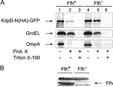
KdpD depends on SRP for targeting to the membrane. A. To test the requirement of SRP, the Ffh-depletion strain WAM121 was induced with arabinose or tightly repressed in the presence of glucose. E. coli strain WAM121 expressing KdpD (pSF51) was grown overnight in LB medium supplemented with arabinose. The cells were washed twice with LB medium, diluted 1:20 into fresh LB medium supplemented with arabinose (Ffh+) or glucose (Ffh-), and grown to an OD600 of 0.4. The cells were then transferred to M9 minimal medium and induced with 1 mM IPTG for 10 min. Cells were labelled with [35S]methionine for 1 min and chased with 500 μg ml−1 cold l-methionine for 2 min and subsequently converted to spheroplasts and incubated with (lanes 2 and 5) or without proteinase K (lanes 1 and 4) at a final concentration of 0.5 mg ml−1 on ice for 1 h. A lysis control was included by adding proteinase K and 2.5% Trition X-100 (lanes 3 and 6). All samples were precipitated with 20% TCA, immunoprecipitated with antisera against KdpD (upper panel), GroEL (middle panel) and OmpA (lower panel), and analysed by SDS-PAGE and visualized by phosphorimaging. B. Immunoblot analysis of Ffh levels in the WAM121 strain expressing KdpD under arabinose (Ffh+) and glucose (Ffh-) conditions. The samples were separated onto 12.5% SDS-PAGE and immunobloted with anti-Ffh antibody. C. To test the requirement of FtsY, the FtsY-depletion strain IY26 was induced with arabinose or tightly repressed in the presence of glucose. E. coli strain IY26 expressing KdpD was grown in LB medium with either arabinose (FtsY+) or glucose (FtsY-) for 4 h. The cells were then transferred to M9 minimal medium and induced with 1 mM IPTG for 10 min. Cells were pulse-labelled for 1 min and chased with cold l-methionine for 2 min and subsequently analysed as above. D. Immunoblot analysis of FtsY levels in the IY26 strain expressing KdpD under arabinose (FtsY+) and glucose (FtsY-) conditions. Immunoblot analysis was done using an FtsY antiserum. ppf, protease protected fragment.
Fig. 3
Fig. 3
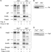
Protease accessibility of the epitope-tagged KdpD-N(HA)–GFP fusion protein in the Ffh-depletion strain WAM121. A. Cells expressing KdpD-N(HA)–GFP were grown in the presence of arabinose (Ffh+) or in the presence of glucose (Ffh-) and pulse-labelled for 1 min and chased for 2 min. Cells were then converted to spheroplasts and treated with or without proteinase K for 1 h, as described for Fig. 2A. The epitope-tagged KdpD-N(HA)–GFP fusion protein was immunoprecipitated with antiserum to HA (for the epitope in the first periplasmic loop) and then analysed as described for Fig. 2A. B. Immunoblot analysis of Ffh levels in the WAM121 strain expressing KdpD-N(HA)–GFP under arabinose (Ffh+) and glucose (Ffh-) conditions. Immunoblot analysis was done using antiserum to Ffh.
Fig. 4
Fig. 4
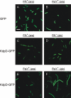
Localization of KdpD–GFP in vivo by fluorescence microscopy. Cells of strains WAM121 and IY26 bearing the GFP plasmid (A and B) or KdpD–GFP fusion plasmid (C-F) were grown in LB medium either in the presence of arabinose (Ffh+ FtsY+) or in the presence of glucose to deplete Ffh (D) or FtsY (F). Cells were induced with 1 mM IPTG for 1 h at 30°C. After induction, transcription was blocked by the addition of rifampicin (1 mg ml−1, final concentration) for 45 min. Cells were inspected under a fluorescence microscope as described in the Experimental procedures section. The bar represents 5 μm.
Fig. 5
Fig. 5
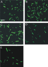
Deletion of the first 50 amino acids of KdpD inhibits its targeting to the membrane. E. coli MC1061 expressing KdpD–GFP (A), KdpD-N–GFP (B), KdpDΔ50–GFP (C), GFP (D) and KdpD-NΔ50–GFP (E) were examined by fluorescence microscopy. After induction, transcription was blocked by the addition of rifampicin (1 mg ml−1, final concentration) for 45 min. The bar represents 5 μm.
Fig. 6
Fig. 6
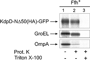
Deletion of the first 50 amino acid residues of KdpD impaired the insertion of KdpD into the membrane. E. coli WAM121 cells were transformed with the plasmid (pMS-NΔ50/GFP) containing the HA epitope-tagged KdpD-N protein (i.e. coding the amino acid residues 51–448 of KdpD) lacking the first 50 amino acid residues of KdpD fused to GFP. The cells were grown in the presence of arabinose as described in Fig. 2A. Cells were induced with 1 mM IPTG for 10 min, pulse-labelled with [35S]methionine for 1 min and chased with non-radioactive methionine for 2 min. The cells were then converted to spheroplasts and treated with or without proteinase K for 1 h. The epitope-tagged KdpD-N protein was immunoprecipitated with antisera to HA (for the epitope in the first periplasmic loop), GroEL and OmpA, and then analysed by SDS-PAGE and visualized by phosphorimaging.
Fig. 7
Fig. 7
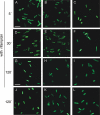
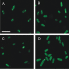
The first 48 amino acids of KdpD target GFP to the membrane. Localization of N48–GFP (A, D, G and J), KdpD–GFP (B, E, H and K), and N25–GFP (C, F, I and L) in wild-type E. coli strain MC1061. Synthesis of the GFP fusion proteins was induced either with IPTG for KdpD–GFP or arabinose for N25–GFP and N48–GFP. After induction for 1 h, rifampicin (1 mg ml−1, final concentration) was added and the localization of the fusion proteins was observed 5 min, 30 min and 2 h post rifampicin addition. The upper panel shows the fluorescence from the fusion proteins 5 min after treatment with rifampicin (A–C). The second panel shows the fluorescence from the fusion proteins 30 min after treatment with rifampicin (D–F). The third panel shows the fluorescence from the fusion proteins 2 h after addition of rifampicin (G–I). The lower panel shows the fluorescence from the fusion proteins without rifampicin after 2 h (J–L). The bars represent 5 μm.
Fig. 8
Fig. 8
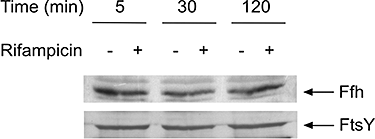
Western blot analysis of Ffh and FtsY levels in cells expressing the fusion proteins in the presence or absence of rifampicin. After induction for 1 h, cells were either treated with rifampicin (1 mg ml−1, final concentration) or without rifampicin for 5, 30 and 120 min as described in Fig. 7. Immunoblotting was done using antiserum against Ffh (upper panel) and FtsY (lower panel), respectively.
Fig. 9
Fig. 9
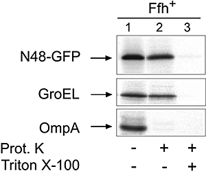
The first 48 amino acids of KdpD do not translocate GFP across the membrane. Cells of strain WAM121 (pSF165) were grown overnight in LB medium supplemented with arabinose. The overnight culture was diluted 1:20 into fresh LB medium supplemented with arabinose and grown to an OD600 of 0.4. The cells were then transferred to M9 minimal medium for 30 min. After induction with IPTG for 10 min, the cells were pulse-labelled with [35S]methionine for 1 min and chased with non-radioactive methionine for 2 min. Samples were prepared and processed as described for Fig. 2A. The N48–GFP protein was immunoprecipitated with antiserum against the N-terminus of KdpD.
Fig. 10
Fig. 10
The amino acids 22–48 of KdpD target GFP to the membrane. Localization of N22–48/GFP in E. coli MC1061 5 min (A), 30 min (B) and 2 h (C) after treatment with rifampicin as described in Fig. 7. D shows the fluorescence from N22–48/GFP without rifampicin after 2 h. The bar represents 5 μm.
Fig. 11
Fig. 11
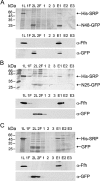
Interaction of the amino acid residues 1–48 of KdpD with His-SRP. Pull-down assays of N48–GFP (A), N25–GFP (B) and GFP (C) with His-SRP. About 5 μg of His-SRP protein (Ffh + 4.5S RNA) was attached to a Ni Sepharose column (lane 1L) which had been pre-equilibrated with 1.5 ml of equilibration buffer as indicated in Experimental procedures. Unbound protein was collected by centrifugation (lane 1F). Purified GFP and GFP fusion proteins (∼5 μg) were incubated with immobilized His-SRP for 1 h at 4°C (lane 2L). Again unbound protein was collected by centrifugation (lane 2F). After three washes (lanes 1–3), bound proteins were eluted with 500 mM imidazole (lanes E1–3) and separated by SDS-PAGE. Gels were silver-stained (upper panels). Bound proteins were identified by Western blotting with anti-Ffh (middle panels) and anti-GFP (lower panels). L, load; F, flow-through; E, elution.
Fig. 12
Fig. 12
The amino acid sequence of the N-terminal signal element that is required for targeting of KdpD to the inner membrane. The minimal peptide of 22–48 is underlined. Hydrophobic amino acids are shaded dark. Marked with a box resembles the sequence of a Walker A motif.

Similar articles

Cited by

References

    1. Alami M, Luke I, Deitermann S, Eisner G, Koch H-G, Brunner J, Müller M. Differential interactions between a twin-arginine signal peptide and its translocase in Escherichia coli. Mol Cell. 2003;12:937–946. - PubMed
    1. Balzer D, Ziegelin G, Pansegrau W, Kruft V, Lanka E. KorB protein of promiscuous plasmid RP4 recognizes inverted sequence repetitions in regions essential for conjugative plasmid transfer. Nucleic Acids Res. 1992;20:1851–1858. - PMC - PubMed
    1. Batey RT, Rambo RP, Lucast L, Rha B, Doudna JA. Crystal structure of the ribonucleoprotein core of the signal recognition particle. Science. 2000;287:1232–1239. - PubMed
    1. Beck K, Wu LF, Brunner J, Müller M. Discrimination between SRP- and SecA/SecB-dependent substrates involves selective recognition of nascent chains by SRP and trigger factor. EMBO J. 2000;19:134–143. - PMC - PubMed
    1. Cormack B, Valdivia R, Falkow S. FACS-optimized mutants of the green fluorescent protein (GFP) Gene. 1996;173:33–38. - PubMed

Publication types

MeSH terms

LinkOut - more resources