. 2008 Jul;9(7):1218-31.
doi: 10.1111/j.1600-0854.2008.00752.x. Epub 2008 Apr 21.

Rab14 regulates apical targeting in polarized epithelial cells

Affiliations

Rab14 regulates apical targeting in polarized epithelial cells

Khameeka N Kitt et al. Traffic. 2008 Jul.

Abstract

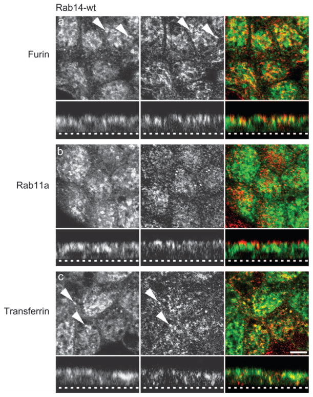
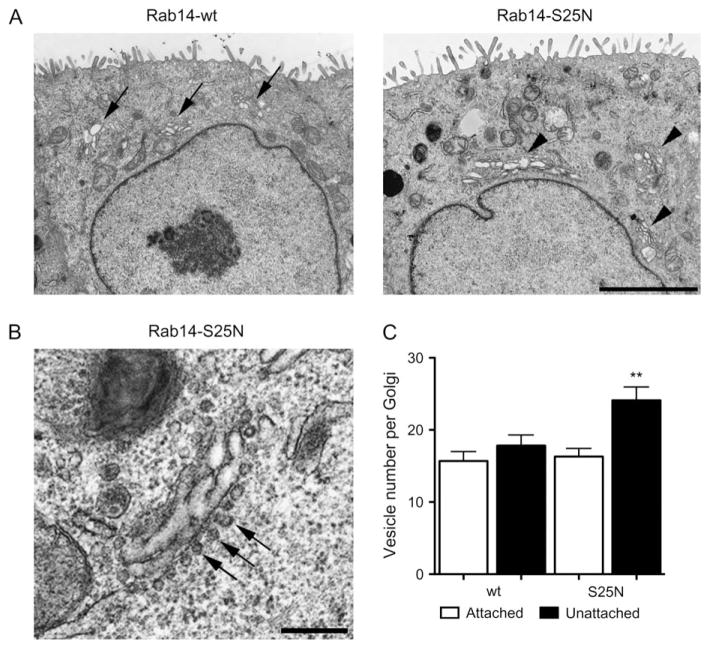
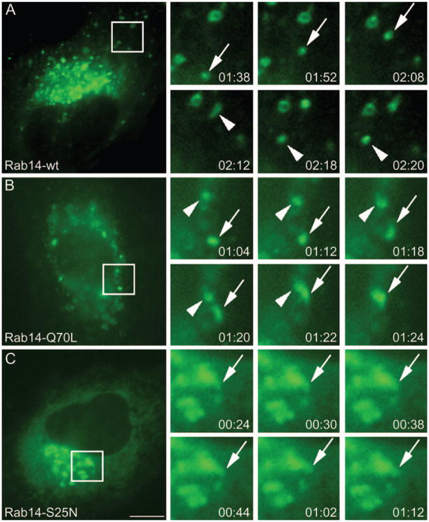
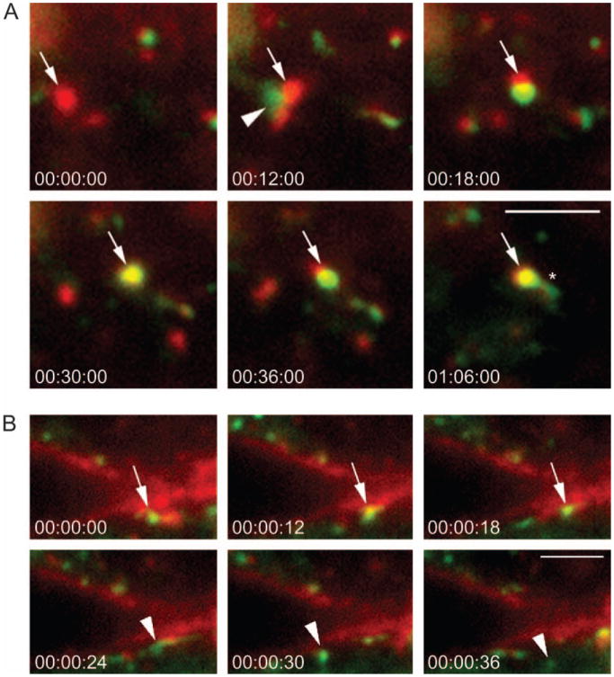
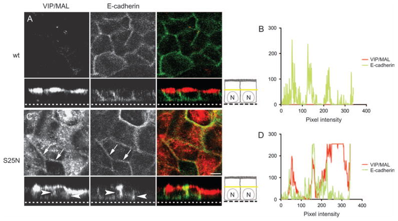
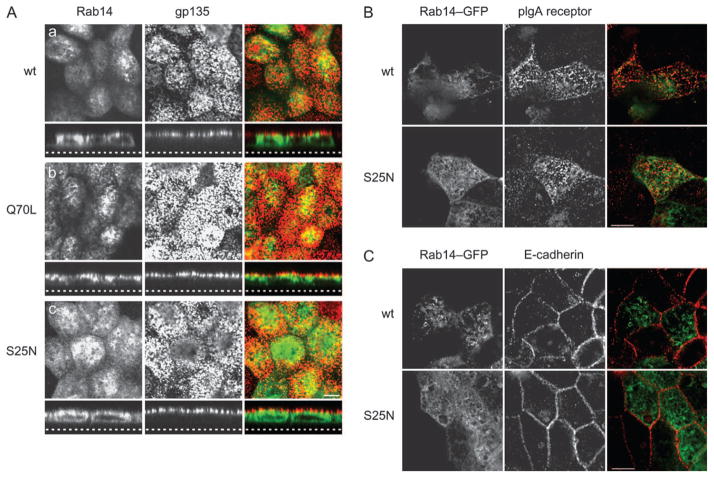
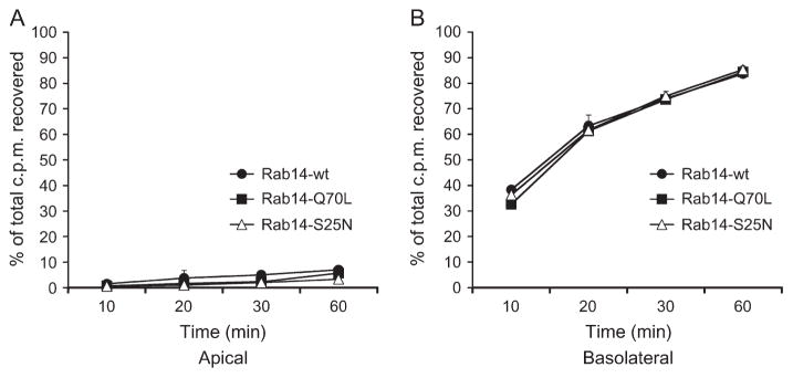
Epithelial cells display distinct apical and basolateral membrane domains, and maintenance of this asymmetry is essential to the function of epithelial tissues. Polarized delivery of apical and basolateral membrane proteins from the trans Golgi network (TGN) and/or endosomes to the correct domain requires specific cytoplasmic machinery to control the sorting, budding and fission of vesicles. However, the molecular machinery that regulates polarized delivery of apical proteins remains poorly understood. In this study, we show that the small guanosine triphosphatase Rab14 is involved in the apical targeting pathway. Using yeast two-hybrid analysis and glutathione S-transferase pull down, we show that Rab14 interacts with apical membrane proteins and localizes to the TGN and apical endosomes. Overexpression of the GDP mutant form of Rab14 (S25N) induces an enlargement of the TGN and vesicle accumulation around Golgi membranes. Moreover, expression of Rab14-S25N results in mislocalization of the apical raft-associated protein vasoactive intestinal peptide/MAL to the basolateral domain but does not disrupt basolateral targeting or recycling. These data suggest that Rab14 specifically regulates delivery of cargo from the TGN to the apical domain.


Figures

Figure 1
Figure 1. Endotubin and VIP/MAL interact with the small GTPase Rab14
A) Endotubin cytoplasmic domain used for bait construction. Motifs critical for apical endosomal targeting are indicated in bold. B) Rab14–GST (wt, Q70L and S25N)-tagged proteins were used to pull down endotubin or VIP/MAL from MDCK cells followed by immunoblotting. Results show that binding of full-length endotubin to Rab14 is not nucleotide dependent (B). However, binding of VIP/MAL is nucleotide dependent (C). None of the Rab14–GST constructs interacts with the basolateral marker, E-cadherin. Input is 5% of total. D) MDCK cells expressing Rab14-wt, -Q70L, or -S25N–GFP (green) and endotubin (red) were visualized by confocal microscopy. Coexpression of Rab14-wt and endotubin resulted in areas of overlap in the apical domain (arrowheads). Rab14-Q70L and endotubin also show extensive colocalization (arrowheads). Coexpression of Rab14-S25N and endotubin resulted in some areas of colocalization as well as a nonpolarized distribution of Rab14-S25N. Dotted line in the z-stack represents the bottom of the filter. Scale bar, 5 μm.
Figure 2
Figure 2. Subcellular localization of Rab14 in polarized epithelial cells
MDCK cells stably expressing Rab14-wt–GFP were labeled with anti-furin (A, red), anti-Rab11a (B, red) or anti-transferrin receptor (C, red) and visualized by confocal microscopy. There is some colocalization of Rab14-wt–GFP with furin (TGN) and transferrin receptor (common endosome) (arrowheads). No colocalization was observed with the ARE marker, Rab11a. Dotted line in the z-stack represents the bottom of the filter. Scale bar, 5 μm.
Figure 3
Figure 3. Subcellular localization of Rab14 in nonpolarized epithelial cells
A) MDCK cells transfected with Rab14-wt–GFP (green), labeled with anti-furin (panel a, red), anti-γ-adaptin (panel b, red) or anti-transferrin receptor (panel c, red) and visualized by deconvolution microscopy. Rab14 and furin (arrows, panel a′) are present in adjacent membrane domains. There is also some colocalization with the transferrin receptor (arrow and arrowheads, panels c′ and c) but no colocalization with γ-adaptin (panel b′). B) Expression of Rab14-wt (B, panel a) or Rab14-Q70L–GFP (B, panel b) did not affect the structure of the TGN, labeled in this study with WGA–Rh. In contrast, expression of Rab14-S25N caused a dramatic expansion and reorganization of the TGN with extensive overlay of the labels (B, panel c, arrows). Scale bars, 10 μm.
Figure 4
Figure 4. Rab14 is not involved in budding/fission of vesicles from the TGN
Low magnification of cells expressing Rab14-wt and Rab14-S25N. Cells expressing Rab14-wt display normal Golgi morphology (arrows, A – left panel) compared with Rab14-S25N-expressing cells (arrowheads, A – right panel) where Golgi membranes are expanded. B) High magnification of a Golgi region in cells expressing Rab14-S25N demonstrates an increase in vesicles lined up at the Golgi membrane (arrows). Scale bar, 0.5 μm. C) Rab14-S25N-expressing cells have a significant increase in unattached vesicles in the peri-Golgi compared with Rab14-wt (**p < 0.005).
Figure 5
Figure 5. Nucleotide states determine motility of Rab14 vesicles
NRK cells expressing Rab14-wt, -Q70L and -S25N–GFP were imaged by live cell fluorescence microscopy. Selected frames from time-lapse images were enlarged and vesicles tracked. Individual frames were taken for 2 min and 20 seconds for each Rab14 construct. (A) Rab14-wt vesicles traffic in and out of the perinuclear region; time lapse shows a vesicle undergoing anterograde transport from the perinuclear region (arrow) and then undergoing retrograde movement (arrowheads). (B) Rab14-Q70L vesicles undergo fusion into the perinuclear region; time lapse shows a vesicle undergoing fusion (arrow) with another vesicle (arrowhead) in the perinuclear region. In contrast, cells expressing (C) Rab14-S25N display no vesicle movement and membrane structures containing Rab14 remain constant (arrows). The time is shown in min:seconds. Scale bar, 10 μm.
Figure 6
Figure 6. Rab14-wt vesicles traffic to early endosomes and plasma membrane
Rab14-wt–GFP (green)-expressing cells were incubated with ricin–TRITC (red) at 4°C and warmed to 37°C. Image acquisition was initiated within 5 min of warming. Individual frames were taken over a 10-min period (see supplementary material videos 4 and 5). A) Rab14-wt–GFP-containing vesicle (arrowhead) docks and fuses with an ricin–TRITC-labeled early endosome (arrow), and subsequently, a Rab14 tubular structure emerges from the vesicle (asterisk in 01:06:00). B) Rab14 vesicle (arrow) is docked at the ricin–TRITC-labeled plasma membrane; additional frames indicate the vesicle fusing with the plasma membrane. The Rab14-wt–GFP-positive vesicle then buds off the plasma membrane and moves into the cytoplasm (arrowhead in 00:00:24). The Rab14-wt–GFP domain then buds off from the plasma membrane (arrowhead in 00:30:00 and 00:36:00). The time is shown in min:seconds:milliseconds. Scale bars, A–B, 2.5 μm.
Figure 7
Figure 7. Inactive Rab14 selectively disrupts targeting of the raft-associated protein VIP/MAL
Cells expressing Rab14-wt or Rab14-S25N and VIP/MAL were labeled and examined by confocal microscopy. The plane of section in the x–y images is indicated by the yellow bar in the cartoon. A) Cells expressing Rab14-wt demonstrate an apical distribution of VIP/MAL, with no labeling in the subapical cytoplasm. No colocalization is observed with E-cadherin. B) Measurement of pixel intensity demonstrates no colocalization with E-cadherin. C) In cells expressing Rab14-S25N, VIP/MAL is retained intracellularly and localizes to the basolateral membrane shown by the colocalization with E-cadherin (arrows and arrowheads). D) Measurement of pixel intensity demonstrates extensive overlay between the two signals. Note: Rab14 is not shown in these images. Dotted line in the z-stack represents the bottom of the filter. Scale bar, 5 μm.
Figure 8
Figure 8. Rab14 does not affect the targeting of the apical proteins, gp135 and pIgA receptor
A) Cells expressing Rab14-wt, Rab14-Q70L and Rab14-S25N have an apical distribution of gp135 (red, a, b and c). B) The distribution of the pIgA receptor and C) E-cadherin was also unaffected by all forms of Rab14. Dotted line in the z-stack represents the bottom of the filter. Scale bars, 5 μm.
Figure 9
Figure 9. Rab14 has no effect on basolateral recycling of transferrin
Polarized epithelial cells expressing wt- (filled circles), Q70L-(filled squares) or S25N-Rab14 (open triangles) were loaded with 125I-labeled transferrin from the basolateral side of the monolayer. Apical (A) and basolateral (B) media were harvested at different time-points to assess basolateral recycling and basolateral-to-apical transcytosis, respectively. Data are presented as mean (±SD) percentage of counts per minute recovered for each condition.

Similar articles

Cited by

References

    1. Mostov K, Su T, ter Beest M. Polarized epithelial membrane traffic: conservation and plasticity. Nat Cell Biol. 2003;5:287–293. - PubMed
    1. Rodriguez-Boulan E, Kreitzer G, Musch A. Organization of vesicular trafficking in epithelia. Nat Rev Mol Cell Biol. 2005;6:233–247. - PubMed
    1. Folsch H, Ohno H, Bonifacino JS, Mellman I. A novel clathrin adaptor complex mediates basolateral targeting in polarized epithelial cells. Cell. 1999;99:189–198. - PubMed
    1. Folsch H, Pypaert M, Schu P, Mellman I. Distribution and function of AP-1 clathrin adaptor complexes in polarized epithelial cells. J Cell Biol. 2001;152:595–606. - PMC - PubMed
    1. Gan Y, McGraw TE, Rodriguez-Boulan E. The epithelial-specific adaptor AP1B mediates post-endocytic recycling to the basolateral membrane. Nat Cell Biol. 2002;4:605–609. - PubMed

Publication types

LinkOut - more resources