. 2008 Apr 21;181(2):351-65.
doi: 10.1083/jcb.200709033.

ErbB2 directly activates the exchange factor Dock7 to promote Schwann cell migration

Affiliations

ErbB2 directly activates the exchange factor Dock7 to promote Schwann cell migration

Junji Yamauchi et al. J Cell Biol. .

Abstract

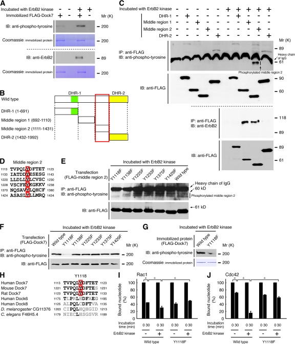
The cellular events that precede myelination in the peripheral nervous system require rapid and dynamic morphological changes in the Schwann cell. These events are thought to be mainly controlled by axonal signals. But how signals on the axons are coordinately organized and transduced to promote proliferation, migration, radial sorting, and myelination is unknown. We describe that the axonal signal neuregulin-1 (NRG1) controls Schwann cell migration via activation of the atypical Dock180-related guanine nucleotide exchange factor (GEF) Dock7 and subsequent activation of the Rho guanine triphosphatases (GTPases) Rac1 and Cdc42 and the downstream c-Jun N-terminal kinase. We show that the NRG1 receptor ErbB2 directly binds and activates Dock7 by phosphorylating Tyr-1118. Dock7 knockdown, or expression of Dock7 harboring the Tyr-1118-to-Phe mutation in Schwann cells, attenuates the effects of NRG1. Thus, Dock7 functions as an intracellular substrate for ErbB2 to promote Schwann cell migration. This provides an unanticipated mechanism through which ligand-dependent tyrosine phosphorylation can trigger the activation of Rho GTPase-GEFs of the Dock180 family.


Figures

Figure 1.
Figure 1.
NRG1 promotes Schwann cell migration through the ErbB2 and 3 heterodimer. (A and B) The migration of primary Schwann cells was measured by using Boyden chambers. Filters were coated with DRG axonal membranes. After incubation for 6 h with normal or conditioned medium from DRG neurons containing 5 μg/ml of control IgG or ErbB3-Fc, Schwann cells were stained with Giemsa solution and the number of migrating cells was counted (16 independent fields). Bar, 50 μm. (C and D) Filters in Boyden chambers were coated with DRG axonal membranes, collagen (type I or IV), fibronectin, or laminin. Schwann cell migration was measured in the presence of normal medium or conditioned medium (eight independant fields). (E–G) In the presence of control IgG or ErbB3-Fc, Schwann cells were incubated with or without 20 ng/ml of NRG1, NT3, or IGF-I in Boyden chambers (eight independent fields). Filters were coated with collagen (type I). (H and I) Schwann cell reaggregates were placed onto DRG neurons and control IgG or ErbB3-Fc was added. After 6 h, DRG axons were stained with an antineurofilament antibody (red), and Schwann cells were stained with an anti-S100β antibody (green). The distance of migration was measured (n = 16). Bar, 100 μm. (J and K) Schwann cells were pretreated in the presence or absence of 10 μM AG825 and then incubated with or without 20 ng/ml NRG1 in Boyden chambers. The number of migrating cells was counted (16 independent fields). Bar, 50 μm. (L and M) Schwann cells were transfected with control, ErbB2, or ErbB3 siRNA and incubated with or without NRG1 in Boyden chambers (16 independent fields). To confirm the effects of siRNAs, the lysates of transfected cells were immunoblotted with an anti-ErbB2, ErbB3, or actin antibody. Error bars show ±SD. Data were evaluated by using one-way ANOVA (*, P < 0.01; ***, P < 0.02).
Figure 2.
Figure 2.
NRG1-induced migration of Schwann cells is dependent on the activation of the Rho GTPases Rac1 and Cdc42. (A) Schwann cells were pretreated with or without 2 ng/ml C. difficile Toxin B or 2 μg/ml C3 exoenzyme, and migration was assayed in Boyden chambers (12 independent fields). (B and C) After the addition of NRG1 for 0–120 min, endogenous Rac1-GTP in the lysates of Schwann cells was affinity precipitated with GST-Pak1-CRIB and immunoblotted with an anti-Rac1 antibody. The levels of Rac1-GTP were normalized to the amount of total Rac1 (n = 3). (D and E) The Rac1 activities were measured at 0–360 min (n = 5). (F–I) Endogenous Cdc42-GTP in the cell lysates was affinity precipitated with GST-Pak1-CRIB. The Cdc42-GTP levels were normalized to the amount of total Cdc42 (n = 3). Error bars show ±SD. Data were evaluated by using one-way ANOVA (*, P < 0.01).
Figure 3.
Figure 3.
JNK acts downstream of Rho GTPases to promote Schwann cell migration. (A) Schwann cells were pretreated in the presence or absence of 10 μM SP600125 or 20 μM JNK inhibitor I and incubated with or without 20 ng/ml NRG1 in Boyden chambers (12 independent fields). (B and C) Schwann cells were stimulated with NRG1 for 0–120 min. The cell lysates were immunoblotted with an anti-(pThr183/pTyr185) JNK antibody that recognizes active JNK. The cell lysates were also immunoblotted with an anti-JNK antibody. The levels of phosphorylated forms were normalized to the amount of total JNK (n = 3). (D and E) JNK phosphorylation was measured at 0–360 min (n = 3). (F) Cells were pretreated with or without 2 ng/ml C. difficile Toxin B. After incubation with NRG1 for 120 min, JNK phosphorylation was assayed (n = 5). Error bars show ±SD. Data were evaluated by using one-way ANOVA (*, P < 0.01).
Figure 4.
Figure 4.
Dock7 is required for migration and the activation of Rac1, Cdc42, and JNK induced by NRG1 in Schwann cells. (A and B) Schwann cells were transfected with control, Dock7-1, or Dock7-2 siRNA and incubated with or without NRG1 in Boyden chambers (12 independent fields). To confirm the effects of siRNA, the lysates of transfected cells were immunoblotted with an anti-Dock7, Rac1, Cdc42, or actin antibody. (C and D) Schwann cells were transfected with control or Dock7-1 siRNA and stimulated with NRG1 for 60 min. The activities of Rac1 and Cdc42 were assayed by affinity precipitation with GST-Pak1-CRIB (n = 3). (E) Cells were transfected with control or Dock7-1 siRNA and JNK phosphorylation was measured (n = 3). Error bars show ±SD. Data were evaluated by using one-way ANOVA (*, P < 0.01).
Figure 5.
Figure 5.
NRG1 activation of the ErbB2 and 3 heterodimer stimulates the GEF activity of Dock7. (A–C) 125 ng of immobilized FLAG-Dock7-DHR-2 was incubated with 16 ng/μl GST-Rac1, Cdc42, or RhoA and 3 μM [3H]GDP in 30 μl of reaction buffer for 0–30 min, and the guanine nucleotide binding activities were measured (n = 10). (D–F) The release of [3H]GDP from GST-Rac1-[3H]GDP, Cdc42-[3H]GDP, or GST-RhoA-[3H]GDP by FLAG–Dock7–DHR-2 was measured (n = 10). Immunoprecipitated FLAG-Dbs-DHPH was used as the positive control for the RhoA-GEF. Dock7–DHR-2, closed circle; control, open circle; Dbs-DHPH, closed square. (G–L) 293T cells were transfected with pCMV–FLAG–Dock7–DHR-2 or pCMV-FLAG-Dbs-DHPH. The cell lysates were affinity precipitated with 20 μg each of nucleotide-free GST-Rho GTPase (Rac1G15A, Cdc42G15A, or RhoAG17) or the wild type (Rac1, Cdc42, or RhoA) and immunoblotted with an anti-FLAG antibody. The total FLAG–Dock7–DHR-2 or FLAG-Dbs-DHPH is also shown. Each GST-Rho GTPase was immobilized in the same experimental conditions, subjected to SDS-PAGE, and stained with Coomassie brilliant blue. (M and O) 293T cells were cotransfected with pCMV-FLAG-Dock7, pCMV-ErbB2, and pCMV-ErbB3 and stimulated with or without NRG1 for 30 min. The expression of ErbB2 and 3 in 293T cells was below the detection level of immunoblotting (not depicted). The release of [3H]GDP from GST-Rac1-[3H]GDP or Cdc42-[3H]GDP by immunoprecipitated FLAG-Dock7 was measured (n = 3). (N and P) Cells were cotransfected with pCMV-FLAG-Dock7, pCMV-ErbB2, and pCMV-ErbB3. The affinity precipitation of the cell lysates with GST-Rac1G15A or Cdc42G15A was performed. The total FLAG-Dock7 is also shown. Error bars show ±SD. Data were evaluated by using one-way ANOVA (*, P < 0.01).
Figure 6.
Figure 6.
ErbB2 directly binds and activates Dock7 by phosphorylating Tyr-1118. (A) 250 ng of immobilized full-length FLAG-Dock7 protein was incubated in 30 μl of reaction buffer containing 20 μM of cold ATP in the presence or absence of 100 ng ErbB2 kinase for 30 min, washed, and immunoblotted with an anti-pTyr or ErbB2 antibody. Immobilized FLAG-Dock7 was also stained with Coomassie brilliant blue. (B) The schematic structures of Dock7 and the domains are illustrated. Red rectangle, Tyr in the middle region 2. (C) 293T cells were transfected with the plasmid encoding DHR-1, middle region 1, middle region 2, or DHR-2 of Dock7. The lysates of transfected cells were immunoprecipitated with an anti-FLAG antibody, incubated with ErbB2 kinase and ATP, and immunoblotted with an anti-pTyr or ErbB2 antibody. A shift in the mobility of the bands for the tyrosine-phosphorylated protein was observed. The cell lysates were also immunoblotted with an anti-FLAG antibody. (D) The amino acid sequences containing six tyrosine residues in the middle region 2 are shown. (E) Cells were transfected with the plasmid encoding each middle region 2 containing one Tyr-to-Phe mutation. The samples, immunoprecipitated with the anti-FLAG antibody, were incubated with ErbB2 kinase and ATP. A shift in the mobility was observed in bands of the tyrosine-phosphorylated protein. The tyrosine phosphorylation of the constructs and their expression are also shown. (F) Cells were transfected with each full-length Dock7 harboring one Tyr-to-Phe mutation in the middle region 2, immunoprecipitated with anti-FLAG antibody, and incubated with ErbB2 kinase and ATP. The tyrosine phosphorylation of the constructs and their expression are also shown. (G) 250 ng of immobilized full-length FLAG-Dock7 or FLAG-Dock7Y1118F was incubated with ErbB2 kinase and ATP. (H) A comparison of the amino acid sequences surrounding the ErbB2 phosphorylation sites (red squares) of mammalian Dock7 with other homologous proteins is shown. Black, conserved amino acids; grey, nonconserved amino acids. (I and J) Immobilized FLAG-Dock7 or the Y1118F mutant was incubated in 30 μl of reaction buffer containing 20 μM of cold ATP in the presence or absence of ErbB2 kinase and washed. The release of [3H]GDP from GST-Rac1–[3H]GDP or Cdc42–[3H]GDP by immobilized proteins was measured (n = 3). Error bars show ±SD. Data were evaluated by using one-way ANOVA (*, P < 0.01).
Figure 7.
Figure 7.
NRG1, acting through the phosphorylation of Dock7 at Tyr-1118, regulates Schwann cell migration. (A) After stimulation with vehicle or 20 ng/ml of NRG1, NT3, or IGF-I for 30 min, Schwann cells were lysed, immunoprecipitated with an anti-Dock7 antibody, and immunoblotted with an anti-(pTyr1118)Dock7 antibody. In some experiments, cells were treated with or without AG825 or ErbB3-Fc. The cell lysates were also immunoblotted with an anti-Dock7 antibody. (B) After stimulation with vehicle or NRG1, immunoprecipitated Dock7 was immunoblotted with an anti-ErbB2 antibody. Immunoblots for ErbB2, ErbB3, and Dock7 are shown. (C) After stimulation with vehicle or NRG1, Schwann cells were costained with the following antibodies: anti-(pTyr1118)Dock7 (green; top) and anti-(pTyr1112)ErbB2 (red; top), anti-(pTyr1118)Dock7 (green; middle) and anti-ErbB2 (red; middle), and anti-Dock7 (green; bottom) and anti-ErbB2 (red; bottom). After stimulation with vehicle or NRG1, increased colocalization (indicated by arrows) was observed (Bar, 25 μm). a–d are magnifications of the boxed areas as indicated (Bar, 10 μm). Dotted lines indicate the outlines of cells. (D and E) Affinity precipitation with GST-Rac1G15A or Cdc42G15A was performed and immunoblotted with an anti-Dock7 antibody. Immunoblots for Dock7 are shown.
Figure 8.
Figure 8.
Effects of the Tyr-to-Phe mutations in the middle region 2 of Dock7 on NRG1-induced migration of Schwann cells. (A and B) pEGFP, pEGFP-siRNA–-resistant wild-type Dock7, or pEGFP-siRNA–resistant Dock7Y1118F was cotransfected with control or Dock7-1 siRNA into Schwann cells. The number of GFP-fluorescent migrating Schwann cells in Boyden chambers was counted. Bar, 100 μm. (C) Expression of GFP-tagged siRNA-sensitive wild-type Dock7 or Dock7-1 siRNA-resistant Dock7 (wild type, Y1118F, Y1138F, Y1225F, Y1233F, Y1375F, or Y1429F) in Schwann cells are shown in the immunoblots. The cell lysates were immunoblotted with an anti-GFP or -actin antibody. (D and E) Schwann cells were cotransfected with pEGFP-siRNA–resistant wild-type or mutated Dock7 together and Dock7-1 siRNA. The number of GFP-fluorescent migrating Schwann cells was counted. Bar, 100 μm. Error bars show ±SD. Data were evaluated by using one-way ANOVA (n = 16; *, P < 0.01; ***, P < 0.02).
Figure 9.
Figure 9.
Tyr-1118 of Dock7 is essential for NRG1-induced migration of reaggregated Schwann cells. (A and B) pEGFP or pEGFP-siRNA–resistant wild-type Dock7 or Dock7Y1118F was cotransfected with control or Dock7-1 siRNA into Schwann cells. Cells were fixed with PFA, blocked, and stained with an anti-neurofilament antibody (red). The immunofluorescence images were merged. Bar, 100 μm. Error bars show ±SD. Data were evaluated by using one-way ANOVA (n = 16; *, P < 0.01; **, P < 0.015; ***, P < 0.02).

Similar articles

Cited by

References

    1. Arthur, W.T., S.M. Ellerbroek, C.J. Der, K. Burridge, and K. Wennerberg. 2002. XPLN, a guanine nucleotide exchange factor for RhoA and RhoB, but not RhoC. J. Biol. Chem. 277:42964–42972. - PubMed
    1. Benninger, Y., T. Thurnherr, J.A. Pereira, S. Krause, X. Wu, A. Chrostek-Grashoff, D. Herzog, K.A. Nave, R.J. Franklin, D. Meijer, et al. 2007. Essential and distinct roles for cdc42 and rac1 in the regulation of Schwann cell biology during peripheral nervous system development. J. Cell Biol. 177:1051–1061. - PMC - PubMed
    1. Brugnera, E., L. Haney, C. Grimsley, M. Lu, S.F. Walk, A.C. Tosello-Trampont, I.G. Macara, H. Madhani, G.R. Fink, and K.S. Ravichandran. 2002. Unconventional Rac-GEF activity is mediated through the Dock180-ELMO complex. Nat. Cell Biol. 4:574–582. - PubMed
    1. Bunge, R.P. 1993. Expanding roles for the Schwann cell: ensheathment, myelination, trophism and regeneration. Curr. Opin. Neurobiol. 3:805–809. - PubMed
    1. Chan, J.R., C. Jolicoeur, J. Yamauchi, J. Elliott, J.P. Fawcett, B.K. Ng, and M. Cayouette. 2006. The polarity protein Par-3 directly interacts with the p75 neurotrophin receptor to regulate myelination. Science. 314:832–836. - PubMed

Publication types