. 2008 Apr 18;133(2):250-64.
doi: 10.1016/j.cell.2008.03.028.

Direct reprogramming of terminally differentiated mature B lymphocytes to pluripotency

Affiliations

Direct reprogramming of terminally differentiated mature B lymphocytes to pluripotency

Jacob Hanna et al. Cell. .

Erratum in

  • Cell. 2008 Jul 25;134(2):365

Abstract

Pluripotent cells can be derived from fibroblasts by ectopic expression of defined transcription factors. A fundamental unresolved question is whether terminally differentiated cells can be reprogrammed to pluripotency. We utilized transgenic and inducible expression of four transcription factors (Oct4, Sox2, Klf4, and c-Myc) to reprogram mouse B lymphocytes. These factors were sufficient to convert nonterminally differentiated B cells to a pluripotent state. However, reprogramming of mature B cells required additional interruption with the transcriptional state maintaining B cell identity by either ectopic expression of the myeloid transcription factor CCAAT/enhancer-binding-protein-alpha (C/EBPalpha) or specific knockdown of the B cell transcription factor Pax5. Multiple iPS lines were clonally derived from both nonfully and fully differentiated B lymphocytes, which gave rise to adult chimeras with germline contribution, and to late-term embryos when injected into tetraploid blastocysts. Our study provides definite proof for the direct nuclear reprogramming of terminally differentiated adult cells to pluripotency.


Figures
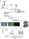
Figure 1
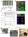
Figure 1. Transgenic inducible expression of Oct4, Sox2, Klf4 and c-Myc in the mouse B cell lineage
(A) Schematic drawing representing the strategy used in this study for reprogramming cells from the B cell lineage. (B) MEFs heterozygous for the ROSA26-M2rtTA and Nanog-GFP alleles were infected with Dox inducible lentiviruses encoding Oct4, Sox2, Klf4 and c-Myc. Typical morphology of colonies derived from infected MEFs cultured with Dox for 12 days. (C) ES-like morphology of Nanog-GFP+ MEF derived iPS lines (MEF-iPS#1 cell line is shown). These cell lines were injected into blastocysts to derive embryonic and adult chimeras (D). Different B cell subsets were isolated from embryonic liver or adult spleen and bone marrow tissues and used for reprogramming experiments. (E) Quantitative RT-PCR assay of viral transgene expression in MEF-iPS#1 chimera derived mouse embryonic fibroblasts (MEF) obtained at day E13.5 and 8 week old adult tail tip fibroblasts (TTF) and B cells subsets from bone marrow (BM) or spleen with (4μg/ml) or without Dox treatment for 7 days as indicated in the figure.
Figure 2
Figure 2. Reprogramming of non-terminally differentiated B cells to pluripotency
(A) 8 week old chimera derived bone marrow B cell subsets (50*10^3 cells per well) and spleen IgM+IgD+ mature B cells (250*10^3 spleen cells per well) were plated on OP9 bone marrow stroma with conditioned media and Dox. After 14 days plates were fixed and stained for AP activity. (B) Images for characteristic colonies at different time point obtained from sorted B220+CD25+ bone marrow cells. (C) PCR analysis for rearrangements in selected iB-iPS. MEFs and splenocytes were used as negative and positive controls, respectively. (D) Immunostaining for ES cell markers on a representative line iB-iPS#8. (E) A chimeric mouse from iB-iPS #8 cell line. Agouti colored hairs originate from injected iPS lines. (F) Southern blot analysis of iB-iPS line #8 grown on MEF feeders and of tail tip biopsy taken from the derived chimera. MEF and spleen B cells were used as negative and positive controls, respectively, for rearrangement detection. Red arrows indicate the two rearranged heavy chain alleles. GL indicates germline fragment representing non-rearranged configuration of the Igh locus. (G) An adult old chimeric pseudo-male mouse from iB-iPS #9 cell line was mated with Balb/C females and repeatedly achieved 100% germline transmission, as indicated by the agouti color of all litters obtained.
Figure 3
Figure 3. Reprogramming of terminally differentiated mature B cells to pluripotency
IgM+IgD+ cells were isolated from spleen of a 10 week old chimera, infected for 24 hours with viral supernatants of retroviruses encoding different combinations of transcription factors and were transferred afterwards to OP9 coated wells and grown in conditioned media in the presence of Dox. “20 factors” indicate pMXs based constructs previously used to reprogram mouse fibroblasts as indicated in the Experimental Procedures section. After 14 days of Dox induction and additional viral transduction as indicated for each well, cells were stained for AP. One out of 3 independent experiments using different combinations of factors is shown. (B) Representative images of colonies at different time points after infection with C/EBPα retrovirus and Dox induction of chimeric spleen derived IgM+IgD+ mature B cell cultures. (C) Representative pictures of changes in cell morphology after passaging picked colonies at day 14 from IgM+IgD+ B cell cultures infected with C/EBPα and treated with Dox.
Figure 4
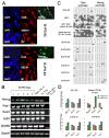
Figure 4. Marker expression and promoter methylation analysis of B-iPS lines
(A) Immunoflorescence staining for ES pluripotency markers Oct4, Nanog (signal obtained from GFP cassette knocked in the endogenous Nanog locus), SSEA1 and AP. (B) RT-PCR analysis of selected ES marker genes in B cells, ES cells, MEF-iPS cells and different iB-iPS and B-iPS lines. (C) Analysis of the methylation state of the Oct4 and Nanog promoters using bisulphate sequencing. Open squares indicate unmethylated and filled squares methylated CpG dinucleotides. (D) Real-time PCR after chromatin immunoprecipitation using antibodies against H3K4me3 and H3K27me3. Shown are the Log2 enrichments for several previously reported “bivalent” loci in ES cells.
Figure 5
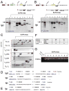
Figure 5. Productive heavy and light chain rearrangements in B-iPS lines
(A) Genomic DNA from B-iPS lines grown on gelatin was digested with EcoRI and analyzed for V(D)J rearrangements at the Igh locus by Southern blotting using a 3′ JH4 probe. (B) Vκ-Jκrearrangements at the Igκ locus were determined by Southern blot analysis of BamHI digested genomic DNA using a 3′Jκ5 probe. (A–B) Any rearrangement occurring in these loci abolishes the asterik marked restriction sites of the enzyme used. After rearrangements, the size of the genomic fragment bound by the probe depends on the next available 5′ restriction site in relation to the abolished asterik marked sites. MEFs served as negative control and CD19+ splenocytes were used as positive controls. GL denotes the position of germline DNA fragment. (C) PCR analysis of Vκ-Jκ and Vλ1-Jλ light chain rearrangements and of VHQ52-DJH, VH7183-DJH, VHGam3.8-DJH VHJ558-DJH and D-JH rearrangements in selected B-iPS. Rearrangements to different J segments are numbered on the left side of the panels. Detailed characterization of the different immunoglobulin gene rearrangements by cloning and sequencing was performed to confirm the presence of productive heavy and light chain rearrangements in the cell lines obtained. Blue arrows indicate “productive rearrangements” and red arrows indicate” non-productive rearrangements”. (D) Sequences from rearranged IgH and Igκ chains from B-iPS line #4 obtained by RT-PCR (Panel C, lane 4). V, D and J segments involved in IgH rearrangement are indicated, as well as V and J segments from the Igκ locus. (E) Sequencing of the Igλrearrangement (Panel C, lane 9) cloned from B-iPS line #9. (F) Summary for non-silent and silent mutations found in predicted variable region of the encoded B cell receptor as detected by sequencing productive heavy and light chain rearrangements of B-iPS line #4. (G) Transgene specific primers were designed and used to detect the presence of C/EBPα provirus in B-iPS lines.
Figure 6
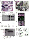
Figure 6. Developmental potential of B-iPS cell lines
(A) Hematoxylin and eosin staining of a representative teratoma derived from B-iPS line #4. (B) Adult chimeric mice obtained from B-iPS lines in 2N Balb/C or B6D2F1 host blastocysts. (C) Southern blot analysis for detection of IgK light chain rearrangements on BamH1 digested genomic DNA (as detailed in Figure 5A) from different tissues obtained from MEF-iPS #1 derived chimeras (used as negative control) and B-iPS #4 cell line. (D) B-iPS#1 line was transduced with lentiviral ubiquitin-EGFP vector before injection in 2N blastocysts. Germline transmission was achieved from one of the chimeras, as we obtained a number of GFP+ embryos, demonstrating the transmission of constitutively expressed EGFP transgene. (E) “All B-iPS cell embryos” were generated by injection of B-iPS cell into 4N blastocysts. Live E14.5 embryos generated from B-iPS#4 cell lines. (F) Highly sensitive 50 cycle PCR analysis on 4N embryos obtained from B-iPS#4 for detection of loss of the amplicon representing germline line configuration of IgH locus in order to prove that the embryos developed entirely from iPS cells. Equimolar mix of 5′ primers DQ52 (recognizes downstream region that is always excised upon any recombination of the locus) and DSF (degenerate primer recognizing D cassettes) with 3′ JH-4 primer. This PCR could yield 4 rearrangement bands (D-JH1,2,3,4) and a germline 2 Kb band as indicated in the locus scheme. (G) Immunological analysis for detection of Igκ and Igλ surface antigen on peripheral blood B cells of chimeras obtained from constitutively GFP labeled B-iPS #4 and #9 (marked with lentiviral ubiquitin-EGFP vector before injection). Presented panels were gated on CD19+ cells and show Igκ and Igλ expression on iPS derived B cell (GFP+) and host B cells (GFP−). Green rectangles highlight B-iPS derived of GFP+ B cells.
Figure 7
Figure 7. Reprogramming efficiency of adult mature B cells into iPS cells
(A) Schematic representation for experiment attempting to measure reprogramming efficiency. 3*10^6 CD19+ adult B cells were infected with retrovirus encoding C/EBPα-NeoR construct and after 24 hours we sorted IgM+IgD+ mature adult B cells and plated them as single cells in 96 wells plates preplated with OP9 stromal cell line. Cells were grown in conditioned medium + Dox + LIF throughout the experiment. On day 6, culture wells were subjected to puromycin and neomycin selections for 5 days, which allowed only the growth of transgenic B cells infected with C/EBPα. On day 20, the wells containing drug resistant cells were screened for Nanog-GFP expression by FACS analysis. Wells that scored positive were subsequently passaged on MEFs in ES media and grown into iPS cell lines. (B) Established cell lines from each experiment were retrospectively confirmed to carry different B cell receptor rearrangements and to contain C/EBPα pro-viral transgenes. (C) Efficiency was determined by dividing the number of GFP+ wells by the number of wells that contained neomycin and puromycin resistant cells.

Similar articles

Cited by

References

    1. Alt F, Rosenberg N, Lewis S, Thomas E, Baltimore D. Organization and reorganization of immunoglobulin genes in A-MULV-transformed cells: rearrangement of heavy but not light chain genes. Cell. 1981;27:381–390. - PubMed
    1. Aoi T, Yae K, Nakagawa M, Ichisaka T, Okita K, Takahashi K, Chiba T, Yamanaka S. Generation of Pluripotent Stem Cells from Adult Mouse Liver and Stomach Cells. Science (Science Express) 2008 - PubMed
    1. Bernstein BE, Mikkelsen TS, Xie X, Kamal M, Huebert DJ, Cuff J, Fry B, Meissner A, Wernig M, Plath K, et al. A bivalent chromatin structure marks key developmental genes in embryonic stem cells. Cell. 2006;125:315–326. - PubMed
    1. Blelloch R, Wang Z, Meissner A, Pollard S, Smith A, Jaenisch R. Reprogramming efficiency following somatic cell nuclear transfer is influenced by the differentiation and methylation state of the donor nucleus. Stem cells (Dayton, Ohio) 2006;24:2007–2013. - PMC - PubMed
    1. Boyer LA, Lee TI, Cole MF, Johnstone SE, Levine SS, Zucker JP, Guenther MG, Kumar RM, Murray HL, Jenner RG, et al. Core transcriptional regulatory circuitry in human embryonic stem cells. Cell. 2005;122:947–956. - PMC - PubMed

Publication types