. 2008 May 1;121(Pt 9):1488-94.
doi: 10.1242/jcs.020792. Epub 2008 Apr 14.

Dissecting the role of PtdIns(4,5)P2 in endocytosis and recycling of the transferrin receptor

Affiliations

Dissecting the role of PtdIns(4,5)P2 in endocytosis and recycling of the transferrin receptor

Namiko Abe et al. J Cell Sci. .

Abstract

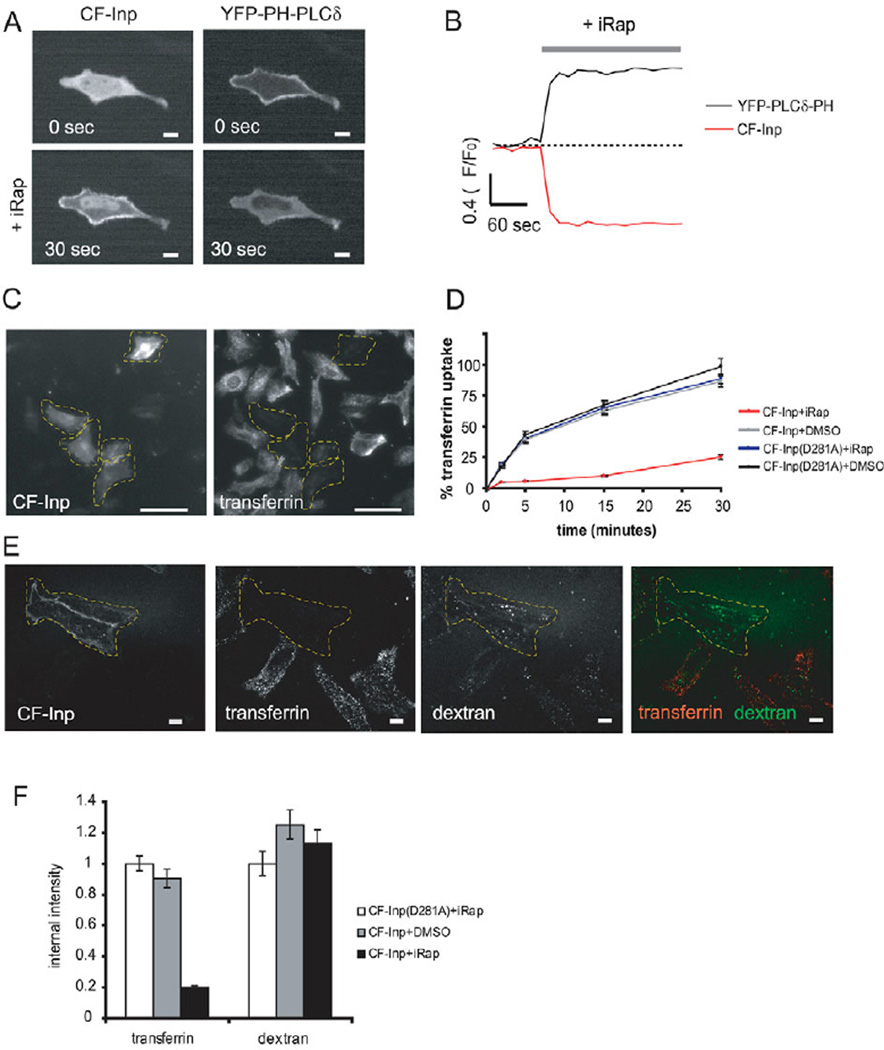
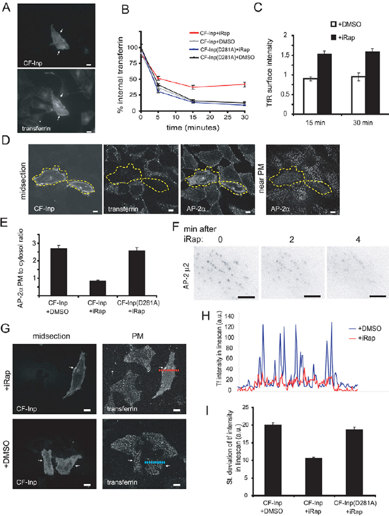
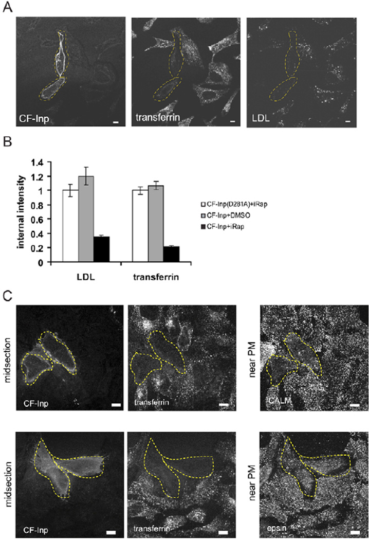
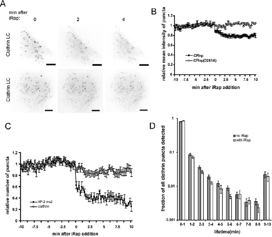
Endocytosis and recycling of membrane proteins are key processes for nutrient uptake, receptor signaling and synaptic transmission. Different steps in these fission and fusion cycles have been proposed to be regulated by physiological changes in plasma membrane (PM) phosphatidylinositol (4,5)-bisphosphate [PtdIns(4,5)P(2)] concentration. Here, we use a chemical enzyme-translocation strategy to rapidly reduce PM PtdIns(4,5)P(2) levels while monitoring clathrin-mediated endocytosis and recycling. PtdIns(4,5)P(2) hydrolysis blocked transferrin receptor endocytosis and led to a marked increase in the concentration of transferrin receptors in the PM, suggesting that endocytosis is more sensitive to changes in PtdIns(4,5)P(2) than recycling. Reduction of PM PtdIns(4,5)P(2) levels led to a near complete dissociation of Adaptor protein 2 (AP-2) from the PM but had only a small effect on clathrin assembly. This argues that receptor-mediated PtdIns(4,5)P(2) reduction preferentially suppresses AP-2-mediated targeting of cargo to endocytic sites rather than the assembly of clathrin coats or recycling of endocytic vesicles.


Figures

Fig. 1
Fig. 1
PM PtdIns(4,5)P2 hydrolysis disrupts endocytosis of the transferrin receptor, but not fluid-phase endocytosis. (A,B) iRap induces rapid translocation of CFP-FKBP-Inp (CF-Inp) from the cytosol to the PM and dissociation of PtdIns(4,5)P2 biosensor YFP-PLC™-PH from the PM. Sample image (A), and corresponding time course of CF-Inp and YFP-PLC™-PH translocation (B). Scale bars: 10 µm. (C,D) Rapid PtdIns(4,5)P2 hydrolysis inhibits transferrin uptake. HeLa cells expressing Lyn-FRB and either CF-Inp or CF-Inp(D281A) were treated with iRap or DMSO for 1 minute before addition of Alexa-Fluor-594-conjugated transferrin. (C) Sample image of CF-Inp-expressing cells treated with iRap and fixed after 15 minutes of Alexa-Fluor-594-conjugated-transferrin uptake. Dashed area indicates the location of Lyn-FRB- and CF-Inp-expressing cells. Scale bars: 50 µm. (D) Time-course of Alexa-Fluor-594-conjugatedtransferrin uptake (significance of difference P<9×10–46 at the 15-minute time point). Error bars represent s.e.m. (E,F) Rapid PtdIns(4,5)P2 hydrolysis does not affect dextran uptake. Following treatment with either DMSO or iRap, cells expressing Lyn-FRB and CF-Inp were assayed for internalization of Alexa-Fluor-594-conjugated transferrin and fluorescein-dextran simultaneously. (E) Representative image of CF-Inp-expressing cells treated with iRap and fixed after Alexa-Fluor- 594-conjugated-transferrin and fluorescein-dextran uptake. Dashed area indicates the location of Lyn- FRB- and CF-Inp-expressing cells. Scale bars: 10 µm. (F) Quantification of transferrin and dextran uptake. For each tracer, the intracellular fluorescence intensity was normalized to that measured in cells expressing Lyn-FRB and CF-Inp( D281A) that were treated with iRap. Error bars represent s.e.m.
Fig. 2
Fig. 2
PM PtdIns(4,5)P2 hydrolysis partially inhibits transferrin receptor recycling to the surface while fully suppressing AP-2α PM localization and targeting of transferrin receptor (TfR) to endocytic sites. (A,B) Rapid PtdIns(4,5)P2 hydrolysis causes partial inhibition of the reinsertion of internalized transferrin receptors back to the cell surface. Cells expressing Lyn-FRB and either CF-Inp or CF-Inp( D281A) were treated with Alexa-Fluor-594- conjugated transferrin for 20 minutes, then kept in Alexa-Fluor-594-conjugated-transferrin-free media for an additional 5, 15 or 30 minutes with iRap or DMSO. (A) Internal Alexa-Fluor-594-conjugated transferrin remaining in CF-Inp-translocated cells after 15 minutes of recycling. Arrows point to Lyn- FRB- and CF-Inp-expressing cells. Scale bars: 10 µm. (B) Time course of internal transferrin loss. Intracellular fluorescence intensity was normalized to that measured in cells expressing Lyn-FRB and CF-Inp(D281A) that were fixed immediately after Alexa-Fluor-594-conjugated-transferrin uptake without recycling (significance of difference P<7×10–11 at the 15-minute time point). Error bars represent s.e.m. (C) Rapid PtdIns(4,5)P2 hydrolysis increases the amount of transferrin receptors expressed at the PM, suggesting that PtdIns(4,5)P2 regulates endocytosis significantly more than it does recycling. Cells expressing Lyn-FRB and CF-Inp were treated with iRap or DMSO for 15 or 30 minutes, then stained with transferrin receptor antibody to label surface transferrin receptors. Receptor expression is normalized to the fluorescence intensity measured in cells without iRap or DMSO treatment. Error bars represent s.e.m. (D) Antibody against AP-2α shows that PtdIns(4,5)P2 depletion causes a dissociation of AP- 2α puncta from the PM. Cells expressing Lyn-FRB and CF-Inp were treated with Alexa-Fluor-594- conjugated transferrin for 5 minutes following a 1-minute treatment with iRap. Dashed area indicates the location of Lyn-FRB- and CF-Inp-expressing cells. Scale bars: 10 µm. (E) Quantitative analysis of the PM dissociation of AP-2α using ratio imaging of the fluorescence intensity near the PM versus that of the midsection. Error bars represent s.e.m. (F) TIRF imaging of AP-2-µ2–YFP fluorescence at the PM better resolves the rapid loss of AP-2-µ2-YFP from the PM upon iRap-induced translocation of CF-Inp. Scale bars: 5 µm. (G-I) The AP-2-adaptor-mediated clustering of transferrin receptors is also lost upon PtdIns(4,5)P2 hydrolysis. Cells expressing Lyn-FRB and either CF-Inp or CF-Inp(D281A) were treated with either DMSO or iRap for 5 minutes, then incubated with Alexa-Fluor-594-conjugated transferrin at 4°C to label transferrin receptors at the surface. Representative images are shown in G. Arrows point to Lyn-FRB- and CF-Inp-expressing cells. Scale bar: 10 µm. (H) Linescan of transferrin intensity from broken lines drawn in G reveals the loss of sharp peaks of transferrin intensity upon PtdIns(4,5)P2 hydrolysis. (I) Mean of the standard deviations of transferrin intensity calculated from each linescan collected. Error bars represent s.e.m.
Fig. 3
Fig. 3
PM PtdIns(4,5)P2 hydrolysis significantly suppresses LDL receptor endocytosis and weakly suppresses PM localization of epsin and CALM adaptors. (A,B) Following treatment with either DMSO or iRap, cells expressing Lyn-FRB and CF-Inp were simultaneously assayed for internalization of DiI-labeled LDL and Alexa-Fluor-488-conjugated transferrin. (A) Representative image of CF-Inp-expressing cells treated with iRap and fixed after transferrin and LDL uptake, showing suppression of the internalization of both receptors in the same cells. Dashed area indicates the location of Lyn-FRB- and CF-Inp-expressing cells. Scale bars: 10 µm. (B) Quantification of transferrin and LDL uptake. For each tracer, the intracellular fluorescence intensity was normalized to that measured in cells expressing Lyn-FRB and CF-Inp(D281A) treated with iRap. Error bars represent s.e.m. (C) Antibody against CALM (top row) or epsin (bottom row) reveals PM-associated punctate localization in untransfected control cells but a more cytosolic distribution in PtdIns(4,5)P2-depleted cells. Cells expressing Lyn-FRB and CF-Inp were treated with Alexa-Fluor-594-conjugated transferrin for 5 minutes following a 1-minute treatment with iRap. Dashed lines indicate the location of Lyn-FRB- and CF-Inp-expressing cells. Scale bars: 10 µm.
Fig. 4
Fig. 4
PtdIns(4,5)P2 hydrolysis has a much smaller effect on clathrin assembly at the PM compared to its effect on dissociating AP-2 adaptors. (A) TIRF imaging of YFP-CLC fluorescence shows only a partial loss of CLC at the PM upon iRap-induced translocation of CFInp. Scale bars: 5 µm. (B) PtdIns(4,5)P2 hydrolysis resulted in only an ~20% reduction in mean clathrin-puncta intensity at the PM. Mean intensity value for each time point was normalized to the maximum mean intensity value measured in each cell. Error bars represent s.e.m. (C) Comparison of the number of puncta at each time point observed for AP-2-µ2–YFP or YFP-CLC after iRap-induced translocation of CF-Inp. Number of puncta was normalized to the maximum number detected in each cell. PtdIns(4,5)P2 hydrolysis caused a more than fourfold greater effect on the number of AP- 2-µ2–YFP-positive puncta compared with that on YFP-CLC-positive puncta. Errors represent s.e.m. (D) Normalized histogram of the lifetimes of YFP-CLC-positive puncta before and after iRap-induced translocation of CF-Inp. There was no significant reduction in the lifetime of CLC-positive puncta in iRap-treated cells. Errors represent s.e.m.

Similar articles

Cited by

References

    1. Brodsky FM, Chen CY, Knuehl C, Towler MC, Wakeham DE. Biological basket weaving: formation and function of clathrin-coated vesicles. Annu. Rev. Cell Dev. Biol. 2001;17:517–568. - PubMed
    1. Boucrot E, Saffarian S, Massol R, Kirchhausen T, Ehrlich M. Role of lipids and actin in the formation of clathrin-coated pits. Exp. Cell Res. 2006;312:4036–4048. - PMC - PubMed
    1. Cremona O, De Camilli P. Phosphoinositides in membrane traffic at the synapse. J. Cell Sci. 2001;114:1041–1451. - PubMed
    1. Di Paolo G, De Camilli P. Phosphoinositides in cell regulation and membrane dynamics. Nature. 2006;443:651–657. - PubMed
    1. Di Paolo G, Moskowitz HS, Gipson K, Wenk MR, Voronov S, Obayashi M, Flavell R, Fitzsimonds RM, Ryan TA, De Camilli P. Impaired PtdIns(4,5)P2 synthesis in nerve terminals produces defects in synaptic vesicle trafficking. Nature. 2004;431:415–422. - PubMed

Publication types