The art of cellular communication: tunneling nanotubes bridge the divide
- PMID: 18386044
- PMCID: PMC2323029
- DOI: 10.1007/s00418-008-0412-0
The art of cellular communication: tunneling nanotubes bridge the divide
Abstract
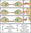
The ability of cells to receive, process, and respond to information is essential for a variety of biological processes. This is true for the simplest single cell entity as it is for the highly specialized cells of multicellular organisms. In the latter, most cells do not exist as independent units, but are organized into specialized tissues. Within these functional assemblies, cells communicate with each other in different ways to coordinate physiological processes. Recently, a new type of cell-to-cell communication was discovered, based on de novo formation of membranous nanotubes between cells. These F-actin-rich structures, referred to as tunneling nanotubes (TNT), were shown to mediate membrane continuity between connected cells and facilitate the intercellular transport of various cellular components. The subsequent identification of TNT-like structures in numerous cell types revealed some structural diversity. At the same time it emerged that the direct transfer of cargo between cells is a common functional property, suggesting a general role of TNT-like structures in selective, long-range cell-to-cell communication. Due to the growing number of documented thin and long cell protrusions in tissue implicated in cell-to-cell signaling, it is intriguing to speculate that TNT-like structures also exist in vivo and participate in important physiological processes.
Figures


Similar articles
-
Intercellular transfer mediated by tunneling nanotubes.Curr Opin Cell Biol. 2008 Aug;20(4):470-5. doi: 10.1016/j.ceb.2008.03.005. Epub 2008 May 2. Curr Opin Cell Biol. 2008. PMID: 18456488 Review.
-
Tunneling nanotubes: a new route for the exchange of components between animal cells.FEBS Lett. 2007 May 22;581(11):2194-201. doi: 10.1016/j.febslet.2007.03.071. Epub 2007 Apr 4. FEBS Lett. 2007. PMID: 17433307 Review.
-
Wiring through tunneling nanotubes--from electrical signals to organelle transfer.J Cell Sci. 2012 Mar 1;125(Pt 5):1089-98. doi: 10.1242/jcs.083279. Epub 2012 Mar 7. J Cell Sci. 2012. PMID: 22399801 Review.
-
Tunneling nanotube (TNT)-like structures facilitate a constitutive, actomyosin-dependent exchange of endocytic organelles between normal rat kidney cells.Exp Cell Res. 2008 Dec 10;314(20):3669-83. doi: 10.1016/j.yexcr.2008.08.022. Epub 2008 Sep 13. Exp Cell Res. 2008. PMID: 18845141
-
The Ways of Actin: Why Tunneling Nanotubes Are Unique Cell Protrusions.Trends Cell Biol. 2021 Feb;31(2):130-142. doi: 10.1016/j.tcb.2020.11.008. Epub 2020 Dec 9. Trends Cell Biol. 2021. PMID: 33309107 Review.
Cited by
-
Exosomes: vesicular carriers for intercellular communication in neurodegenerative disorders.Cell Tissue Res. 2013 Apr;352(1):33-47. doi: 10.1007/s00441-012-1428-2. Epub 2012 May 19. Cell Tissue Res. 2013. PMID: 22610588 Free PMC article. Review.
-
Tubular bridges for bronchial epithelial cell migration and communication.PLoS One. 2010 Jan 28;5(1):e8930. doi: 10.1371/journal.pone.0008930. PLoS One. 2010. PMID: 20126618 Free PMC article.
-
Direct Cell-Cell Communication via Membrane Pores, Gap Junction Channels, and Tunneling Nanotubes: Medical Relevance of Mitochondrial Exchange.Int J Mol Sci. 2022 May 30;23(11):6133. doi: 10.3390/ijms23116133. Int J Mol Sci. 2022. PMID: 35682809 Free PMC article. Review.
-
Mitochondrial Transfer and Regulators of Mesenchymal Stromal Cell Function and Therapeutic Efficacy.Front Cell Dev Biol. 2020 Dec 7;8:603292. doi: 10.3389/fcell.2020.603292. eCollection 2020. Front Cell Dev Biol. 2020. PMID: 33365311 Free PMC article. Review.
-
Intercellular cytosolic transfer correlates with mesenchymal stromal cell rescue of umbilical cord blood cell viability during ex vivo expansion.Cytotherapy. 2012 Oct;14(9):1064-79. doi: 10.3109/14653249.2012.697146. Epub 2012 Jul 10. Cytotherapy. 2012. PMID: 22775077 Free PMC article.
References
-
- {'text': '', 'ref_index': 1, 'ids': [{'type': 'DOI', 'value': '10.1016/j.tips.2005.06.001', 'is_inner': False, 'url': 'https://doi.org/10.1016/j.tips.2005.06.001'}, {'type': 'PMC', 'value': 'PMC1350964', 'is_inner': False, 'url': 'https://pmc.ncbi.nlm.nih.gov/articles/PMC1350964/'}, {'type': 'PubMed', 'value': '15978680', 'is_inner': True, 'url': 'https://pubmed.ncbi.nlm.nih.gov/15978680/'}]}
- Ambudkar SV, Sauna ZE, Gottesman MM, Szakacs G (2005) A novel way to spread drug resistance in tumor cells: functional intercellular transfer of P-glycoprotein (ABCB1). Trends Pharmacol Sci 26:385–387 - PMC - PubMed
-
- {'text': '', 'ref_index': 1, 'ids': [{'type': 'DOI', 'value': '10.1016/j.tcb.2004.07.001', 'is_inner': False, 'url': 'https://doi.org/10.1016/j.tcb.2004.07.001'}, {'type': 'PubMed', 'value': '15308205', 'is_inner': True, 'url': 'https://pubmed.ncbi.nlm.nih.gov/15308205/'}]}
- Baluška F, Hlavacka A, Volkmann D, Menzel D (2004a) Getting connected: actin-based cell-to-cell channels in plants and animals. Trends Cell Biol 14:404–408 - PubMed
-
- {'text': '', 'ref_index': 1, 'ids': [{'type': 'DOI', 'value': '10.1038/428371a', 'is_inner': False, 'url': 'https://doi.org/10.1038/428371a'}, {'type': 'PubMed', 'value': '15042068', 'is_inner': True, 'url': 'https://pubmed.ncbi.nlm.nih.gov/15042068/'}]}
- Baluška F, Volkmann D, Barlow PW (2004b) Cell bodies in a cage. Nature 428:371 - PubMed
-
- {'text': '', 'ref_index': 1, 'ids': [{'type': 'DOI', 'value': '10.1093/aob/mch109', 'is_inner': False, 'url': 'https://doi.org/10.1093/aob/mch109'}, {'type': 'PMC', 'value': 'PMC4242365', 'is_inner': False, 'url': 'https://pmc.ncbi.nlm.nih.gov/articles/PMC4242365/'}, {'type': 'PubMed', 'value': '15155376', 'is_inner': True, 'url': 'https://pubmed.ncbi.nlm.nih.gov/15155376/'}]}
- Baluška F, Volkmann D, Barlow PW (2004c) Eukaryotic cells and their Cell Bodies: Cell Theory revised. Ann Bot (Lond) 94:9–32 - PMC - PubMed
-
- None
- Bard J (1992) Morphogenesis: the cellular and molecular processes of developmental anatomy. Cambridge Univ Press, Cambridge, U.K
Publication types
MeSH terms
Substances
LinkOut - more resources
Full Text Sources
Other Literature Sources

