. 2008 Jun;19(6):2661-72.
doi: 10.1091/mbc.e07-12-1263. Epub 2008 Apr 2.

Novel interactions of ESCRT-III with LIP5 and VPS4 and their implications for ESCRT-III disassembly

Affiliations

Novel interactions of ESCRT-III with LIP5 and VPS4 and their implications for ESCRT-III disassembly

Soomin Shim et al. Mol Biol Cell. 2008 Jun.

Abstract

The AAA+ ATPase VPS4 plays an essential role in multivesicular body biogenesis and is thought to act by disassembling ESCRT-III complexes. VPS4 oligomerization and ATPase activity are promoted by binding to LIP5. LIP5 also binds to the ESCRT-III like protein CHMP5/hVps60, but how this affects its function remains unclear. Here we confirm that LIP5 binds tightly to CHMP5, but also find that it binds well to additional ESCRT-III proteins including CHMP1B, CHMP2A/hVps2-1, and CHMP3/hVps24 but not CHMP4A/hSnf7-1 or CHMP6/hVps20. LIP5 binds to a different region within CHMP5 than within the other ESCRT-III proteins. In CHMP1B and CHMP2A, its binding site encompasses sequences at the proteins' extreme C-termini that overlap with "MIT interacting motifs" (MIMs) known to bind to VPS4. We find unexpected evidence of a second conserved binding site for VPS4 in CHMP2A and CHMP1B, suggesting that LIP5 and VPS4 may bind simultaneously to these proteins despite the overlap in their primary binding sites. Finally, LIP5 binds preferentially to soluble CHMP5 but instead to polymerized CHMP2A, suggesting that the newly defined interactions between LIP5 and ESCRT-III proteins may be regulated by ESCRT-III conformation. These studies point to a role for direct binding between LIP5 and ESCRT-III proteins that is likely to complement LIP5's previously described ability to regulate VPS4 activity.


Figures
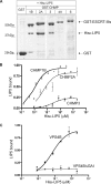
Figure 1.
Figure 1.
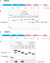
LIP5 binds to ESCRT-III proteins. (A) Interaction of LIP5 with a subset of ESCRT-III proteins. GST and GST-ESCRT-III proteins immobilized on glutathione-Sepharose beads were incubated with E. coli lysate containing His6-LIP5. Bound material was separated on a SDS-PAGE gel and stained with Coomassie blue. Where necessary, lanes were rearranged as indicated by white lines. Immunoblotting with an anti-His6 antibody confirmed that no His6-LIP5 bound to GST-CHMP4A or GST-CHMP6 (not shown). (B) Solid phase assay of LIP5 binding to GST-CHMP1B, 2A, and 3. His6-LIP5 bound to immobilized GST-CHMP proteins was detected with NTA-HRP and TMB colorimetric substrate. EC50 values determined by nonlinear regression analysis ranged from 10 to 20 nM for CHMP1B (▴, solid line), from 49–60 nM for CHMP2A (○, dotted line), and from 0.3–1 μM for CHMP3 (*, alternating dashed line) in several independent experiments. Error bars, the SD from one experiment run in duplicate. Absorbance data were normalized to the Bmax for CHMP1B. (C) Binding of His6-LIP5 to GST-VPS4B(E235Q) and GST-VPS4B(E235Q, ΔGAI) β domain mutant. EC50 for VPS4B (E235Q) (■, solid line) was 60 nM. LIP5 binding to VPS4B(E235Q, ΔGAI) did not change as a function of LIP5 added (▴, dotted line). Error bars, the SD from one experiment run in duplicate, and the absorbance data were normalized to the Bmax for VPS4B(E235Q).
Figure 2.
Figure 2.
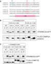
C-terminal sequences in CHMP2A and CHMP1B are required for LIP5 binding. (A) Predicted CHMP2A secondary structure obtained using a neural network based algorithm (http://www.compbio.dundee.ac.uk/∼www-jpred/). Pink and blue boxes correspond to predicted α-helices with pI higher than 8 and lower than 6, respectively. (B) Effects of deleting N- and C-terminal sequences from CHMP2A on LIP5 binding. GST and GST-CHMP2A proteins with the indicated sequences immobilized on beads were incubated with E. coli lysate containing His6-LIP5. Bound material was analyzed by staining with Coomassie blue. (C) Predicted CHMP1B secondary structure. (D) Effects of deleting N- and C-terminal sequences from CHMP1B on LIP5 binding. GST and GST-CHMP1B proteins with the indicated sequences immobilized on beads were incubated with E. coli lysate containing His6-LIP5. Bound material was analyzed by staining with Coomassie blue (top panel) and by immunoblotting with an anti-His6 antibody (bottom panel). Immunoblotting was needed because His6-LIP5 migrates similarly to GST-CHMP1B(106-199).
Figure 3.
Figure 3.
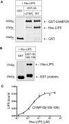
CHMP2A and CHMP1B α6 region is responsible for LIP5 binding. (A) Effect of CHMP2A L216A mutation on interaction with LIP5. GST and GST-CHMP2A proteins immobilized on beads were incubated with E. coli lysate containing His6-LIP5. Bound material was analyzed by staining with Coomassie blue. Where necessary, gel lanes were rearranged as shown by a white line. (B) Binding of His6-LIP5 to GST-CHMP1B(169-199). This CHMP1B fragment contains α6 and surrounding sequences but does not include α5. (C) Solid phase assay of His6-LIP5 binding to GST-CHMP1B(169-199) carried out as described in Figure 1. The EC50 of 25 nM is similar to that of His6-LIP5 for full-length CHMP1B. Absorbance data were normalized to the Bmax for full-length CHMP1B measured in parallel.
Figure 4.
Figure 4.
LIP5 associates preferentially with polymerized CHMP2A and CHMP1B in transfected mammalian cells. (A) Cosedimentation of LIP5 with CHMP2A. HEK293T cells cotransfected with GFP-LIP5 or GFP-LIP5ΔN, and the indicated FLAG-CHMP2A constructs were solubilized in 1% Triton X-100 and centrifuged. The distribution of CHMP2A and LIP5 in the resulting supernatant (S) and pellet (P) was visualized by immunoblotting. LIP5ΔN is equivalent to a deletion in Vta1 that impairs binding to Vps60 (Azmi et al., 2006). Experiments with LIP5ΔN were performed separately from those with full-length LIP5 and are therefore shown in a separate box. (B) Cosedimentation of LIP5 with CHMP1B. The same experiments performed with FLAG-CHMP1B constructs.
Figure 5.
Figure 5.
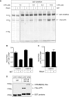
Binding sites for VPS4 and LIP5 in CHMP2A and CHMP1B overlap. (A) VPS4A MIT domain reduces LIP5 binding to GST-CHMP2A. GST-CHMP2A was incubated with His6-LIP5 alone or together with 300 μM His6-VPS4A MIT domain or ribonuclease A as indicated. Bound LIP5 was visualized by staining with colloidal Coomassie blue and quantified by infrared fluorescence scanning. The bound MIT domain can be seen as an increased intensity in the dye front. (B) Quantitation of the effect of MIT domain on binding of 3.2 μM LIP5 to CHMP2A or (in parallel experiments) CHMP1B. (C) Quantitation of lack of effect of the same concentration (300 μM) of ribonuclease A on binding of 1.6 μM LIP5 to GST-CHMP2A. (D) LIP5 and VPS4B(E235Q) bind similarly to GST-CHMP1B(169-199). Material retained on GST or GST-CHMP1B(169-199) after incubation with the indicated 5 μM protein is shown on a gel stained with Coomassie Blue.
Figure 6.
Figure 6.
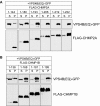
Effects of C-terminal deletions suggest existence of secondary binding site for VPS4B in CHMP2A and CHMP1B. (A) HEK293T cells cotransfected with VPS4B(E235Q)-GFP and indicated FLAG-CHMP2A constructs were solubilized in 1% Triton X-100 and centrifuged. The resulting distribution of VPS4B(E235Q) and CHMP2A between supernatant (S) and pellet (P) was visualized by immunoblotting. (B) Same experiment but with FLAG-CHMP1B constructs.
Figure 7.
Figure 7.
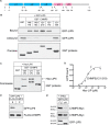
Conserved acidic residues in α5 are part of secondary VPS4-binding site. (A) Sequences of predicted α5 and surrounding sequences in CHMP2 and CHMP1 proteins. Highly conserved acidic residues near the center of the helix are colored red; less conserved pairs of acidic residues in CHMP2A are colored blue. Pairs of alanine replacements in CHMP2A studied below are designated mut a, mut b, and mut c as indicated. The conserved central pair of acidic residues was also mutated in CHMP1B and designated as mut a. (B) Effect of double alanine mutants on cosedimentation of VPS4B(E235Q) with full-length (1-222) or α6-deleted (1-206) CHMP2A in cotransfected HEK293 cells. Cells were solubilized in 1% Triton X-100 and centrifuged. The resulting distribution of VPS4B(E235Q) and CHMP2A between supernatant (S) and pellet (P) was visualized by immunoblotting. (C) Effect of mut a on cosedimentation of VPS4B(E235Q) with full-length (1-199) or α6-deleted α1–α5 (1-181) CHMP1B.
Figure 8.
Figure 8.
Unique properties of CHMP5-LIP5 complex: LIP5 binds to CHMP5 α5 preferentially in the soluble fraction. (A) Predicted secondary structure of CHMP5. (B) GST and GST-CHMP5 proteins immobilized on beads were incubated with HEK293T cell lysate containing GFP-LIP5. Bound and unbound fractions were analyzed by immunoblotting with anti-GFP antibody. GST proteins were visualized by staining the immunoblot with Ponceau red. (C) GST and GST-CHMP5 proteins on beads were incubated with E. coli lysate containing His6-LIP5, and the bound material was analyzed by staining with Coomassie blue. Where necessary, lanes were rearranged as indicated by white lines. (D) Binding of His6-LIP5 to immobilized GST-CHMP5(121-219), detected, and analyzed as in Figure 1B. EC50 values ranged from 1 to 2 nM. Error bars, SD from one experiment performed in duplicate. (E) LIP5 does not cosediment with CHMP5. HEK293T cells cotransfected with GFP-LIP5 and FLAG-CHMP5 were solubilizated in 1% Triton X-100 and centrifuged. The resulting supernatant (S) and pellet (P) were analyzed by immunoblotting. (F) Coimmunoprecipitation of CHMP5-myc with LIP5-GFP from cotransfected HEK293T cells. LIP5-GFP or LIP5ΔN-GFP was immunoprecipitated from the soluble lysate of cotransfected cells. Bound proteins and lysate were analyzed by immunoblotting.
Figure 9.
Figure 9.
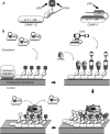
Model showing proposed engagement of LIP5 with ESCRT-III proteins and VPS4. (A) Binding sites for LIP5 and Vps4 in individual ESCRT-III subunits. Sites defined in this study in CHMP2A and CHMP1B are shown at left and in CHMP5 at right. The schematic structure of the ESCRT-III subunits is based on the crystal structure of CHMP3 (Muziol et al., 2006). (B) Proposed model of ESCRT-III assembly, disassembly, and interaction with LIP5 and VPS4. Most ESCRT-III proteins are closed monomers in the cytoplasm and do not bind to LIP5, although CHMP5 interacts differently with LIP5 and can bind in the cytoplasm. As ESCRT-III complexes assemble on the endosomal membrane, individual subunits open and expose sequences at their C-termini for binding to LIP5 and/or VPS4. How these two proteins share their overlapping binding sites remains to be determined, but their separate ability to bind each other (via domains that are not engaged with the ESCRT-III proteins, the β domain in VPS4 (Scott et al., 2005a; Vajjhala et al., 2006) and the VSL domain in LIP5 (Azmi et al., 2006) is likely to reinforce their association. Once some threshold is reached (perhaps assembly of VPS4 rings), we propose that VPS4 engages its secondary contact site in α5 of the ESCRT-III proteins. Based on analogy to other AAA+ proteins, this may allow VPS4 to unfold individual ESCRT-III subunits and release them into the cytoplasm where they revert to their closed and monomeric state.

Comment in

  • Mol Biol Cell. 19:2349.

Similar articles

Cited by

References

    1. Azmi I., Davies B., Dimaano C., Payne J., Eckert D., Babst M., Katzmann D. J. Recycling of ESCRTs by the AAA-ATPase Vps4 is regulated by a conserved VSL region in Vta1. J. Cell Biol. 2006;172:705–717. - PMC - PubMed
    1. Azmi I. F., Davies B. A., Xiao J., Babst M., Xu Z., Katzmann D. J. ESCRT-III family members stimulate Vps4 ATPase activity directly or via Vta1. Dev. Cell. 2008;14:50–61. - PubMed
    1. Babst M. A protein's final ESCRT. Traffic. 2005;6:2–9. - PubMed
    1. Babst M., Katzmann D. J., Estepa-Sabal E. J., Meerloo T., Emr S. D. Escrt–III: an endosome-associated heterooligomeric protein complex required for mvb sorting. Dev. Cell. 2002;3:271–282. - PubMed
    1. Babst M., Sato T. K., Banta L. M., Emr S. D. Endosomal transport function in yeast requires a novel AAA-type ATPase, Vps4p. EMBO J. 1997;16:1820–1831. - PMC - PubMed

Publication types

Substances

LinkOut - more resources