. 2008 Mar 28;29(6):729-41.
doi: 10.1016/j.molcel.2008.01.013.

SUMO-2/3 modification and binding regulate the association of CENP-E with kinetochores and progression through mitosis

Affiliations

SUMO-2/3 modification and binding regulate the association of CENP-E with kinetochores and progression through mitosis

Xiang-Dong Zhang et al. Mol Cell. .

Abstract

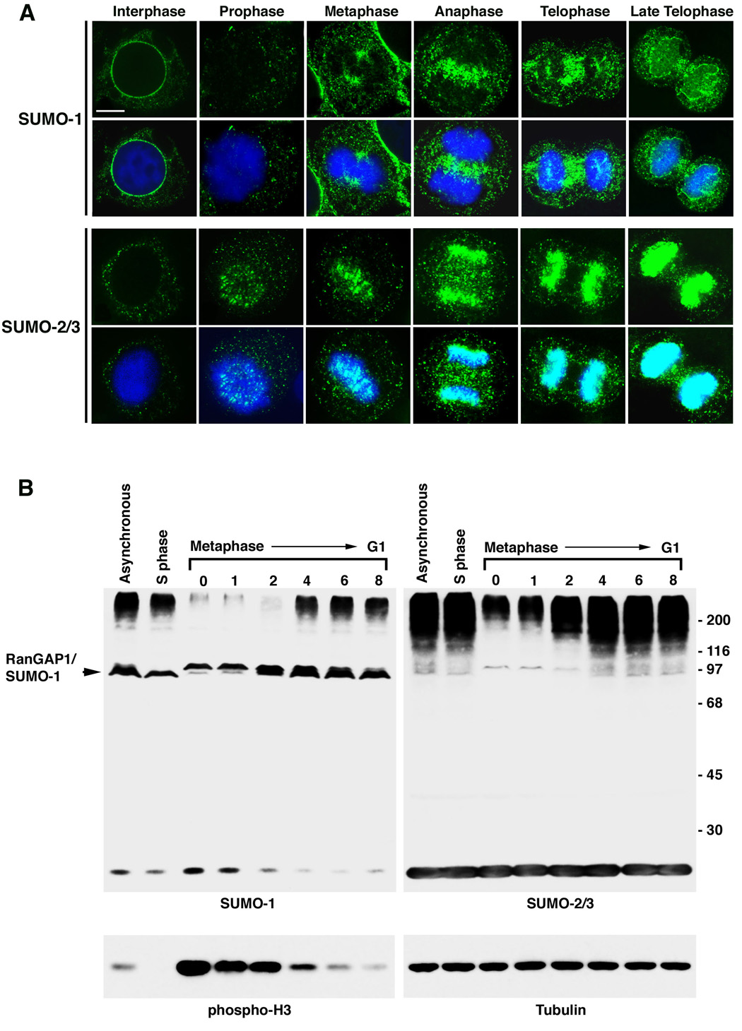
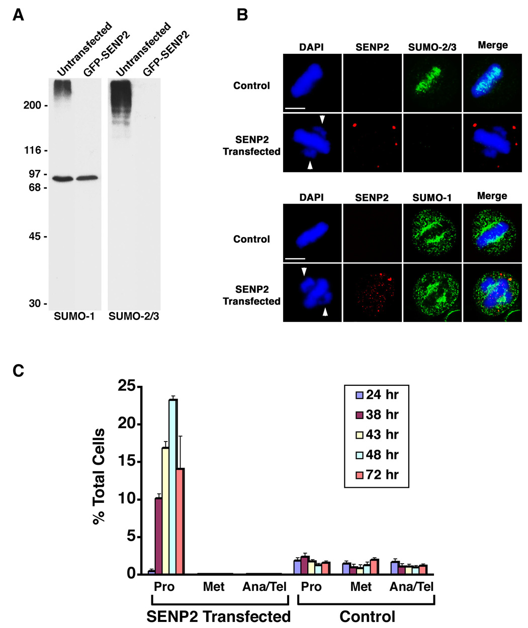
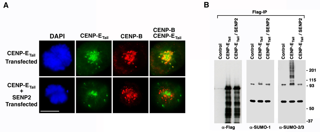
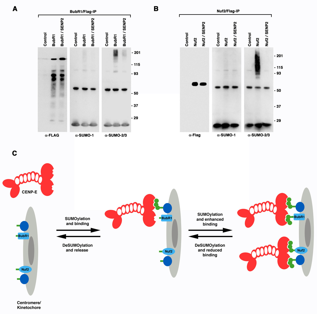
SUMOylation is essential for cell-cycle regulation in invertebrates; however, its functions during the mammalian cell cycle are largely uncharacterized. Mammals express three SUMO paralogs: SUMO-1, SUMO-2, and SUMO-3 (SUMO-2 and SUMO-3 are 96% identical and referred to as SUMO-2/3). We found that SUMO-2/3 localize to centromeres and condensed chromosomes, whereas SUMO-1 localizes to the mitotic spindle and spindle midzone, indicating that SUMO paralogs regulate distinct mitotic processes in mammalian cells. Consistent with this, global inhibition of SUMOylation caused a prometaphase arrest due to defects in targeting the microtubule motor protein CENP-E to kinetochores. CENP-E was found to be modified specifically by SUMO-2/3 and to possess SUMO-2/3 polymeric chain-binding activity essential for kinetochore localization. Our findings indicate that SUMOylation is a key regulator of the mammalian cell cycle, with SUMO-1 and SUMO-2/3 modification of different proteins regulating distinct processes.


Figures

Figure 1
Figure 1
SUMO-1 and SUMO-2/3 conjugates localize to distinct sub-cellular domains during mitosis and are differentially regulated. A. HeLa cells were permeabilized with digitonin, fixed with formaldehyde and analyzed by immunofluorescence confocal microscopy using SUMO-1 or SUMO-2/3 specific mAbs. Indicated cell cycle stages were defined by DNA staining with DAPI. Bar equals 5 µm. B. HeLa cells were arrested in S phase using thymidine, or synchronized in mitosis by a thymidine and nocodazole block followed by release from nocodazole for the indicated times (hr). Cell lysates were separated by SDS-PAGE and analyzed by immunoblotting with SUMO-1 or SUMO-2/3 specific mAbs. As markers for mitosis and protein loading, lysates were probed with antibodies against phospho-histone H3 and α-tubulin.
Figure 2
Figure 2
SUMO-2/3 modified proteins distinct from topoisomerase IIα localize to the centromere and inner kinetochore plate of mitotic chromosomes. A. HeLa cells were permeabilized with digitonin and fixed with formaldehyde. Cells were labeled with SUMO-2/3 mAbs and CENP-B or CENP-C antibodies and analyzed by immunofluorescence confocal microscopy. Bar equals 10 µm. B. Purified mitotic chromosomes were separated by SDS-PAGE and analyzed by immunoblotting with SUMO-2/3, SUMO-1 and Topoisomerase IIα (Topo IIα) specific mAbs. C. HTETOP cells were cultured in the absence of tetracycline (−Tet) to allow topoisomerase IIα expression, or in the presence of tetracycline (+Tet) to inhibit expression. Cells were permeabilized with digitonin, fixed with formaldehyde and analyzed by immunofluorescence microscopy using topoisomerase IIα or SUMO-2/3 specific mAbs. Bar equals 10 µm.
Figure 3
Figure 3
Over-expression of SENP2 inhibits SUMOylation and causes a prometaphase arrest. A. GFP-SENP2 transfected HeLa cells were purified by flow cytometry 48 hr after transfection and lysates were analyzed by immunoblotting with SUMO-1 or SUMO-2/3 specific mAbs. Equal numbers of untransfected cells were analyzed as controls. B. Myc-tagged SENP2 and control transfected HeLa cells were permeabilized with digitonin and fixed with formaldehyde 48 hr after transfection. Cells were double labeled with anti-Myc antibodies and SUMO-2/3 or SUMO-1 specific mAbs. Arrows indicate unaligned chromosome pairs at spindle poles. Bars equal 10 µm. C. The fraction of total cells present at each stage of mitosis following transfection with Myc-tagged SENP2 (n ≥ 135 cells for each time point) or empty vector (Control) (n ≥ 633 cells for each time point) was determined by DAPI staining and fluorescence microscopy (Pro = prophase and prometaphase; Met = metaphase; Ana/Tel = anaphase and telophase). The plotted values are the means plus standard deviation (SD) from a minimum of 3 experiments.
Figure 4
Figure 4
Inhibition of SUMOylation blocks CENP-E association with kinetochores and results in persistent activation of the spindle checkpoint. HeLa cells were transfected with Myc-tagged SENP2 or empty vector. Cells were fixed with paraformaldehyde and permeabilized with Triton X-100 48 hr after transfection and analyzed by immunofluorescence microscopy following labeling with DAPI and the indicated antibodies. Bars equal 10 µm. A. Analysis with anti-Myc and Aurora B antibodies. B. Analysis with anti-Myc and Hec1 antibodies. C. Analysis with anti-Myc and CENP-E antibodies. D. Analysis with anti-Myc and BubR1 antibodies.
Figure 5
Figure 5
Localization of the CENP-E tail domain to kinetochores correlates with its SUMO-2/3 modification. A. HeLa cells were transfected with a plasmid coding for FLAG-tagged CENP-E tail domain alone or together with a plasmid coding for Myc-tagged SENP2. Cells were fixed with paraformaldehyde and permeabilized with Triton X-100 48 hr after transfection and analyzed by immunofluorescence microscopy after labeling with DAPI and anti-FLAG and CENP-B antibodies. Bar equals 10 µm. B. 293 cells were transfected with empty plasmid (Control), plasmid coding for FLAG-tagged CENP-E tail domain alone, or plasmid coding for FLAG-tagged CENP-E tail domain together with plasmid coding for Myc-tagged SENP2. The FLAG-tagged CENP-E tail domain was immunopurified in the presence of 1% Empigen BB and analyzed by immunoblotting with FLAG, SUMO-1 or SUMO-2/3 specific antibodies as indicated.
Figure 6
Figure 6
The association of CENP-E with kinetochores is dependent on non-covalent binding to SUMO-2/3 polymeric chains. A. Sequence alignment of a putative SIM within the CENP-E tail domain and known SIMs in PML and Daxx. Two isoleucine residues, at positions 2307 and 2308, were mutated to lysines to generate the SIM mutant CENP-E tail domain. B. Wild type (WT) and SIM mutant CENP-E tail domains were expressed in vitro in the presence of [35S]methionine and incubated with glutathione sepharose beads containing GST, SUMO-1, SUMO-2, SUMO-1 polymeric chains or SUMO-2 polymeric chains. Bound proteins were analyzed by SDS-PAGE and autoradiography. Input represents 50% of the total protein used in each binding assay. Relative binding activity was quantified by phosphorimage analysis. The relative binding activity of the SIM mutant was normalized to that of WT. Error bars indicate SD from 3 experiments. C. HeLa cells were transfected with FLAG-tagged wild type (WT) and SIM mutant CENP-E tail domain constructs and analyzed by immunofluorescence microscopy 48 hr after transfection. Cells were double labeled with anti-FLAG and CENP-B antibodies. Shown are prophase cells expressing wild type and SIM mutant CENP-E tail domains. Bar equals 10 µm. D. Late telophase and post-mitotic cells expressing FLAG-tagged SIM mutant CENP-E tail domain were analyzed by immunofluorescence microcopy with anti-FLAG antibodies (left two panels). Midbody localization of endogenous CENP-E was detected in untransfected cells with anti-CENP-E antibodies (right two panels). DNA was detected using DAPI. Bar equals 10 µm. E. HeLa cell lysates were prepared 48 hr following transfection with FLAG-tagged wild type (WT) and SIM mutant CENP-E tail domain constructs and analyzed by immunoblotting with anti-FLAG and tubulin antibodies. F. The fraction of cells present at each stage of mitosis 48 hr after transfection with FLAG-tagged wild type (WT) (n ≥ 105 cells) or SIM mutant (n ≥ 273 cells) CENP-E tail domains was determined by DAPI staining and fluorescence microscopy (Pro = prophase and prometaphase; Met = metaphase; Ana/Tel = anaphase and telophase; Multi-Nuc = multi-nucleated cells). The plotted values are the means plus SD from 3 experiments.
Figure 7
Figure 7
BubR1 and Nuf2 are specifically modified by SUMO-2/3 in vivo, suggesting a model for SUMO-2/3 dependent targeting of CENP-E to kinetochores. A. 293 cells were transfected with empty plasmid (Control), FLAG-tagged BubR1 plasmid alone, or FLAG-tagged BubR1 and Myc-tagged SENP2 plasmids together. FLAG-tagged BubR1 was immunopurified and analyzed by immunoblotting with FLAG, SUMO-1 or SUMO-2/3 specific antibodies as indicated. B. SUMOylation of Nuf2 was analyzed as described for BubR1. C. A proposed model for the roles of SUMO-2/3 modification and polymeric chain binding in regulating the association of CENP-E with kinetochores. CENP-E is depicted as a dimer containing SUMO-2/3 polymeric chain interacting motifs (notches) within the carboxyl-terminal tail domain. The kinetochore/centromere is depicted as having associated proteins, including BubR1 and Nuf2, with sites for covalent SUMO-2/3 modification (green bars). The relative levels of SUMOylation and deSUMOylation at the kinetochore define the stability of CENP-E association. Although not depicted, CENP-E may also be covalently modified by SUMO-2/3 polymeric chains. SUMOylation of CENP-E could further contribute to kinetochore association by mediating interactions with other SIM-containing proteins at the kinetochore.

Similar articles

Cited by

References

    1. Ayaydin F, Dasso M. Distinct in vivo dynamics of vertebrate SUMO paralogues. Mol Biol Cell. 2004;15:5208–5218. - PMC - PubMed
    1. Azuma Y, Arnaoutov A, Dasso M. SUMO-2/3 regulates topoisomerase II in mitosis. J Cell Biol. 2003;163:477–487. - PMC - PubMed
    1. Bachant J, Alcasabas A, Blat Y, Kleckner N, Elledge SJ. The SUMO-1 isopeptidase Smt4 is linked to centromeric cohesion through SUMO-1 modification of DNA topoisomerase II. Mol Cell. 2002;9:1169–1182. - PubMed
    1. Carpenter AJ, Porter AC. Construction, characterization, and complementation of a conditional-lethal DNA topoisomerase IIalpha mutant human cell line. Mol Biol Cell. 2004;15:5700–5711. - PMC - PubMed
    1. Chan GK, Schaar BT, Yen TJ. Characterization of the kinetochore binding domain of CENP-E reveals interactions with the kinetochore proteins CENP-F and hBUBR1. J Cell Biol. 1998;143:49–63. - PMC - PubMed

Publication types

LinkOut - more resources