

Hindlimb movement in the cat induced by amplitude-modulated stimulation using extra-spinal electrodes
- PMID: 18369283
- PMCID: PMC2823076
- DOI: 10.1088/1741-2560/5/2/002
Hindlimb movement in the cat induced by amplitude-modulated stimulation using extra-spinal electrodes
Abstract
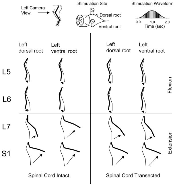
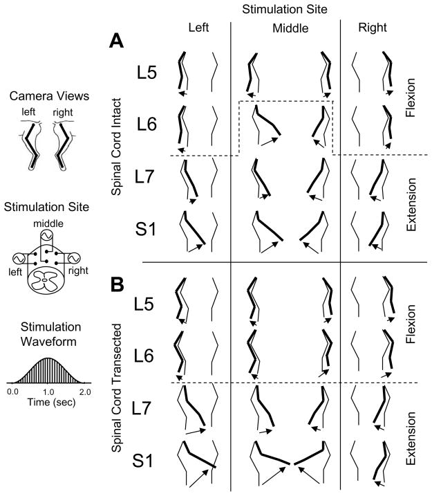
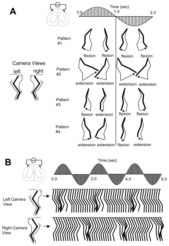
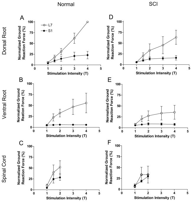
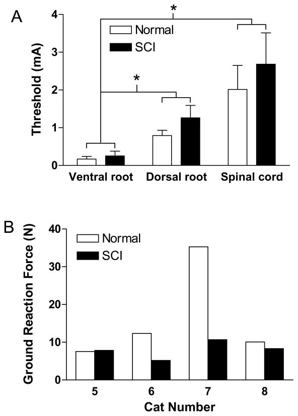
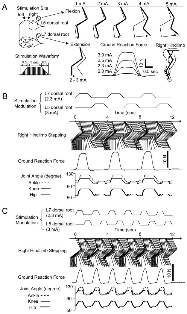
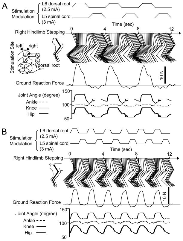
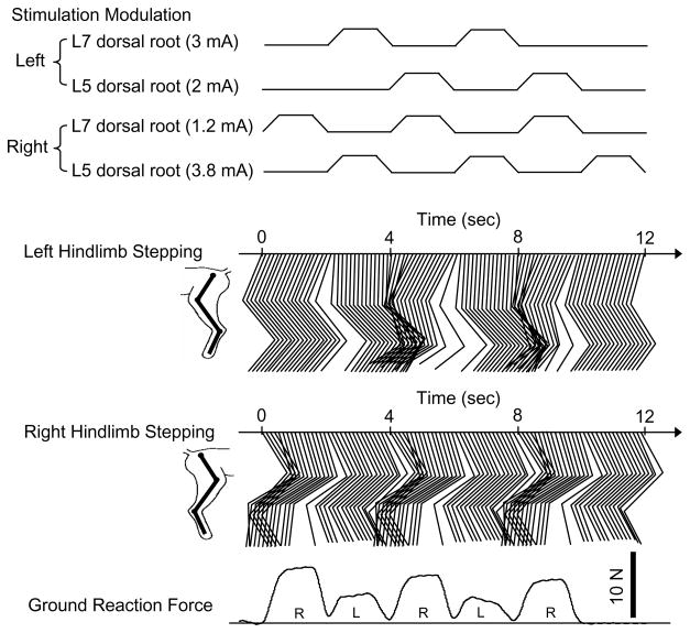
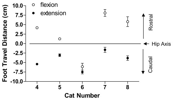
Hindlimb movement in the cat induced by electrical stimulation with an amplitude-modulated waveform of the dorsal surface of the L5-S1 spinal cord or the L5-S1 dorsal/ventral roots was investigated before and after acute spinal cord transection at the T13-L1 level. Stimulation of the spinal cord or dorsal/ventral root at the same spinal segment induced similar movements including coordinated multi-joint flexion or extension. The induced movements changed from flexion to extension when the stimulation was moved from rostral (L5) to caudal (S1) spinal segments. Stimulation of a dorsal or ventral root on one side induced only ipsilateral hindlimb movement. However, stimulation on the dorsal surface of the spinal cord along the midline or across the spinal cord induced bilateral movements. The extension induced by stimulation of L7 dorsal root produced the largest ground reaction force that was strong enough to support body weight. Dorsal root stimulation induced a larger ground reaction force than ventral root stimulation and produced a more graded recruitment curve. Stepping at different speeds could be generated by combined stimulation of the rostral (L5) and the caudal (L6/L7) spinal segments with an appropriate timing between the different stimulation channels. Acute transection of the spinal cord did not change the responses indicating that the induced movements did not require the involvement of the supraspinal locomotor centers. The methods and the stimulation strategy developed in this study might be utilized to restore locomotor function after spinal cord injury.
Figures
Similar articles
-
Multi-joint movement of the cat hindlimb evoked by microstimulation of the lumbosacral spinal cord.Exp Neurol. 2003 Oct;183(2):620-7. doi: 10.1016/s0014-4886(03)00210-3. Exp Neurol. 2003. PMID: 14552903
-
Controlled unilateral isometric force generated by epidural spinal cord stimulation in the rat hindlimb.IEEE Trans Neural Syst Rehabil Eng. 2012 Jul;20(4):549-56. doi: 10.1109/TNSRE.2012.2190424. Epub 2012 Jun 15. IEEE Trans Neural Syst Rehabil Eng. 2012. PMID: 22717526
-
Selective activation of muscle groups in the feline hindlimb through electrical microstimulation of the ventral lumbo-sacral spinal cord.IEEE Trans Rehabil Eng. 2000 Mar;8(1):11-21. doi: 10.1109/86.830944. IEEE Trans Rehabil Eng. 2000. PMID: 10779103
-
Transplants and neurotrophic factors increase regeneration and recovery of function after spinal cord injury.Prog Brain Res. 2002;137:257-73. doi: 10.1016/s0079-6123(02)37020-1. Prog Brain Res. 2002. PMID: 12440372 Review.
-
A review of methods for achieving upper limb movement following spinal cord injury through hybrid muscle stimulation and robotic assistance.Exp Neurol. 2020 Jun;328:113274. doi: 10.1016/j.expneurol.2020.113274. Epub 2020 Mar 5. Exp Neurol. 2020. PMID: 32145251 Review.
Cited by
-
Fast inference of spinal neuromodulation for motor control using amortized neural networks.J Neural Eng. 2022 Oct 18;19(5):10.1088/1741-2552/ac9646. doi: 10.1088/1741-2552/ac9646. J Neural Eng. 2022. PMID: 36174534 Free PMC article.
-
Prediction of isometric forces from combined epidural spinal cord and neuromuscular electrical stimulation in the rat lower limb.Res Sq [Preprint]. 2023 Oct 4:rs.3.rs-3377679. doi: 10.21203/rs.3.rs-3377679/v1. Res Sq. 2023. Update in: Sci Rep. 2024 Jul 9;14(1):15871. doi: 10.1038/s41598-024-66773-9. PMID: 37886495 Free PMC article. Updated. Preprint.
References
-
- Agnew WF, McCreery DB. Neural Prostheses: Fundamental Studies. Englewood Cliffs: Prentice Hall; 1990.
-
- Aoyagi Y, Mushahwar VK, Stein RB, Prochazka A. Movements elicited by electrical stimulation of muscles, nerves, intermediate spinal cord, and spinal roots in anesthetized and decerebrate cats. IEEE Trans Neural Sys Rehabil Eng. 2004;12:1–11. - PubMed
-
- Barthelemy D, Leblond H, Provencher J, Rossignol S. Non-locomotor and locomotor hindlimb responses evoked by electrical microstimulation of the lumbar cord in spinalized cats. J Neurophysiol. 2006;96:3273–3292. - PubMed
-
- Barthelemy D, Leblond H, Rossignol S. Characteristics and mechanisms of locomotion induced by intraspinal microstimulation and dorsal root stimulation in spinal cats. J Neurophysiol. 2007;97:1986–2000. - PubMed
-
- Bras H, Cavallari P, Jankowska E, Kubin L. Morphology of midlumbar interneurones relaying information from group II muscle afferents in the cat spinal cord. J Comp Neurol. 1989;290:1–15. - PubMed
Publication types
MeSH terms
Grants and funding
LinkOut - more resources
Full Text Sources
Medical
Miscellaneous