Xeroderma pigmentosum-variant patients from America, Europe, and Asia
- PMID: 18368133
- PMCID: PMC2562952
- DOI: 10.1038/jid.2008.48
Xeroderma pigmentosum-variant patients from America, Europe, and Asia
Abstract
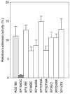
Xeroderma pigmentosum-variant (XP-V) patients have sun sensitivity and increased skin cancer risk. Their cells have normal nucleotide excision repair, but have defects in the POLH gene encoding an error-prone polymerase, DNA polymerase eta (pol eta). To survey the molecular basis of XP-V worldwide, we measured pol eta protein in skin fibroblasts from putative XP-V patients (aged 8-66 years) from 10 families in North America, Turkey, Israel, Germany, and Korea. Pol eta was undetectable in cells from patients in eight families, whereas two showed faint bands. DNA sequencing identified 10 different POLH mutations. There were two splicing, one nonsense, five frameshift (3 deletion and 2 insertion), and two missense mutations. Nine of these mutations involved the catalytic domain. Although affected siblings had similar clinical features, the relation between the clinical features and the mutations was not clear. POLH mRNA levels were normal or reduced by 50% in three cell strains with undetectable levels of pol eta protein, indicating that nonsense-mediated message decay was limited. We found a wide spectrum of mutations in the POLH gene among XP-V patients in different countries, suggesting that many of these mutations arose independently.
Conflict of interest statement
CONFLICT OF INTEREST
The authors state no conflict of interest.
Figures


Similar articles
-
Identification of a novel nonsense mutation in POLH in a Chinese pedigree with xeroderma pigmentosum, variant type.Int J Med Sci. 2013 Apr 21;10(6):766-70. doi: 10.7150/ijms.6095. Print 2013. Int J Med Sci. 2013. PMID: 23630442 Free PMC article.
-
Rare exon 10 deletion in POLH gene in a family with xeroderma pigmentosum variant correlating with protein expression by immunohistochemistry.G Ital Dermatol Venereol. 2020 Jun;155(3):349-354. doi: 10.23736/S0392-0488.16.05158-0. G Ital Dermatol Venereol. 2020. PMID: 32635709
-
Correlation of phenotype/genotype in a cohort of 23 xeroderma pigmentosum-variant patients reveals 12 new disease-causing POLH mutations.Hum Mutat. 2014 Jan;35(1):117-28. doi: 10.1002/humu.22462. Hum Mutat. 2014. PMID: 24130121
-
Molecular genetics of Xeroderma pigmentosum variant.Exp Dermatol. 2003 Oct;12(5):529-36. doi: 10.1034/j.1600-0625.2003.00124.x. Exp Dermatol. 2003. PMID: 14705792 Review.
-
The accurate bypass of pyrimidine dimers by DNA polymerase eta contributes to ultraviolet-induced mutagenesis.Mutat Res. 2024 Jan-Jun;828:111840. doi: 10.1016/j.mrfmmm.2023.111840. Epub 2023 Nov 7. Mutat Res. 2024. PMID: 37984186 Review.
Cited by
-
Reproductive Health in Xeroderma Pigmentosum: Features of Premature Aging.Obstet Gynecol. 2019 Oct;134(4):814-819. doi: 10.1097/AOG.0000000000003490. Obstet Gynecol. 2019. PMID: 31503159 Free PMC article.
-
Strict sun protection results in minimal skin changes in a patient with xeroderma pigmentosum and a novel c.2009delG mutation in XPD (ERCC2).Exp Dermatol. 2009 Jan;18(1):64-8. doi: 10.1111/j.1600-0625.2008.00763.x. Epub 2008 Jul 7. Exp Dermatol. 2009. PMID: 18637129 Free PMC article.
-
Xeroderma pigmentosum and acute myeloid leukemia: a case report.J Med Case Rep. 2021 Aug 26;15(1):437. doi: 10.1186/s13256-021-02969-1. J Med Case Rep. 2021. PMID: 34446105 Free PMC article.
-
Identification of a novel nonsense mutation in POLH in a Chinese pedigree with xeroderma pigmentosum, variant type.Int J Med Sci. 2013 Apr 21;10(6):766-70. doi: 10.7150/ijms.6095. Print 2013. Int J Med Sci. 2013. PMID: 23630442 Free PMC article.
-
Human DNA polymerase η is pre-aligned for dNTP binding and catalysis.J Mol Biol. 2012 Jan 27;415(4):627-34. doi: 10.1016/j.jmb.2011.11.038. Epub 2011 Nov 29. J Mol Biol. 2012. PMID: 22154937 Free PMC article.
References
-
- Arlett CF, Harcourt SA, Broughton BC. The influence of caffeine on cell survival in excision-proficient and excision-deficient xeroderma pigmentosum and normal human cell strains following ultraviolet-light irradiation. Mutat Res. 1975;33:341–6. - PubMed
-
- Bienko M, Green CM, Crosetto N, Rudolf F, Zapart G, Coull B, et al. Ubiquitin-binding domains in Y-family polymerases regulate translesion synthesis. Science. 2005;310:1821–4. - PubMed
-
- Bootsma D, Kraemer KH, Cleaver JE, Hoeijmakers JHJ. Nucleotide excision repair syndromes: xeroderma pigmentosum, Cockayne syndrome, and trichothiodystrophy. In: Vogelstein B, Kinzler KW, editors. The Genetic Basis of Human Cancer. 2. New York: McGraw-Hill; 2002. pp. 211–37.
-
- Boudsocq F, Ling H, Yang W, Woodgate R. Structure-based interpretation of missense mutations in Y-family DNA polymerases and their implications for polymerase function and lesion bypass. DNA Repair (Amst) 2002;1:343–58. - PubMed
Publication types
MeSH terms
Substances
Grants and funding
LinkOut - more resources
Full Text Sources
Medical
Research Materials

