. 2008 Apr 1;180(7):4848-57.
doi: 10.4049/jimmunol.180.7.4848.

Age-related dysregulation of CD8+ T cell memory specific for a persistent virus is independent of viral replication

Affiliations

Age-related dysregulation of CD8+ T cell memory specific for a persistent virus is independent of viral replication

Anna Lang et al. J Immunol. .

Abstract

The immune system devotes substantial resources to the lifelong control of persistent pathogens, which were hypothesized to play an important role in immune aging. Specifically, the presence of latent herpesviruses has been correlated with immune exhaustion and shorter lifespan in octogenarians. But neither the causality nor the mechanistic link(s) were established, and the relative roles of persistent antigenic stimulation and of virus-independent homeostatic disturbances in T cell aging remain unresolved. We longitudinally analyzed expansion, contraction, and long-term maintenance of CD8(+) T cells responding to localized infection with a latent virus, HSV-1. Young mice exhibited the expected expansion and contraction of HSV-1-specific cells and the stable maintenance of memory T cells into advanced adulthood. However, upon entry into senescence, many (>40%) animals exhibited an accumulation in Ag-specific cells (memory inflation) which in some animals was comparable to that observed in acute infection. Inflation occurred to the same extent in control mice and mice continuously treated with the anti-HSV drug famciclovir, which inhibits viral replication and was able to reduce expression of the glycoprotein B. Age-related inflation was also found long after infection with an acute virus. The inflating cells largely maintained Ag-specific function, and exhibited typical central memory phenotype, with no signs of Ag-specific activation. They exhibited increased expression of CD122 and CD127, akin to the Ag-independent T cell clonal expansions found in old specific pathogen-free laboratory mice. This collectively suggests that, in this model, the inflating cells may be selected for high responsiveness to environmental cytokines largely in an Ag-independent manner.


Conflict of interest statement

The authors report no financial conflict of interest nor commercial affiliations.

Figures
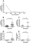
Figure 1
Figure 1. CD8+ T-cell memory is stable in adult but dysregulated in old animals following ocular HSV-1 infection
A. Stable maintenance of gB-8p specific memory CD8+ T-cells in adult animals. A cohort of young B6 mice (n = 9) was infected with 106 PFU HSV-1 (i.c. infection). Blood samples were taken at day 8, and at 1.5, 3, and 4 months p.i. and stained with anti-CD8+ and gB-8p:Kb tetramer. The values show the average percent of tetramer+ cells within CD8+ T-cells ± SEM. The data is representative of three independent experiments. B-E. Memory inflation occurs with advanced age. B. A cohort of young B6 mice (n=50) was infected as in A and followed longitudinally. Blood samples were taken from adult (4 m.p.i.) and old mice (18-24 m.p.i.), and stained as in A. The values show the percent of tetramer+ cells within CD8+ T-cells (each individual animal is represented by a dot, the dash reflects the average). The increase in the average percent of tetramer+ CD8+ T-cells in the old was significant (p<0.0001, paired Student’s t-test). C. Mice were infected as in A and select animals were sacrificed as adults (n = 12) or old (n = 17) and the frequency of gB-specific memory CD8+ T-cells was determined in their spleens as above. The increase in memory cell frequency was significant in the old (p=0.036, Student’s t-test) D and E. Absolute numbers of tetramer+ CD8+ T-cells in blood (D) and spleens (E) of mice from part C and D, respectively. Numbers were obtained from absolute counts and percentages of tetramer+ CD8+ T-cells.
Figure 2
Figure 2. Functional responses of HSV-specific memory CD8+ T-cells in adult and old mice
A. Most gB-8p-specific memory CD8+ T-cells produce IFNγ. Splenocytes from latently infected adult (n=7, 10 m.p.i., 12 mo) and old (n=16, 18-24 m.p.i., 20-26 mo) mice were stimulated for 6 hours with 10-6 M gB-8p peptide in presence of brefeldin A, and then stained using gB-8p:Kb tetramer, CD8 (surface) and/or intracellular IFNγ. The X values show the percent of tetramer+ cells, and the Y values shows the percent of IFNγ+ cells within total CD8+ T-cell pool of individual mice. The values for IFNγ show the net IFNγ (background IFNγ staining from unstimulated wells was subtracted; background was < 0.2%). Excellent correlation of the tetramer and IFNγ staining was observed (R2 = 0.982). B. Cytotoxic activity of memory CD8+ T-cells from mice at various times post HSV-1 infection. Splenocytes harvested from mice in A were cultured for 5 days in presence of gB-8p:Kb – pulsed stimulators. On day 5 of culture the cytotoxic function of HSV-specific CTLs was tested in a standard 51Cr release assay. The CTLs (effectors) were mixed with gB-8p:Kb – pulsed EL-4 cells that were intracellularly labeled with 51Cr (targets). Background lysis values obtained with peptide-negative targets (<8%) were subtracted. The X axis values show the percent of tetramer+ cells at day 0 of culture, and the Y axis values show the percent of specific lysis at effector-to- target ratio of 80:1. The numbers on the graph indicate the IDs of specific mice as discussed in the results.
Figure 3
Figure 3. Expanded gB-8p-specific memory cells in old HSV-1 infected mice uniformly exhibit central memory phenotype
Splenocytes (shown) or lymphocytes isolated from blood (not shown) from old HSV-1 infected mice with age-related expansions of gB-8p-specific memory CD8+ T-cells were stained with CD8, tetramer and a panel of surface markers (CD44, CD62L, Ly6C, CD127, CD122, CD43, CD27). The staining of tetramer+ (top panel) and tetramer- (bottom panel) CD8+ T-cells from a representative mouse is shown. The same phenotype was observed in CD8+ T-cells isolated from blood (not shown).
Figure 4
Figure 4. A-C. Famvir blocks viral replication and decreases the extent of gB transcription
Famvir treatment during acute infection abrogates viral replication and lowers the latent viral load to undetectable level. Young mice were infected i.c. with HSV-1 as described in Materials and Methods. Some mice (n=5) received Famvir continuously in the drinking water starting on day (-7). A. On days 3 and 5 p.i. TGs of Famvir-treated (n=5) and untreated (n=5) mice were isolated and tested for presence of infectious virus by plaque assay. Dashed line represents the level of reliable detection. B. On day 30 p.i. the TGs from untreated (n=7) or Famvir-treated (n=10) mice were isolated and subjected to a reactivation assay as described in Materials and Methods. The total titer of the virus reactivated from TG culture from each mouse is shown. No virus was detected in the cultures from Famvir-treated mice (n.d., not detected). Dashed line represents the level of reliable detection. C. Splenocytes from mice in B were stained with anti-CD8 and gB-8p:Kb tetramer. Dashed line represents levels found in control, uninfected animals. D-E. Famvir decreases the extent of gB transcription and prevents viral DNA synthesis during reactivation from latency. TGs were isolated from latently infected (4 m.p.i.) mice and subjected to standard TG explant reactivation assay in vitro. Quantitative real-time PCR was performed to detect the level of gB transcription (D) and viral DNA load (E). RNA and DNA were isolated from TGs ex-vivo (n = 5), or after 5 day TG explant culture in control media (n= 5) or media containing 1 mM Famvir (n = 5), as described in Materials and Methods. Mice whose TGs were cultured in presence of Famvir were given Famvir in their drinking water for 7 days prior to organ harvest to allow for drug’s immediate access to TGs upon initiation of explant culture. The data represents the total number of gB transcripts (D) or viral genome copies (E) per individual mouse TG. The data is representative of 2 independent experiments.
Figure 5
Figure 5. Effect of viral reactivation on development of age-related memory CD8+ T-cell expansions
A-C. Continuous Famvir treatment of latently infected mice does not prevent the development of age-related memory CD8+ T-cell expansions. Young mice (n=16) were infected i.c. with HSV-1 and their gB-8p-specific CD8+ T-cell responses were followed longitudinally. Half of the cohort was continuously treated with Famvir starting on day 14 p.i. The same mice were screened for frequency of their HSV-specific memory CD8+ T-cells as adults and old. Age-related memory inflation was observed both in control and Famvir-treated group, and there were no significant differences in the frequency (A) or absolute numbers (B) of gB-8p-specific memory CD8+ T-cells in the old mice from either group. Mice undergoing memory inflation had the same latent viral load as the memory inflation-free mice, as determined by real-time PCR at the time of sacrifice at 20 m.p.i. (C). The data is representative of two independent experiments. D-E. Continuous Famvir treatment of latently infected mice affects the size and phenotype of memory CD8+ T-cell response in brain. Mice were infected with HSV-1 as adults and continuously treated with Famvir from day 14 p.i. as in A. At 22 m.p.i. the mice were sacrificed, and lymphocytes isolated from their brains were stained with anti-CD8 and tetramer (D) and CD127 (E). F. Age-associated CD8+ memory inflation in old mice infected with acute virus. Young mice (2-4 mo) were infected with WNV. Peripheral blood lymphocytes were stained with anti-CD8 and NS4b:Db tetramer in adult (n = 17) or in old mice (18-22 m.p.i., n = 50).
Figure 6
Figure 6. Clonal composition and expression of IL7Rα and IL15Rβ by expanded gB-8p-specific memory CD8+ T-cells in old mice
A. CDR3 length analysis of the BV segments involved in gB-8p-specific CD8+ T cell response in mice undergoing late-age memory inflation. CD8+ T-cells from mice exhibiting old-age memory inflation were purified using magnetic bead separation, the RNA was extracted and the diversity of CDR3 lengths of BV8 (8.1, 8.2 and 8.3) and BV10 segments was determined as described in Materials and Methods. CDR3 profiles from three representative mice are shown. B. Elevated expression of IL7Rα (CD127) and IL15Rβ (CD122) on expanded gB-8p-specific memory cells in old mice. Splenocytes from old latently infected mice (n=6) undergoing old-age memory inflation were stained with CD8, CD44, CD62L, tetramer, CD127 and CD122. The percentage of CD122+ and CD127+ cells as well as the mean fluorescent intensity (MFI) of CD127 was compared between central memory phenotype (CD44+ CD62L+) tetramer+ (top panels) or tetramer- (bottom panel) CD8+ T-cells. The values represent the average ± SD (first two rows) or the p-value (obtained from paired Student’s t-test comparing the values from the tetramer+ and tetramer- fraction of cells of individual mice), and the data is representative of two independent experiments.

Similar articles

Cited by

References

    1. Miller RA. The aging immune system: primer and prospectus. Science. 1996;273:70–74. - PubMed
    1. Linton PJ, Dorshkind K. Age-related changes in lymphocyte development and function. Nat Immunol. 2004;5:133–139. - PubMed
    1. Cambier J. Immunosenescence: a problem of lymphopoiesis, homeostasis, microenvironment, and signaling. Immunol Rev. 2005;205:5–6. - PubMed
    1. Surh CD, Sprent J. Regulation of naive and memory T-cell homeostasis. Microbes Infect. 2002;4:51–56. - PubMed
    1. Fry TJ, Mackall CL. The many faces of IL-7: from lymphopoiesis to peripheral T cell maintenance. J Immunol. 2005;174:6571–6576. - PubMed

Publication types