. 2008 Jun;19(6):2465-75.
doi: 10.1091/mbc.e07-12-1215. Epub 2008 Mar 19.

Deficiency of zonula occludens-1 causes embryonic lethal phenotype associated with defected yolk sac angiogenesis and apoptosis of embryonic cells

Affiliations

Deficiency of zonula occludens-1 causes embryonic lethal phenotype associated with defected yolk sac angiogenesis and apoptosis of embryonic cells

Tatsuya Katsuno et al. Mol Biol Cell. 2008 Jun.

Abstract

Zonula occludens (ZO)-1/2/3 are the members of the TJ-MAGUK family of membrane-associated guanylate kinases associated with tight junctions. To investigate the role of ZO-1 (encoded by Tjp1) in vivo, ZO-1 knockout (Tjp1(-/-)) mice were generated by gene targeting. Although heterozygous mice showed normal development and fertility, delayed growth and development were evident from E8.5 onward in Tjp1(-/-) embryos, and no viable Tjp1(-/-) embryos were observed beyond E11.5. Tjp1(-/-) embryos exhibited massive apoptosis in the notochord, neural tube area, and allantois at embryonic day (E)9.5. In the yolk sac, the ZO-1 deficiency induced defects in vascular development, with impaired formation of vascular trees, along with defective chorioallantoic fusion. Immunostaining of wild-type embryos at E8.5 for ZO-1/2/3 revealed that ZO-1/2 were expressed in almost all embryonic cells, showing tight junction-localizing patterns, with or without ZO-3, which was confined to the epithelial cells. ZO-1 deficiency depleted ZO-1-expression without influence on ZO-2/3 expression. In Tjp1(+/+) yolk sac extraembryonic mesoderm, ZO-1 was dominant without ZO-2/3 expression. Thus, ZO-1 deficiency resulted in mesoderms with no ZO-1/2/3, associated with mislocalization of endothelial junctional adhesion molecules. As a result, angiogenesis was defected in Tjp1(-/-) yolk sac, although differentiation of endothelial cells seemed to be normal. In conclusion, ZO-1 may be functionally important for cell remodeling and tissue organization in both the embryonic and extraembryonic regions, thus playing an essential role in embryonic development.


Figures

Figure 1.
Figure 1.
Generation of ZO-1–deficient mice. (A) Restriction maps of the wild type allele, the targeting vector, and the targeted allele of the ZO-1 gene. The targeting vector contained the pgk-neo cassette in its middle portion to delete the two to three exons in the targeted allele. The position of the probe for Southern blotting is indicated as a bar (Probe). Ev, EcoRV; Ps, PstI; EI, EcoRI; B, BsrDI; and Pv, PvuII. (B) Genotypic analysis by Southern blotting of EcoRI-digested genomic DNA from wild-type (+/+), heterozygous (+/−), and homozygous (−/−) mice for the ZO-1 gene allele. Southern blotting with the probe indicated in A yields 21- and 8-kb bands from the wild-type and targeted allele, respectively. (C) Loss of ZO-1 protein in the embryonic extract of Tjp1−/− mice examined by immunoblotting. Anti-ZO-1 immunoblotting of the embryonic protein extracts of ZO-1–deficient mice. Embryonic protein extracts (10 μg) from Tjp1+/+ (+/+), Tjp1+/− (+/−), and Tjp1−/− (−/−) mice were immunoblotted with anti-ZO-1 mAb. In the wild-type and heterozygous embryonic extracts, the ZO-1 band is detected, whereas in the homozygous extract, this band is not detected.
Figure 2.
Figure 2.
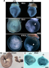
Macroscopic analysis of Tjp1+/+ and Tjp1−/− embryos at E8.5-E10.5. (A) Photographs showing freshly dissected Tjp1+/+ and Tjp1−/− embryos. Slight growth retardation was obvious in Tjp1−/− embryos compared with Tjp1+/+ embryos at E8.5 (E8.5). From E9.5 onward, Tjp1−/− embryos were markedly smaller than Tjp1+/+ embryos, and almost all failed to turn, a process that occurs in Tjp1+/+ embryos around E9.0 (E9.5). Despite the obvious presence of an allantois (arrowhead) at E9.5 and E10.5, chorioallantoic fusion did not occur in the Tjp1−/− embryos. During E8.5 to E10.5, the Tjp1−/− embryo proper was approximately the same size, although the yolk sac was enlarged to a normal size, compared with Tjp1+/+ litter embryos. The large vitelline vessels (arrows) were detected in Tjp1+/+ yolk sac at E9.5 and E10.5. (B) PECAM-1 stained embryo proper. Although the growth was critically retarded, vasculogenesis, and angiogenesis occurred in the Tjp1+/+ and Tjp1−/− embryo proper. (C) PECAM-1–stained embryonic yolk sac. Numerous large, branching vessels were present in Tjp1+/+ yolk sacs as revealed by PECAM-1 staining, showing the process of angiogenesis. In contrast, the arrangement of branching vessels was not detected extraembryonically in Tjp1−/− yolk sacs, without signs of angiogenesis. Bars, 1 mm.
Figure 3.
Figure 3.
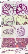
Histological analysis of transverse sections of Tjp1+/+ and Tjp1−/− embryos at E9.0. (A) Caudal regions. The posterior of Tjp1+/+ and Tjp1−/− embryos proper showed a normal neural tube (nt), dorsal aorta (da), and hindgut (hg), and an abnormal morphology of notochord (nc) in Tjp1−/− embryos. (B) Primitive heart. No obvious change was discerned between Tjp1+/+ and Tjp1−/− embryos. (C) Allantois. Although Tjp1−/− allantois was associated with apoptosis, a well-defined vascular network was developed similar to the Tjp1+/+ allantois. (D) Placentas. A transverse sectional analysis of Tjp1+/+ placentas showed the presence of immature embryonic erythrocytes (arrows) and unnucleated maternal erythrocytes (arrowheads), whereas the labyrinth layer of Tjp1−/− embryos lacked immature embryonic nucleated erythrocytes and embryonic blood vessels derived from the mesoderm. Left bottom, high-magnification images of the boxed regions. (E) Yolk sac. In the Tjp1+/+ yolk sac, the vessels were well formed, but in the Tjp1−/− yolk sac, the vessels were dramatically enlarged. Specific structures are as follows: en, extraembryonic endoderm; me, extraembryonic mesoderm; i.e., immature erythrocyte; and ec, endothelial cell. Bars, 100 μm.
Figure 4.
Figure 4.
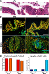
Apoptosis in Tjp1+/+ and Tjp1−/− embryos at E8.5 to E9.5. (A) Hematoxylin- and eosin-stained transverse sectional images of embryos at E9.0 around the notochord. In Tjp1−/− embryos, the border of the notochord (nc) seemed to be disorganized. Tjp1−/− embryos showed the presence of apoptotic or necrotic cells (arrows) at the notochord, neural tube, hindgut, and mesenchym around them. Specific structures are as follows: nt, neural tube; hg, hind gut; and nc, notochord. Bar, 20 μm. (B) Immunofluorescence images for Ki-67, caspase-3, and DAPI. At E8.5, almost no changes were detected between Tjp1+/+ and Tjp1−/− embryos. At E9.5, in Tjp1−/− embryos, caspase-3–positive apoptotic cells scattered beyond the normal bordered layer of the notochord or neural tube, whereas little apoptotic cells were detected in litter Tjp1+/+ embryos. (C) Quantification of Ki-67 and caspase-3–positive cells (mean and SE [n = 6]). Caspase-3 signals suggested that in Tjp1−/− embryos, the apoptotic cells were significantly increased compared with Tjp1+/+ embryos. Bars, 50 μm.
Figure 5.
Figure 5.
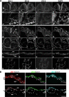
Immunolabeling of ZO-1, ZO-2, and ZO-3 in E8.5 wild-type Tjp1+/+ embryos. (A) a–d, caudal parts of embryos. a′-d′, high magnification of the boxed region of caudal parts of embryos, each corresponding to a–d, respectively. ZO-1 and ZO-2 localized at cell–cell junctions in the mesenchymal cells (arrows). e–h, cephalic parts of embryos. i–l, primitive heart. m–p, yolk sac. q–t, allantois. Specific. structures are as follows: ng, neural groove; fg, foregut; nt, neural tube; so, somite; en, extraembryonic endoderm; and me, extraembryonic mesoderm. Bars, 50 μm (a–d and e–t), 20 μm (a′–d′). (B) Immunofluorescence images of frozen sections of Tjp1+/+ and Tjp1−/− yolk sacs at E9.5 stained for ZO-1, ZO-2, and ZO-3, respectively. In the wild-type Tjp1+/+ yolk sac, ZO-1 was expressed in the extraembryonic endoderm and mesoderm, whereas in the homozygous Tjp1−/− yolk sac, signals for ZO-1 became undetectable. ZO-2 and ZO-3 were expressed only in extraembryonic endoderm in Tjp1+/+ embryos. Note that the ZO-2 signal was apically concentrated in the Tjp1−/− extraembryonic endoderm. Specific structures are as follows: en, extraembryonic endoderm; and me, extraembryonic mesoderm. Bars, 50 μm.
Figure 6.
Figure 6.
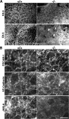
Analysis of yolk sac extraembryonic vascular development. (A) Immunolabeling for PECAM-1 in whole-mount yolk sacs of E8.5 and E9.5 embryos of Tjp1+/+ and Tjp1−/− mice. The immunostaining revealed whole patterns of vasculate not differing between Tjp1+/+ and Tjp1−/− embryos at E8.5, although at E9.5, PECAM-1–stained vasculates were quite different. Tjp1−/− extraembryonic mesoderm lacked the fine branched arrangements of vessels constituted by angiogenesis. The connection sites between extraembryonic endoderm and mesoderm were indicated by arrowheads. (B) High-resolution immunofluorescence micrographs labeled for PECAM-1, VE-cadherin, and JAM-A. It was noted that the immunofluorescently labeled patterns for JAM-A, but not for PECAM-1 and VE-cadherin, differed between E8.5 Tjp1+/+ and Tjp1−/− yolk sac. Bars, 100 μm (A) and 25 μm (B).
Figure 7.
Figure 7.
Paracellular barrier assay of the Tjp1+/+ and Tjp1−/− yolk sac at E9.5. (A) Biotinylation from the outside of yolk sacs in Tjp1+/+ and Tjp1−/− embryos. Conjugated biotin was detected with avidin-Texas Red, which did not penetrate the paracellular barrier. (B) Biotinylation from the inside of yolk sac in Tjp1+/+ and Tjp1−/− embryos. When biotin was subcutaneously injected into the excoelomic cavity (*), it was spread beyond the extraembryonic mesodermal cell sheets, showing no barrier function in the extraembryonic mesoderm. Bar, 50 μm.
Figure 8.
Figure 8.
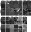
Whole-mount immunofluorescence micrographs of extraembryonic endoderm and mesoderm for cell–cell adhesion-related proteins in Tjp1+/+ and Tjp1−/− mice at E 9.5. In Tjp1+/+ and Tjp1−/− extraembryonic endoderm, the immunofluorescence patterns for various types of cell adhesion-related proteins revealed the normal formation of the epithelial cell sheets with TJs. It is noteworthy that the ZO-2 signal was concentrated in cell–cell adhesion sites of the Tjp1−/− extraembryonic endoderm. Furthermore, the immunofluorescence patterns of TJ-related proteins such as occludin, claudin-6, and JAM-A were also concentrated in cell–cell adhesion sites of the Tjp1−/− extraembryonic endoderm. In contrast, in extraembryonic mesoderm, the immunofluorescence patterns of afadin and α/β-catenin substantiated cell sheet formation but without concentration of TJ-proper proteins, such as occludin, claudin, and JAM-A. Note that in ZO-1−/− extraembryonic mesoderm, the signals for ZO-1/2/3 were not detected. Bar, 50 μm.

Similar articles

Cited by

References

    1. Adachi M., Inoko A., Hata M., Furuse K., Umeda K., Itoh M., Tsukita Sh. Normal establishment of epithelial tight junctions in mice and cultured cells lacking expression of ZO-3, a tight-Junction MAGUK Protein. Mol. Cell Biol. 2006;26:9003–9015. - PMC - PubMed
    1. Argraves W. S., Drake C. J. Genes critical to vasculogenesis as defined by systematic analysis of vascular defects in knockout mice. Anat. Rec. A Discov. Mol. Cell Evol. Biol. 2005;286:875–884. - PubMed
    1. Balda M. S., Gonzalez-Mariscal L., Matter K., Cereijido M., Anderson J. M. Assembly of the tight junction: the role of diacylglycerol. J. Cell Biol. 1993;123:293–302. - PMC - PubMed
    1. Baumer S., Keller L., Holtmann A., Funke R., August B., Gamp A., Wolburg H., Wolburg-Buchholz K., Deutsch U., Vestweber D. Vascular endothelial cell–specific phosphotyrosine phosphatase (VE-PTP) activity is required for blood vessel development. Blood. 2006;107:4754–4762. - PubMed
    1. Bazzoni G., Martinez-Estrada O. M., Orsenigo F., Cordenonsi M., Citi S., Dejana E. Interaction of junctional adhesion molecule with the tight junction components ZO-1, cingulin, and occludin. J. Biol. Chem. 2000;275:20520–20526. - PubMed

Publication types

MeSH terms

LinkOut - more resources