

The E3 ubiquitin ligase EDD is an adverse prognostic factor for serous epithelial ovarian cancer and modulates cisplatin resistance in vitro
- PMID: 18349819
- PMCID: PMC2275489
- DOI: 10.1038/sj.bjc.6604281
The E3 ubiquitin ligase EDD is an adverse prognostic factor for serous epithelial ovarian cancer and modulates cisplatin resistance in vitro
Erratum in
- Br J Cancer. 2008 Jun 3;98(11):1880. Murali, Rajmohan [added]
Abstract
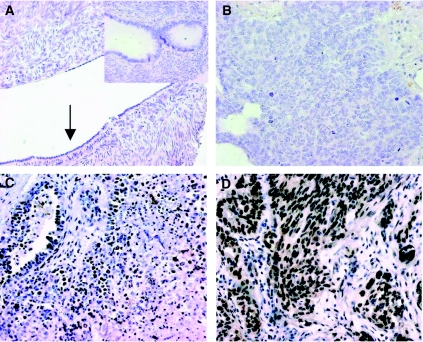
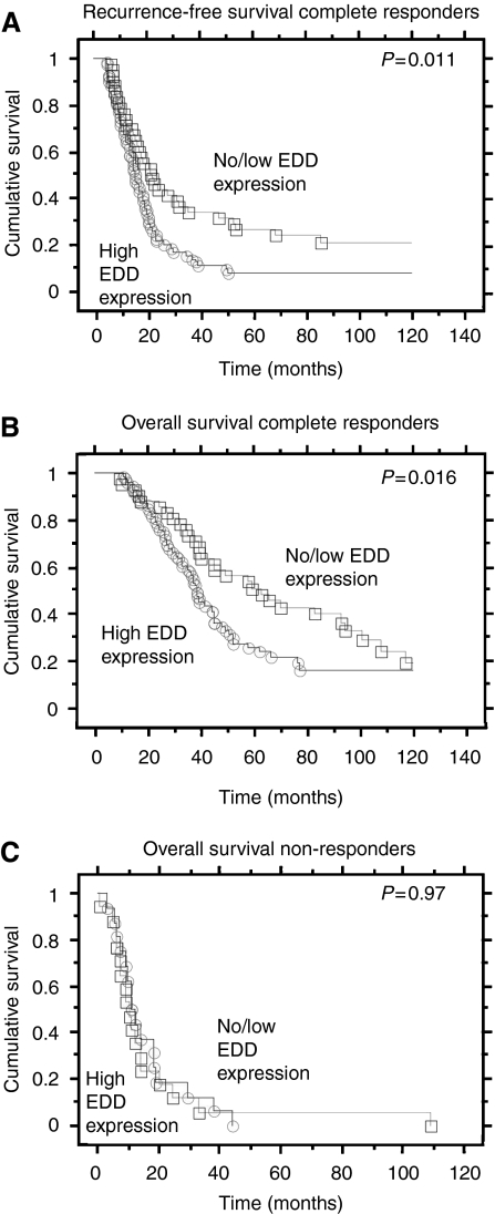
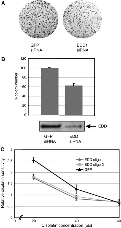
Despite a high initial response rate to first-line platinum/paclitaxel chemotherapy, most women with epithelial ovarian cancer relapse with recurrent disease that becomes refractory to further cytotoxic treatment. We have previously shown that the E3 ubiquitin ligase, EDD, a regulator of DNA damage responses, is amplified and overexpressed in serous ovarian carcinoma. Given that DNA damage pathways are linked to platinum resistance, the aim of this study was to determine if EDD expression was associated with disease recurrence and platinum sensitivity in serous ovarian cancer. High nuclear EDD expression, as determined by immunohistochemistry in a cohort of 151 women with serous ovarian carcinoma, was associated with an approximately two-fold increased risk of disease recurrence and death in patients who initially responded to first-line chemotherapy, independently of disease stage and suboptimal debulking. Although EDD expression was not directly correlated with relative cisplatin sensitivity of ovarian cancer cell lines, sensitivity to cisplatin was partially restored in platinum-resistant A2780-cp70 ovarian cancer cells following siRNA-mediated knockdown of EDD expression. These results identify EDD as a new independent prognostic marker for outcome in serous ovarian cancer, and suggest that pathways involving EDD, including DNA damage responses, may represent new therapeutic targets for chemoresistant ovarian cancer.
Figures
Similar articles
-
EDD enhances cell survival and cisplatin resistance and is a therapeutic target for epithelial ovarian cancer.Carcinogenesis. 2014 May;35(5):1100-9. doi: 10.1093/carcin/bgt489. Epub 2013 Dec 30. Carcinogenesis. 2014. PMID: 24379240 Free PMC article.
-
Checkpoint kinase 2 (Chk2) supports sensitivity to platinum-based treatment in high grade serous ovarian cancer.Gynecol Oncol. 2014 Jun;133(3):591-8. doi: 10.1016/j.ygyno.2014.03.557. Epub 2014 Mar 20. Gynecol Oncol. 2014. PMID: 24657486
-
Ovarian serous carcinomas acquire cisplatin resistance and increased invasion through downregulation of the high-temperature-required protein A2 (HtrA2), following repeated treatment with cisplatin.Med Oncol. 2017 Nov 22;34(12):201. doi: 10.1007/s12032-017-1058-3. Med Oncol. 2017. PMID: 29168038
-
Predicting Prognosis and Platinum Resistance in Ovarian Cancer: Role of Immunohistochemistry Biomarkers.Int J Mol Sci. 2023 Jan 19;24(3):1973. doi: 10.3390/ijms24031973. Int J Mol Sci. 2023. PMID: 36768291 Free PMC article. Review.
-
A Potential Role for HUWE1 in Modulating Cisplatin Sensitivity.Cells. 2021 May 20;10(5):1262. doi: 10.3390/cells10051262. Cells. 2021. PMID: 34065298 Free PMC article. Review.
Cited by
-
A whole-genome RNAi screen identifies an 8q22 gene cluster that inhibits death receptor-mediated apoptosis.Proc Natl Acad Sci U S A. 2011 Oct 25;108(43):E943-51. doi: 10.1073/pnas.1100132108. Epub 2011 Sep 26. Proc Natl Acad Sci U S A. 2011. PMID: 21949371 Free PMC article.
-
Prognostic biomarkers in endometrial and ovarian carcinoma.Virchows Arch. 2014 Mar;464(3):315-31. doi: 10.1007/s00428-013-1509-y. Epub 2014 Feb 7. Virchows Arch. 2014. PMID: 24504546 Review.
-
Chemotherapeutic drugs: Cell death- and resistance-related signaling pathways. Are they really as smart as the tumor cells?Transl Oncol. 2021 May;14(5):101056. doi: 10.1016/j.tranon.2021.101056. Epub 2021 Mar 6. Transl Oncol. 2021. PMID: 33684837 Free PMC article. Review.
-
Significance of the E3 ubiquitin protein UBR5 as an oncogene and a prognostic biomarker in colorectal cancer.Oncotarget. 2017 Nov 20;8(64):108079-108092. doi: 10.18632/oncotarget.22531. eCollection 2017 Dec 8. Oncotarget. 2017. PMID: 29296225 Free PMC article.
-
EDD enhances cell survival and cisplatin resistance and is a therapeutic target for epithelial ovarian cancer.Carcinogenesis. 2014 May;35(5):1100-9. doi: 10.1093/carcin/bgt489. Epub 2013 Dec 30. Carcinogenesis. 2014. PMID: 24379240 Free PMC article.
References
-
- Agarwal R, Kaye SB (2003) Ovarian cancer: strategies for overcoming resistance to chemotherapy. Nat Rev Cancer 3: 502–516 - PubMed
-
- Auersperg N, Wong AST, Choi KC, Kang SK, Leung PCK (2001) Ovarian surface epithelium: biology, endocrinology and pathology. Endocr Rev 22: 255–288 - PubMed
-
- Bali A, O'Brien PM, Edwards LS, Sutherland RL, Hacker NF, Henshall SM (2004) Cyclin D1, p53 and p21Waf1/Cip1 expression is predictive of poor clinical outcome in serous epithelial ovarian cancer. Clin Cancer Res 10: 5168–5177 - PubMed
-
- Birrer MJ, Johnson ME, Hao K, Wong KK, Park DC, Bell A, Welch WR, Berkowitz RS, Mok SC (2007) Whole genome oligonucleotide-based array comparative genomic hybridization analysis identified fibroblast growth factor 1 as a prognostic marker for advanced-stage serous ovarian adenocarcinomas. J Clin Oncol 25: 2281–2287 - PubMed
-
- Blumenthal M, Kardosh A, Dubeau L, Borok Z, Schonthal AH (2003) Suppression of the transformed phenotype and induction of differentiation-like characteristics in cultured ovarian tumor cells by chronic treatment with progesterone. Mol Carcinog 38: 160–169 - PubMed
Publication types
MeSH terms
Substances
LinkOut - more resources
Full Text Sources
Medical
Miscellaneous
