. 2008 Mar 24;180(6):1073-86.
doi: 10.1083/jcb.200706009. Epub 2008 Mar 17.

Orchestration of the S-phase and DNA damage checkpoint pathways by replication forks from early origins

Affiliations

Orchestration of the S-phase and DNA damage checkpoint pathways by replication forks from early origins

Julie M Caldwell et al. J Cell Biol. .

Abstract

The S-phase checkpoint activated at replication forks coordinates DNA replication when forks stall because of DNA damage or low deoxyribonucleotide triphosphate pools. We explore the involvement of replication forks in coordinating the S-phase checkpoint using dun1Delta cells that have a defect in the number of stalled forks formed from early origins and are dependent on the DNA damage Chk1p pathway for survival when replication is stalled. We show that providing additional origins activated in early S phase and establishing a paused fork at a replication fork pause site restores S-phase checkpoint signaling to chk1Delta dun1Delta cells and relieves the reliance on the DNA damage checkpoint pathway. Origin licensing and activation are controlled by the cyclin-Cdk complexes. Thus, oncogene-mediated deregulation of cyclins in the early stages of cancer development could contribute to genomic instability through a deficiency in the forks required to establish the S-phase checkpoint.


Figures
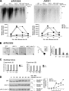
Figure 1.
Figure 1.
The number of stalled chromosomal replication forks early in S phase is reduced in dun1Δ and chk1Δ dun1Δ cells in response to HU and correlates with a defect in S-phase checkpoint activation. (A, top) Cells were released from G1 into medium containing 200 mM HU. Chromosomal DNA was subjected to alkaline gel electrophoresis. RIs were monitored by Southern blot analysis using a probe for the indicated replication origin. (A, bottom) Relative intensity of RI signal normalized to the amount of DNA loaded. Seven independent experiments showed similar trends and one typical experiment is shown for each ARS. (B) Cells were treated as in A. (B, left) Southern blot analysis of chromosomal DNA prepared 45 min after release into HU and subjected to 2D gel electrophoresis was performed using a probe for ARS305. Arrows designate signal that represents replication fork–containing structures. (B, middle) Illustration of restriction fragment structures and the corresponding Southern blot patterns. (B, right) Mean ratio of signal for replication fork–containing structures to linear DNA as a percentage of the wild-type (WT) ratio for three experiments. Error bars represent one standard deviation from the mean. (C) Budding index. 100 cells from A and B were scored for buds. Additional data that support this trend were observed in other replicates (Tables S2 and S3, available at http://www.jcb.org/cgi/content/full/jcb.200706009/DC1). (D, left) Cells were released from G1 into medium containing 200 mM HU. Rad53p phosphorylation was monitored by Western analysis. The Molecular masses (in kilodaltons) are indicated to the right of each panel. (D, right) The ratio of phosphorylated to unphosphorylated Rad53p signal.
Figure 2.
Figure 2.
Chk1p is activated in dun1Δ cells in late S phase and G2 during an unperturbed cell cycle. (A, top) Cells were released from G1. Western blot analysis was performed to monitor HA-Chk1p protein migration. (A, bottom) dun1Δ cells were treated as in the top panel, except cells were released into medium containing nocodazole. The asterisks emphasize the time points at which HA-Chk1p phosphorylation was observed. The Molecular masses (in kilodaltons) are indicated to the right of each panel. (B) Cells were plated on YPD medium containing either 10 or 50 mM HU and incubated at 30°C for 24 h, and the number of cells per colony was counted for 50 colonies. Bars represent the mean number of cells per colony. Error bars represent one standard deviation from the mean.
Figure 3.
Figure 3.
Deletion of RPD3, which encodes a histone deacetylase, suppresses the HU sensitivity of chk1Δ dun1Δ cells. (A) Cells were spotted onto YPD medium containing the indicated concentration of HU (mM). (B) Analyses of replication fork–containing structures were performed as in Fig. 1 B, using a probe for the late chromosomal origin ARS603 (top) or the early chromosomal origin ARS305 (bottom). (C, top)Rad53p phosphorylation in response to HU treatment is restored to wild-type levels and kinetics in chk1D dun1D cells by additional replication origins that are activated in early S phase. Rad53p phosphorylation was monitored as in Fig. 1 D, except that the cells were released at 18°C. (C, bottom left) Ratio of phosphorylated to nonphosphorylated Rad53p. (C, bottom right) Budding index.
Figure 4.
Figure 4.
Suppression of the HU sensitivity of chk1Δ dun1Δ cells by an episomal replication origin activated early in S phase. (A and B) Cells containing the indicated number of copies of pARS (pRS416, pRS415), p2μ (pRS426, pRS425), p12, or p12ARS were spotted onto YPD medium containing the indicated concentration of HU (in millimoles). (C and D) Analyses of replication fork–containing structures were performed as in Fig. 1 B, using probes for the indicated plasmid or ARS305. All cells analyzed in D contained pRS416. (E)Rad53p phosphorylation in response to HU treatment is restored to wild-type levels and kinetics in chk1D dun1D cells by additional replication origins that are activated in early S phase. Rad53p phosphorylation was monitored as in Fig. 3 C. The Molecular masses (in kilodaltons) are indicated to the right of each panel. (E, bottom left) Ratio of phosphorylated to nonphosphorylated Rad53p. (E, bottom right) Budding index.
Figure 5.
Figure 5.
The timing of origin activation and of the episomal replication fork pause is critical to restore viability to chk1Δ dun1Δ cells. Targeting the acetylase Gcn5p–Gcn4p complex to the late activating origin ARS1412 restored the S-phase checkpoint in chk1D dun1D cells with constitutive expression of Gcn4p. Cells containing a high copy vector expressing Gcn4p and the indicated episomal origin were grown in SC-ura-leu media and spotted onto YPD medium containing the indicated concentration of HU (in millimoles). (bottom) Five representative chk1Δ dun1Δ transformants containing a high copy vector expressing Gcn4p and p12-Ac show results typical for 43/43 chk1Δ dun1Δ transformants containing the same constructs. p12, episome with late activating origin that failed to suppress chk1Δ dun1Δ; p12-Ac, same episome containing binding sites for the Gcn4p–Gcn5p acetylase complex.
Figure 6.
Figure 6.
Replication fork pause plays a role in suppression of HU sensitivity of chk1Δ dun1Δ cells. (A) Cells containing the indicated episome were spotted onto YPD medium containing the indicated concentration of HU (in millimoles). (B) Suppression of checkpoint mutant HU sensitivity by an episomal replication origin is independent of spindle dynamics regulation.Cells containing either p2μ (pRS426) or pARS (pRS416) were spotted onto YPD medium containing the indicated concentration of HU (in millimoles). (C) Cells were released from G1 into medium containing 50 mM HU, fixed, and stained with DAPI and anti-tubulin to visualize nuclei and spindles. For each strain, 100 cells for each time point were scored as having either short (<3 μm) or elongated (≥3 μm) spindles. Data points indicate the percentage of cells with elongated spindles. (D–F) The episomal replication fork pause is critical to restore viability to chk1Δ dun1Δ cells.Cells containing the indicated episomes were spotted onto YPD medium containing the indicated concentration of HU (in millimoles). (A and D, bottom) Schematics of episomes used in these studies.
Figure 7.
Figure 7.
The timing of the episomal replication fork pause is critical to restore viability to chk1Δ dun1Δ cells. (A) Cells containing the indicated episome were spotted onto YPD medium containing the indicated concentration of HU (in millimoles). (B) chk1Δ dun1Δ cells containing the indicated episome were treated as in Fig. 1 B, except that they were released at 18°C. Southern blot analysis of chromosomal DNA prepared at the indicated times after release into HU and subjected to 2D gel electrophoresis was performed using a probe for pARS (pRS416). (C) Restriction fragment shapes that correspond to the observed Southern blot pattern. (D, left) In a separate experiment, cells were treated as in Fig. 3 C and processed for Western analysis to monitor Rad53p phosphorylation. The Molecular masses (in kilodaltons) are indicated to the right of each panel. (D, right) The ratio of phosphorylated to unphosphorylated Rad53p signal and budding index.
Figure 8.
Figure 8.
Model. A deficiency of replication forks from early origins results in activation of the DNA damage checkpoint. RFCL is the checkpoint clamp loader. Ddc2, also known as Lcd1 and in mammals as Atrip, localizes Mec1 to DNA damage lesions (Nyberg et al., 2002; see text for details).

Similar articles

Cited by

References

    1. Alcasabas, A.A., A.J. Osborn, J. Bachant, F. Hu, P.J. Werler, K. Bousset, K. Furuya, J.F. Diffley, A.M. Carr, and S.J. Elledge. 2001. Mrc1 transduces signals of DNA replication stress to activate Rad53. Nat. Cell Biol. 3:958–965. - PubMed
    1. Allen, J.B., Z. Zhou, W. Siede, E.C. Friedberg, and S.J. Elledge. 1994. The SAD1/RAD53 protein kinase controls multiple checkpoints and DNA damage-induced transcription in yeast. Genes Dev. 8:2401–2415. - PubMed
    1. Alvino, G.M., D. Collingwood, J.M. Murphy, J. Delrow, B.J. Brewer, and M.K. Raghuraman. 2007. Replication in hydroxyurea: it's a matter of time. Mol. Cell. Biol. 27:6396–6406. - PMC - PubMed
    1. Aparicio, J.G., C.J. Viggiani, D.G. Gibson, and O.M. Aparicio. 2004. The Rpd3-Sin3 histone deacetylase regulates replication timing and enables intra-S origin control in Saccharomyces cerevisiae. Mol. Cell. Biol. 24:4769–4780. - PMC - PubMed
    1. Aylon, Y., B. Liefshitz, and M. Kupiec. 2004. The CDK regulates repair of double-strand breaks by homologous recombination during the cell cycle. EMBO J. 23:4868–4875. - PMC - PubMed

Publication types

MeSH terms

LinkOut - more resources