. 2008 Jul;106(1):96-106.
doi: 10.1111/j.1471-4159.2008.05339.x. Epub 2008 Jul 1.

A novel calcium-binding protein is associated with tau proteins in tauopathy

Affiliations

A novel calcium-binding protein is associated with tau proteins in tauopathy

Irving E Vega et al. J Neurochem. 2008 Jul.

Abstract

Tauopathies are a group of neurological disorders characterized by the presence of intraneuronal hyperphosphorylated and filamentous tau. Mutations in the tau gene have been found in kindred with tauopathy. The expression of the human tau mutant in transgenic mice induced neurodegeneration, indicating that tau plays a central pathological role. However, the molecular mechanism leading to tau-mediated neurodegeneration is poorly understood. To gain insights into the role that tau plays in neurodegeneration, human tau proteins were immunoprecipitated from brain lysates of the tauopathy mouse model JNPL3, which develops neurodegeneration in age-dependent manner. In the present work, a novel EF-hand domain-containing protein was found associated with tau proteins in brain lysate of 12-month-old JNPL3 mice. The association between tau proteins and the novel identified protein appears to be induced by the neurodegeneration process as these two proteins were not found associated in young JNPL3 mice. Consistently, the novel protein co-purified with the pathological sarkosyl insoluble tau in terminally ill JNPL3 mice. Calcium-binding assays demonstrated that this protein binds calcium effectively. Finally, the association between tau and the novel calcium-binding protein is conserved in human and enriched in Alzheimer's disease brain. Taken together, the identification of a novel calcium-binding protein associated with tau protein in terminally ill tauopathy mouse model and its confirmation in human brain lysate suggests that this association may play an important physiological and/or pathological role.


Figures

Fig. 1
Fig. 1
Identification of a novel tau-associated protein. (a) Tau proteins were immunoprecipitated (IP) from post-nuclear brain extract of 12-month-old JNPL3 transgenic mice (JNPL3, lanes 3–4) and age-matched non-transgenic (NT, lanes 1–2) mice, using the human specific anti-Tau antibody Tau13 (lanes 2 and 4). Protein A beads (B) alone were used as negative control (lanes 1 and 3). The immunoprecipitated proteins were resolved on SDS–PAGE and visualized by silver staining. Those proteins enriched in Tau13 immunoprecipitates were identified by in-gel trypsin digestion and tandem mass spectrometry analysis. The identified proteins are pointed out. The asterisk points out to protein bands corresponding to cross-linked Tau13 antibody. (b) The EFHD2 cDNA was cloned in a bacterial expression vector and the protein was expressed as a GST-fusion protein. The immunoreactivity of the anti-EFHD2M antibody against a peptide of the mouse EFHD2 protein was tested by western blot analysis. Three different amounts of GST and GST-EFHD2 were resolved by SDS–PAGE [20 ng (lanes 1 and 4), 40 ng (lanes 2 and 5), and 80 ng (lanes 3 and 6)]. (c) The expression of EFHD2 protein in different organs of a 6-month-old NT mouse was determined by western blot analysis. The tissues used in the analysis were brain (lane 1), spinal cord (lane 2), liver (lane 3), lung (lane 4), heart (lane 5), kidney (lane 6), spleen (lane 7), and skeletal muscle (lane 8). As loading control, the nitrocellulose membrane was stained with Ponceau-S to detect differences in overall loading. (d) Brain lysate from NT mice was resolved on SDS–PAGE and western blot analysis was performed using anti-human EFHD2 antibody (anti-EFHD2). The anti-EFHD2 antibodies were pre-absorbed at 25°C with GST (lanes 1 and 2) or GST-EFHD2 (lanes 3 and 4).
Fig. 2
Fig. 2
EFHD2 has calcium-binding activity. (a) The recombinant GST (lane 1) and GST-EFHD2 (lane 2) proteins were purified from bacterial extract, using glutathione-conjugated sepharose beads. The amount of protein used for the in vitro calcium-binding assay was verified by resolving the purified recombinant proteins on SDS–PAGE and visualized them by coomassie staining. (b) The same amount of recombinant proteins (1 μg) was used for the in vitro calcium-binding assay. The experiment was carried out as explained in the Materials and methods section. The radioactivity associated with the beads was determined by scintillation counter. Glutathione-conjugated sepharose beads (1) and GST bound to beads (2) were used as negative controls. GST-EFHD2 proteins bound to beads (3) showed the highest radioactivity after incubation with radioactive 45Ca and extensive washing of the beads. The associated radioactivity detected on GST-EFHD2 (3) was reduced by co-incubation with 10 mM CaCl2 (4) and EDTA (5). The bars represent the mean ± SD of three different experiments performed in triplicate.
Fig. 3
Fig. 3
EFHD2 co-immunoprecipitated hyperphosphorylated tau. (a) The hTauP301L (IP-Tau, lane 4) and EFHD2 (IP-EFHD2, lane 6) proteins were immunoprecipitated from post-nuclear brain extract of 12-month-old JNPL3 mice. As control, post-nuclear brain extract from age-matched NT mice (lanes 1–2) and Protein A-conjugated beads alone (lanes 1, 3, and 5) were used. Additionally, EFHD2 proteins were immunoprecipitated from 12-month-old JNPL3 mice (lane 6), using anti-EFHD2M antibodies. The immunoprecipitated proteins were visualized by western blot, using the indicated antibodies. (b) hTauP301L was immunoprecipitated from 3-month-old JNPL3 mice (IP-Tau, lane 4). As control, post-nuclear brain extract from age-matched NT mice (lanes 1–2) and Protein A-conjugated beads alone (lanes 1 and 3) were also used. The immunoprecipitated proteins were visualized by western blot analysis, using the indicated antibodies.
Fig. 4
Fig. 4
EFHD2 co-purified with sarkosyl insoluble tau. (a) The expression of EFHD2 proteins in different brain regions of 12-month-old JNPL3 and age-matched NT mice was determined by western blot analysis, using the anti-EFHD2M antibody. The brain regions selected were brainstem (lanes 1 and 8), cerebellum (lanes 2 and 9), amygdala (lanes 3 and 10), striatum (lanes 4 and 11), hippocampus (lanes 5 and 12), cortex (lanes 6 and 13), and prefrontal cortex (lanes 7 and 14) from NT and JNPL3 mice. A non-specific cross-reacting band was used as loading control (LC). The arrows point out to the lower molecular weight EFHD2 species. (b) The intensity of the lower molecular weight EFHD2 band (arrow in a) was determined by densitometry. (c–d) Sarkosyl insoluble preparation of brain extracts from NT and JNPL3 mice at 12 months (c) and 3 months (d) of age. (c) The brain lysate from a 12-month-old JNPL3 and NT mice were fractionate. Lanes 1, 3, 5, and 7 represent the soluble fractions, while the sarkosyl insoluble fractions are in lanes 2, 4, 6, and 8. The graph represents the quantification of EFHD2 protein in the sarkosyl insoluble fraction in NT (white) and JNPL3 (black) brains. The bars represent mean ± SD of three different experiments. (d) The brain lysate from a 3-month-old JNPL3 and NT mice were fractionated. The lanes 1 and 3 represent the soluble fraction, while lanes 2 and 4 contain the sarksosyl insoluble fraction. hTauP301L and EFHD2 proteins were visualized by western blot analysis, using the indicated antibodies.
Fig. 5
Fig. 5
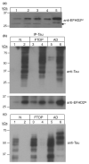
EFHD2 and tau are associated in human tauopathy. (a) The expression of EFHD2 proteins was determined in human post-nuclear brain lysate (lanes 1–4) from normal aging (N; lane 1), FTDP-17 (FTDP; lane 2), Alzheimer's disease (AD) (lane 3), and PSP (lane 4) diagnosed cases. Post-nuclear brain extract from JNPL3 mice was used as positive control (lane 5). EFHD2 was visualized by western blot analysis, using the indicated antibody. The arrow points out to the lower molecular weight EFHD2 species. (b) Human tau proteins were immunoprecipitated (IP-Tau) from normal aging (N, lane 2), FTDP-17 (FTDP, lane 4), and AD (lane 6) post-nuclear brain lysates. As negative control, Protein A-conjugated beads alone (lanes 1, 3, and 5) were used. The immunoprecipitated proteins were visualized by western blot analysis, using the indicated antibodies. (c) The presence of sarkosyl-insoluble tau from diagnosed human tauopathy cases was determined by western blot. Lanes 1, 3, and 5 represent the soluble fractions, while lanes 2, 4, and 6 represent sarkosyl-insoluble fraction. Temporal cortex from normal aging (N), FTDP-17 (FTDP), and AD diagnosed cases was used. Tau proteins were visualized by western blot, using the indicated antibody.

Similar articles

Cited by

References

    1. Ahlijanian MK, Barrezueta NX, Williams RD, et al. Hyperphosphorylated tau and neurofilament and cytoskeletal disruptions in mice overexpressing human p25, an activator of cdk5. Proc. Natl Acad. Sci. USA. 2000;97:2910–2915. - PMC - PubMed
    1. Avramidou A, Kroczek C, Lang C, Schuh W, Jäck H-M, Mielenz D. The novel adaptor protein Swiprosin 1 enhances BCR signals and contributes to BCR-induced apoptosis. Cell Death Differ. 2007;14:1936–1947. - PubMed
    1. Ballatore C, Lee VM-Y, Trojanowski JQ. Tau-mediated neurodegeneration in Alzheimer's disease and related disorders. Nat. Rev. Neurosci. 2007;8:663–672. - PubMed
    1. Berger Z, Roder H, Hanna A, et al. Accumulation of pathological tau species and memory loss in a conditional model of tauopathy. J. Neurosci. 2007;27:3650–3662. - PMC - PubMed
    1. Cruz JC, Tseng HC, Goldman JA, Shih H, Tsai LH. Aberrant Cdk5 activation by p25 triggers pathological events leading to neurodegeneration and neurofibrillary tangles. Neuron. 2003;40:471–483. - PubMed

Publication types

MeSH terms