Comparative Study
. 2008 Apr;11(4):429-39.
doi: 10.1038/nn2074. Epub 2008 Mar 16.

Compartment-specific transcription factors orchestrate angiogenesis gradients in the embryonic brain

Affiliations
Comparative Study

Compartment-specific transcription factors orchestrate angiogenesis gradients in the embryonic brain

Anju Vasudevan et al. Nat Neurosci. 2008 Apr.

Abstract

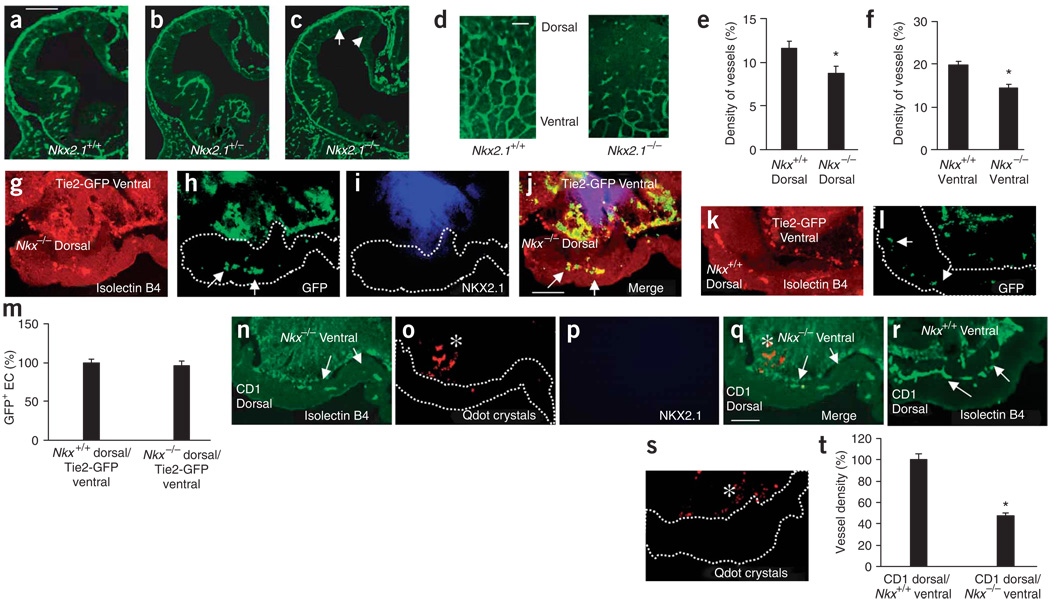
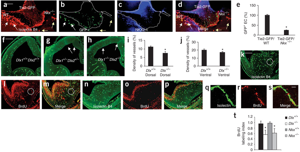
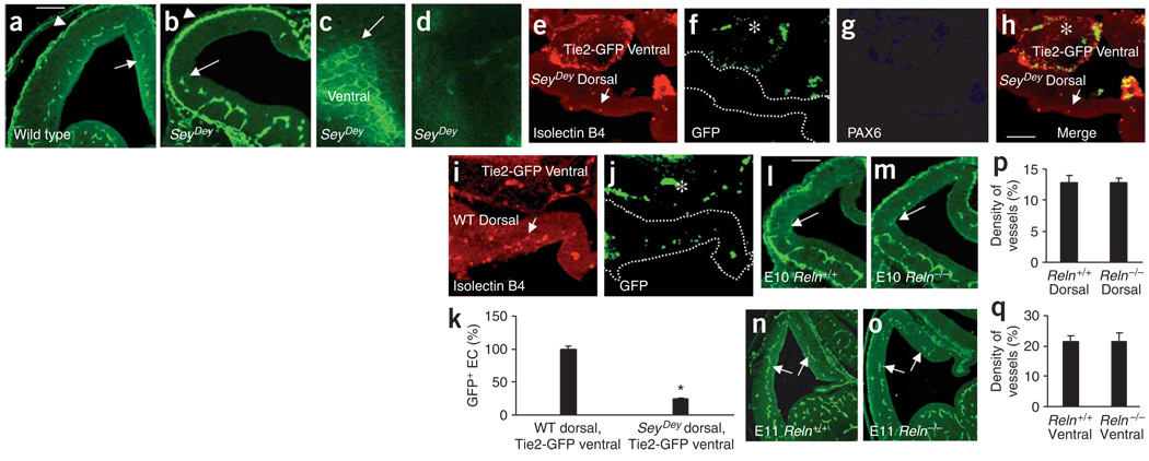
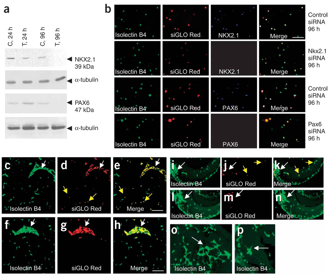
Prevailing notions of cerebral vascularization imply that blood vessels sprout passively into the brain parenchyma from pial vascular plexuses to meet metabolic needs of growing neuronal populations. Endothelial cells, building blocks of blood vessels, are thought to be homogeneous in the brain with respect to their origins, gene expression patterns and developmental mechanisms. These current notions that cerebral angiogenesis is regulated by local environmental signals contrast with current models of cell-autonomous regulation of neuronal development. Here we demonstrate that telencephalic angiogenesis in mice progresses in an orderly, ventral-to-dorsal gradient regulated by compartment-specific homeobox transcription factors. Our data offer new perspectives on intrinsic regulation of angiogenesis in the embryonic telencephalon, call for a revision of the current models of telencephalic angiogenesis and support novel roles for endothelial cells in brain development.


Figures

Figure 1
Figure 1
Angiogenesis gradients in the embryonic telencephalon. (a–c) Isolectin-B4+ vessels in the E9 ventral telencephalon (arrows, a). The periventricular vessels extend into the dorsal telencephalon by E10 and E11 (b,c, arrows) producing a ventrolateral-to-dorsomedial gradient. Pial vessels (arrowheads) fully encircle the telencephalon by E9. (d) Isolectin-B4+ pial vessels covering a E10 dorsal telencephalon, cut open (at arrowheads) and mounted with the ventricular surface up. The basal vessel appears on the telencephalic floor (thick arrow). The dorsal telencephalon is unlabeled (thin arrow). (e–g) Isolectin B4–labeled periventricular vessels in an E11 whole mount appear in a single 20-µm focal plane (e) from which thin vessels emerge at right angles toward the pial surface (f) and contact the pial vessels (arrow in g), which appear in a different focal plane (g). (h,i) Isolectin B4–labeled prominent basal vessel (white star) in E11 ventral telencephalon whole mount unfurls into a periventricular vessel lattice. The broken line (i) indicates the advancing vessel front: the medial telencephalon has no periventricular vessels (white arrow). (j–l) Diagramatic representation of periventricular vessel development. The periventricular vessel network (red) originates from the basal vessel (red asterisk in j) in the telencephalon (peacock green) and grows in ventral-to-dorsal and lateral-to-medial directions. Dotted circle in j is expanded in k (purple, telencephalon; blue cross-hatching, basal forebrain) for a two-dimensional view of the periventricular network (yellow dotted circle) and the basal vessel (red asterisk in k). The boxed area in k represents i, with the medial aspects of the telencephalon devoid of periventricular vessels. (l) Ventral-to-dorsal and lateral-to-medial gradients of periventricular angiogenesis (broken red line with directional arrow). Blue dotted line, pial vessels. Scale bars: a, 100 µm (applies to a–d); e, 50 µm (applies to e–g); h, 100 µm; i, 50 µm.
Figure 2
Figure 2
Dorsal periventricular vessels originate from the ventral telencephalon. (a) Isolectin-B4+ pial and periventricular vessels in the ventral telencephalon (arrow) of an E10 telencephalon explant cultured for 2 h. (b) After 24 h in culture, periventricular vessels enter the dorsal telencephalon (arrow). Arrowheads, pial vessels; LV, lateral ventricle. (c) Periventricular (arrow) and pial (arrowhead) vessels in E11 dorsal explant cultured for 2 h after removing the ventral telencephalon. Curved white arrow, ventral limit of the dorsal explant. (d) PAX6 immunohistochemistry confirms dorsal molecular identity of the explant. (e) Merged c and d. (f) There are pial vessels (arrowheads) but virtually no periventricular vessels in an E10 dorsal explant cultured for 24 h after removing the ventral telencephalon. Arrows indicate the few periventricular vessels. (g) PAX6 labeling confirms the explant’s dorsal molecular identity. (h) Merged f and g, showing the vessel-poor PAX6+ regions. White asterisks: cranial mesenchyme, other non-CNS tissues. (i) Periventricular vessel density (mean ± s.d.) in E10 dorsal explants cultured without the ventral explants (n = 30) is less than that in E11 dorsal-only (n = 25) or E10 whole explants (dorsal + ventral, n = 30). Data normalized to E10 whole explants. *P < 0.0001; E10 whole explants versus E10 dorsal explants. (j) An E10 whole explant cultured for 24 h after removing pial membranes (arrowhead) by microdissection. Isolectin B4 labeled periventricular vessels in the dorsal telencephalon (arrows) even in regions without intact pial membranes. (k) 6-hydroxydopamine-induced pial vessel degeneration in an E10 whole explant. Periventricular vessels appear in ventral and dorsal telencephalon. (l,m) Intact pial membrane in control (l) and disrupted pial membranes in 6-hydroxydopamine-treated (m) explants. Scale bars: a, 100 µm (applies to a–h,j,k); l, 25 µm (applies also to m).
Figure 3
Figure 3
Ventral-to-dorsal migration of endothelial cells. (a) When an E10 dorsal explant is cultured with an E11 ventral explant, isolectin-B4+ periventricular vessels (arrow) appear in the dorsal explant. (b) Pre-explantation Qdot labeling (asterisks) verifies the explant’s ventral origins. (c) Merged a and b. (d–g) When an E10 wild-type dorsal explant was cultured with an E11 Tie2-GFP ventral explant, isolectin-B4+ periventricular vessels (d) appeared in the E10 dorsal explant (arrow). (e) The E10 dorsal periventricular vessels contained GFP+ ventral endothelial cells (arrows). (f) PAX6 labeling confirms the explant’s dorsal identity. (g) Merged d–f. (h,i) E10 dorsal Tie2-GFP explant cultured B500 µm from E11 Tie2-GFP ventral explant for 24 h (h, bright field). Sections of the dorsal explant show GFP+ endothelial cells (arrows, i) in the endogenous pial vessels only. (j) Periventricular vessel density (mean ± s.d.) in E10 dorsal explants cultured with E11 ventral explants for 24 h approaches that of E10 whole explants (dorsal + ventral; n = 30), whereas E10 dorsal explants cultured without the ventral explant show significantly reduced vessel density (n = 30). Vessel densities were normalized to E10 whole explants. *P < 0.0001, E10 dorsal explants versus E10 dorsal explants cultured with E11 ventral explants. (k) Merged e and f showing GFP+ and PAX6+ endothelial cells in the dorsal explant. (l,m) GFP+ endothelial cell (l) from boxed area in k shows labeling with PAX6 (m). (n) Merged l and m. Scale bars: a, 100 µm (applies to a–g,i,k); n, 10 µm (applies to l–n).
Figure 4
Figure 4
Endothelial cells express homeobox transcription factors. (a) NKX2.1 immunohistochemistry (red) in an E11 Tie2-GFP telencephalon labels medial ganglionic eminence (MGE) cells and highlights MGE borders (arrows). Lateral ganglionic eminence (LGE) shows some labeling. (b) A GFP+ endothelial cell in an E11 Tie2-GFP ventral telencephalon expresses NKX2.1 (c) No NKX2.1 labeling in the E11 Nkx2.1−/− telencephalon, confirming antibody specificity. (d) Immunoblot of NKX2.1 in nuclear and cytosolic fractions (NF and CF, respectively) of cultured E13 mouse brain endothelial cells. (e) Cultured E13 mouse brain endothelial cell labeled with isolectin B4, NKX2.1 and DAPI, showing nuclear (white) and cytoplasmic (yellow) NKX2.1. (f) DLX2 immunohistochemistry (red) in E11 Tie2-GFP telencephalon labels lateral ganglionic eminence (LGE) cells. (g) GFP+ endothelial cell in E11 Tie2-GFP ventral telencephalon expresses DLX2. (h) No DLX2 in E11 Dlx1−/−Dlx2−/− telencephalon, confirming antibody specificity. (i) Immunoblot of DLX2 in nuclear and cytosolic fractions (NF and CF, respectively) of cultured E13 mouse brain endothelial cells. (j) Cultured E13 mouse brain endothelial cell labeled with isolectin B4, DLX2 and DAPI, showing nuclear (white) and cytoplasmic (yellow) DLX2 (k) PAX6 immunohistochemistry (red) in E11 Tie2-GFP mouse telencephalon labels dorsal (arrow) telencephalon. (l) GFP+ endothelial cells in E11 Tie2-GFP dorsal telencephalon express PAX6. (m) Decreased PAX6 staining in E11 SeyDey mutant telencephalon. (n) Immunoblot of PAX6 in nuclear and cytosolic fractions (NF and CF, respectively) of cultured E13 mouse brain endothelial cells. (o) Cultured E13 mouse brain endothelial cells labeled with isolectin B4, PAX6 and DAPI, showing PAX6 in the nucleus (white) and cytoplasm (yellow). (p) Ventral (Dlx1/2, medium blue region; Nkx2.1, overlap of medium blue and dark blue regions) and dorsal (Pax6, red) homeobox gene expression defines telencephalic periventricular vessel domains. (q) FACS analysis of GFP+ endothelial cells from Tie2-GFP brains. PE, phycoerythrin. (r) Relative expression (mean ± s.d.) of Nkx2.1, Dlx1, Dlx2 and Pax6 mRNA in GFP+ endothelial cells from E17 (EC1) or E13 (EC2) brains. Data normalized to E13. Scale bars: c, 100 µm (applies to a,c,f,h,k,m); e, 10 µm (applies to b,e,g,j,l,o).
Figure 5
Figure 5
Nkx2.1 and Dlx1/2 regulate telencephalic angiogenesis. (a–c) Isolectin-B4+ blood vessels in E11 Nkx2.1+/+ (a), Nkx2.1+/− (b) and Nkx2.1−/− (c) telencephalon, showing fewer periventricular vessels in Nkx2.1−/−, especially in the dorsal and medial regions (arrows). (d) Impaired angiogenesis in E11 Nkx2.1 −/− dorsal telencephalon whole mount. (e,f) Periventricular vessel density (mean ± s.d.) was significantly reduced in the dorsal (e, n = 15, *P = 0.0066) and ventral (f, n = 15, *P = 0.0025) E11 Nkx2.1−/− telencephalon. (g–j) Cocultured E10 Nkx2.1−/− dorsal and E11 Tie2-GFP ventral explants show isolectin-B4+ vessels in the Nkx2.1−/− dorsal explant (g) that contain GFP+ endothelial cells (h, arrows), confirming the vessels’ ventral origin. (i) NKX2.1 labeling confirms E11 explant’s ventral identity. (j) Merged g–i. (k,l) Cocultured E10 Nkx2.1+/+ dorsal and E11 Tie2-GFP ventral explants show isolectin-B4+ vessels (k) in the Nkx2.1+/+ dorsal explant, which contain GFP+ endothelial cells (l, arrows), confirming the vessels’ ventral origins. (m) Comparable vascularization of Nkx2.1−/− and Nkx2.1+/+ dorsal explants (n = 8 each) cultured with Tie2-GFP ventral explants (n = 8). (n) Cocultured CD1 wild-type E10 dorsal and E11 Nkx2.1−/− ventral explants show isolectin-B4+ vessels in the dorsal explant (white arrows). (o) Pre-explantation Qdot labeling (asterisk) identifying the Nkx2.1−/− ventral explant. (p) No NKX2.1 labeling in the Nkx2.1−/− ventral explant. (q) Merged n–p. (r) Cocultured CD1 wild-type E10 dorsal and E11 Nkx2.1+/+ ventral explants show isolectin-B4+ vessels in the dorsal explant (arrows). (s) Pre-explantation Qdot labeling (white asterisk) identifying Nkx2.1+/+ ventral explant. (t) Periventricular vessel density (mean ± s.d.) in the E10 CD1 wild-type dorsal explant cultured with E11 Nkx2.1−/− ventral explant (n = 7) decreased by 53% (*P < 0.0001) compared with culturing with Nkx2.1+/+ ventral explants (n = 7). Scale bars: a, 100 µm (applies to a–c,g–l); d, 50 µm; q, 100 µm (applies to n–s).
Figure 6
Figure 6
Ventral homeobox genes regulate endothelial cell migration and proliferation. (a) E11 Tie2-GFP ventral explant was cultured with wild-type CD1 and Nkx2.1−/− ventral explants. Isolectin-B4+ vessels entered the Tie2-GFP and the CD1 explants (white arrows) in greater numbers than the Nkx2.1−/− explant (yellow arrows). (b) GFP+ endothelial cells (arrows) in the isolectin-B4+ vessels. (c) NKX2.1 labeled the Tie2-GFP and CD1 but not Nkx2.1−/− explant. (d) Merged a–c. (e) Decrease of ∼75% (n = 7, *P < 0.0001) in GFP+ cell density in the Nkx2.1−/− explant versus CD1 explant. (f–h) Isolectin-B4+ vessels in E11 Dlx1+/+Dlx2+/+ (f), Dlx1+/−Dlx2+/− (g) and Dlx1−/−Dlx2−/− (h) telencephalon. Periventricular vessel numerical density (mean ± s.d.) is significantly decreased in the dorsal (i, n = 15, *P = 0.0034) and ventral (j, n = 15, *P = 0.049) telencephalon of Dlx1−/−Dlx2−/− mice (Dlx−/−) compared to Dlx1+/+Dlx2+/+ littermates (Dlx+/+). (k–m) Isolectin B4 (k) and BrdU (l) labeling in E12 Nkx2.1+/+ telencephalon, and isolectin B4 and BrdU double labeling in the dorsal and ventral telencephalon (m). (n–p) Isolectin B4 (n) and BrdU (o) labeling in E12 Nkx2.1−/− telencephalon. Isolectin B4 and BrdU double labeling (p) is decreased compared to that in the Nkx2.1+/+ telencephalon. (q–s) Isolectin B4–labeled vessels (q), BrdU labeled nuclei (r) and isolectin B4–BrdU double labeling (s) from circled regions in k–m. (t) Significant reduction in ventral BrdU labeling index (mean ± s.d.; *P < 0.0001) in the Dlx1−/−Dlx2−/− (Dlx−/−; n = 14) and Nkx2.1−/− (n = 14) versus wild-type littermates. Scale bars: a, 100 µm (applies to a–d); f, 100 µm (applies to f–h); k, 100 µm (applies to k-p); s, 12 µm (applies to q–s).
Figure 7
Figure 7
Telencephalic angiogenesis in SeyDey mutant mice. (a,b) Isolectin-B4+ vessels in E11 wild-type (a) and SeyDey mutant (b) littermates. Periventricular vessel gradient ‘stops’ at the ventral-dorsal boundary in SeyDey mutant sections (white arrow, b) and whole mounts (c, white arrow). (d) Dorsal telencephalon whole mount from a SeyDey mutant, without periventricular vessels. (e) E10 SeyDey mutant dorsal explant cultured with E11 Tie2-GFP ventral explant shows virtually no isolectin B4 labeling (arrow, the few labeled vessels). (f) GFP+ endothelial cells in the Tie2-GFP ventral explant (asterisk) only. (g) PAX6 is absent in the SeyDey mutant dorsal explant. (h) Merged e–g. (i) E11 Tie2-GFP ventral explant cultured with E10 SeyDey wild-type dorsal explant shows isolectin-B4+ vessels (arrows). (j) GFP+ endothelial cells in the ventral (asterisk) and dorsal explant. (k) GFP+ cell numbers (mean ± s.d.; percentage of total GFP+ cells) were significantly reduced (*P = 0.0001) in the cultured SeyDey mutant versus SeyDey wild-type dorsal explant (n = 8 each). (l–o) Isolectin-B4+ periventricular blood vessel distribution is unaffected in the reeler mutant at E10 (l,m) and E11 (n,o). The periventricular vessels approach the ventral-dorsal boundary at E10 (l,m; arrows) and extend to medial edge of dorsal telencephalon (n,o; arrows) at E11. (p,q) Isolectin-B4+ vessel density (mean ± s.d.) in the dorsal (p, n = 18) and ventral (q, n = 18) telencephalon is not significantly different between E11 reeler mutants and wild-type littermates. Data normalized to wild-type values. Scale bars, 100 µm: a applies to a–d; h applies to e–j; l applies to l–o.
Figure 8
Figure 8
Cell autonomous regulation of endothelial cell migration by Nkx2.1 and Pax6. (a) Virtually complete knockdown of NKX2.1 and PAX6 in endothelial cells after 96 h of NKX2.1 siRNA or PAX6 siRNA transfection (T) versus control (C) transfections. (b) Immunohistochemistry for NKX2.1 and PAX6 confirms the knockdown. A siGLO Red transfection indicator ascertains transfection and identifies transfected cells. Transfected endothelial cells are isolectin-B4+ and siGLO-Red+. However, Nkx2.1 siRNA–transfected cells are NKX2.1, whereas control siRNA–transfected cells are NKX2.1+. PAX6 expression is downregulated in Pax6 siRNA–transfected cells but not in control siRNA–transfected cells. (c,d) Control siRNA transfected endothelial cells transplanted into E11 CD1 wild-type ventral telencephalon and double-labeled with isolectin B4 (c) and siGLO Red (d) migrated from the transplantation site (arrow in c) into the explant (yellow arrows). (e) Merged c and d. (f,g) Virtually all of the Nkx2.1 siRNA–transfected endothelial cells, identified by isolectin B4 (f) and siGLO Red (g) labeling, were restricted to the site of transplantation (arrow in f). (h) Merged f and g. (i–k) Control siRNA–transfected endothelial cells transplanted into E11 CD1 wild-type dorsal telencephalon explants and double labeled with isolectin B4 (i) and siGLO Red (j) migrated from the transplantation site (white arrows) into the explant (i–k, yellow arrows). (l–m) Virtually all the Pax6 siRNA–transfected endothelial cells, identified by isolectin B4 (l) and siGLO Red (m) labeling, were restricted to the site of transplantation. (o,p) Control siRNA–transfected endothelial cells in ventral (o) and dorsal (p) explants form intricate vessel patterns (white arrows). Such patterns were never seen after Nkx2.1 or Pax6 siRNA–transfected endothelial cell transplantation. Scale bars, 100 µm: e applies to c–e,i–n; h applies to f–h,o–p.

Similar articles

Cited by

References

    1. Risau W. Mechanisms of angiogenesis. Nature. 1997;386:671–674. - PubMed
    1. Kurz H. Physiology of angiogenesis. J. Neurooncol. 2000;50:17–35. - PubMed
    1. Strong LH. The early embryonic pattern of internal vascularization of the mammalian cerebral cortex. J. Comp. Neurol. 1964;123:121–138. - PubMed
    1. Carmeliet P, Tessier-Lavigne M. Common mechanisms of nerve and blood vessel wiring. Nature. 2005;436:193–200. - PubMed
    1. Hogan KA, Ambler CA, Chapman DL, Bautch VL. The neural tube patterns vessels developmentally using the VEGF signaling pathway. Development. 2004;131:1503–1513. - PubMed

Publication types

MeSH terms

LinkOut - more resources