Review
. 2008 Mar 13;57(5):634-60.
doi: 10.1016/j.neuron.2008.01.002.

Genetic dissection of neural circuits

Affiliations
Review

Genetic dissection of neural circuits

Liqun Luo et al. Neuron. .

Abstract

Understanding the principles of information processing in neural circuits requires systematic characterization of the participating cell types and their connections, and the ability to measure and perturb their activity. Genetic approaches promise to bring experimental access to complex neural systems, including genetic stalwarts such as the fly and mouse, but also to nongenetic systems such as primates. Together with anatomical and physiological methods, cell-type-specific expression of protein markers and sensors and transducers will be critical to construct circuit diagrams and to measure the activity of genetically defined neurons. Inactivation and activation of genetically defined cell types will establish causal relationships between activity in specific groups of neurons, circuit function, and animal behavior. Genetic analysis thus promises to reveal the logic of the neural circuits in complex brains that guide behaviors. Here we review progress in the genetic analysis of neural circuits and discuss directions for future research and development.


Figures
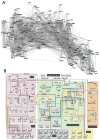
Figure 1
Figure 1. Neural and Gene Networks
(A) Complete wiring diagram of connections among 302 neurons in C. elegans, reconstructed from serial-section EM. Depicted are individual neurons and their connections. For more details see http://www.wormatlas.org/handbook/nshandbook.htm/nswiring.htm. Courtesy of D. Chklovskii. (B) Diagram of gene interaction network that orchestrates early endomesoderm development of sea urchin embryos. Depicted are individual genes and their regulatory relationships. For more details see http://sugp.caltech.edu/endomes/. Courtesy of E. Davidson.
Figure 2
Figure 2. Methods for Targeting Gene Expression
Box provides a glossary for the symbols in (A)–(G). See text for more details. (A) Simple transgenic method to express the coding sequence of target gene of interest under the control of the enhancer/promoter of a gene whose expression is to be mimicked. (B) Bacterial artificial chromosome (BAC)-mediated transgenic expression. (C) Integrase-mediated, site-directed integration of a transgene at a defined chromosomal locus. (D) Knockin of target gene of interest at the endogenous locus of a gene whose expression is to be mimicked. (E) Enhancer trap method, which allows target gene of interest to be under the control of enhancer elements near its chromosomal integration site. (F) Enhancer bashing to create subset expression patterns of an endogenous gene. (G) Restriction of transgene expression is likely due to trapping of repressor elements and chromatin structures local to integration sites. (H) Transgenic mouse expressing GFP under the control of the BAC for the connective tissue growth factor (ctgf). In the cerebral cortex a subpopulation of layer 6b neurons are labeled. The axons of these neurons span all cortical layers and their function is unknown. H2, cell bodies; H3, axonal projections. For more details see http://www.gensat.org/. Courtesy N. Heintz.
Figure 3
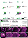
Figure 3. Binary and Intersectional Methods of Gene Expression
Box below provides a glossary for the symbols in (A)–(G′). (A) Yeast transcription factor Gal4 binds to UAS and activates target gene T expression in cells where promoter A is active. The same scheme applies to other transcription factor/binding site-based binary expression systems. (B) Cre/loxP-mediated recombination removes the transcription stop, allowing target gene T to be expressed in cells that are active for both promoters A and C. Promoter C is often constitutive for general application; if promoter C is also specific, it can provide intersectional restrictions with promoter A. Cre can be replaced with a taxoxifen-inducible CreER to allow control of timing and amount of recombination. The same scheme also applies to other site-directed recombination systems, such as Flp/FRT. (C) Combination of Cre/loxP and Flp/FRT recombination systems allow target gene of interest to be expressed in cells that are active for both promoters A and B (and C). (D) The combination of Gal4/UAS and Flp/FRT allows the target gene of interest to be expressed in cells that are active for both promoters A and B. Gal4/UAS can be replaced with other binary expression systems; Flp/FRT can be replaced by other recombination systems. (E) Intersectional method that utilizes the reconstitution of N- and C-terminal parts of Gal4. (F) Target gene is expressed in cells that are active for promoter A but not promoter B, as Gal80 inhibits Gal4 activity. (G and G′) Tetracycline-inducible transcription of target gene T. Dox, doxycycline, a tetracycline analog. (H–I) Examples of restricting gene expression in genetically identified single cells using the MARCM method (see text) in Drosophila. Three olfactory projection neurons (PNs) from three individual flies that send dendrites to the DL1 glomerulus (H0) exhibit stereotyped axon termination patterns in higher olfactory centers, the mushroom body (MB), and particularly, the lateral horn (LH) (H1–H3). Likewise, three PNs that send dendrites to the VA1lm glomerulus (I0) exhibit stereotyped axon terminations (I1–I3) distinct from those of DL1 PNs. Green: mCD8-GFP that labels dendritic and axonal projections of single PNs; magenta: mAB nc82 staining that stains the neuropil structure. Modified from Marin et al., 2002.
Figure 4
Figure 4. Virus-Mediated Gene Expression
(A) Generation of helper virus-free viral vectors. The native viral genome includes coding sequences for genes that are pathogenic (pink) as well as genes required to produce viral proteins, including those which contribute to replication of genetic material (dark blue) and to the structure of the viral particle (green). These genes are flanked by cis-acting elements (magenta) that provide the origin of replication and signal for encapsidation. The packaging construct includes only genes required for replication and structural genes. The vector includes only the cis elements (magenta), which are required to incorporate it into the vector particles, plus the transgene cassette (light blue) that contains transcriptional regulatory elements (e.g., promoters) and coding sequences for transgenes. To produce recombinant vectors, packaging cells are transfected with the packaging construct and the vector construct. Replicated vector genomes are incorporated into virus particles, resulting in the generation of recombinant viral vector. After Verma and Weitzman (2005). (B) Pseudotyping of viral vectors allows modification of viral tropism. Viral vectors are pseduotyped by modifying the packaging construct and replacing structural genes from the native virus (green) with structural genes from some other virus (red). For pseudotyping nonenveloped viruses, such as AAV, the relevant structural protein is capsid. For enveloped viruses such as lentivirus and HSV, the relevant structural proteins are envelope proteins. Selective infection can be achieved by pseudotyping with an envelope or capsid that interacts with cell surface receptors that are present only on a specific subset of cells.
Figure 5
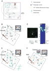
Figure 5. Methods for Functional Circuit Mapping in Brain Slices
(A) The spatial ranges of circuit mapping techniques. The boxes indicate the lengthscales accessible to different methods. Red box, 20 μm, 3D electron microscopy reconstructions; green box, 200 μm, paired recordings; blue box, 1000 μm, laser scanning photostimulation with glutamate uncaging and optical probing; purple box, potentially the entire brain, axon tracing and ChR2-assisted circuit mapping. The reconstruction is a layer 2/3 pyramidal neuron superposed on a schematic of the cat visual cortex (J. Hirsh, USC). Dendrites are in red; axons, in black. (B) Glutamate uncaging mapping. The schematic shows a brain slice in which synaptic responses are recorded in a single neuron (red). Neurons are excited by photolysis of caged glutamate, typically by using a UV laser that is scanned over the brain slice (blue line). If glutamate is photoreleased near the soma (but not on distal dendrites or axons), it evokes action potentials. Postsynaptic whole-cell currents (or potentials) recorded in the recorded neuron are used to generate a map in a computer. This so-called “synaptic input map” is a quantitative representation of the spatial distribution of synaptic input to the recorded neuron. (C and D) The use of glutamate uncaging mapping to measure the spatial distribution of excitatory inputs impinging onto genetically defined GABAergic interneurons (X. Xu and E.C., unpublished data). (C) Morphology of the recorded neuron in neocortical layer 2/3. GFP fluorescence is overlaid with Cy3 streptavidin labeling intracellularly injected biocytin (top). (Bottom) Firing pattern of the recorded neuron. (D) Synaptic input map showing hotspots of input from layer 4 and layer 2/3. Traces to the right are examples of excitatory postsynaptic currents evoked following stimulation at sites 1 and 2. (E) ChR2-assisted circuit mapping. A specific subpopulation of neurons is targeted for expression of ChR2 (green). ChR2-positive neurons (2) and axons (3) are excited by a blue laser that is scanned over the brain slice (blue lines), whereas ChR2-negative neurons are not perturbed (1). Postsynaptic whole-cell currents (or potentials) are used to generate a map in a computer. ChR2-assisted circuit mapping has genetic specificity because ChR2 expression is necessary for exciting action potentials. Furthermore, since severed axons can be excited (3), connectivity between distal brain regions can be studied even in a brain slice. (F) Optical probing. All neurons are bulk-loaded with Ca2+ indicator. One neuron is stimulated with brief bursts of action potentials. Postsynaptic neurons that fire action potentials can be detected using [Ca2+] imaging.
Figure 6
Figure 6. Strategies for Imaging Genetically Specified Neuronal Populations with [Ca2+] Indicators
(A) All neurons are labeled nondiscriminately by bulk-loading with a [Ca2+] indicator (diffuse green). A genetically specified set of neurons express a fluorescent protein (yellow). (B) [Ca2+] imaging in mice expressing GFP in GABAergic interneurons. (Top) Image showing neurons bulk-loaded with [Ca2+] indicator. GFP fluorescence is overlaid in green. (Bottom) Responses of GFP-negative and GFP-positive (GABAergic) neurons to oriented bars. Modifed from Sohya et al., 2007. (C) A genetically specified subpopulation of neurons express a protein (such as tetracysteine motifs; blue) that makes them susceptible to labeling by modified versions of [Ca2+] indicators (such as biarsenicals; green). (D) A genetically specified subpopulation of neurons express a genetically encoded [Ca2+] indicator (green). (E–G) Imaging odor-evoked activity in Kenyon cells of the Drosophila mushroom body using genetically encoded [Ca2+] indicators (G-CaMP1.3) in vivo. (E) G-CaMP fluorescence showing the mushroom body. (F) Two responses to the same odor (difference image; 2 s after odor onset minus baseline). Two Kenyon cells show strong activity. (G) Time course of G-CaMP responses. Modified from Wang et al., 2004.

Similar articles

Cited by

References

    1. Adamantidis AR, Zhang F, Aravanis AM, Deisseroth K, de Lecea L. Neural substrates of awakening probed with optogenetic control of hypocretin neurons. Nature. 2007;450:420–424. - PMC - PubMed
    1. Adrian E. The Mechanisms of Nervous Action. Philadelphia: University of Pennsylvania Press; 1932.
    1. Allen ND, Cran DG, Barton SC, Hettle S, Reik W, Surani MA. Transgenes as probes for active chromosomal domains in mouse development. Nature. 1988;333:852–855. - PubMed
    1. Anderson JC, Martin KA, Whitteridge D. Form, function, and intracortical projections of neurons in the striate cortex of the monkey Macacus nemestrinus. Cereb Cortex. 1993;3:412–420. - PubMed
    1. Arenkiel BR, Peca J, Davison IG, Feliciano C, Deisseroth K, Augustine GJ, Ehlers MD, Feng G. In vivo light-induced activation of neural circuitry in transgenic mice expressing channelrhodopsin-2. Neuron. 2007;54:205–218. - PMC - PubMed

Publication types

LinkOut - more resources