. 2008 May;1783(5):737-47.
doi: 10.1016/j.bbamcr.2008.01.028. Epub 2008 Feb 15.

Allosteric inhibition of the nonMyristoylated c-Abl tyrosine kinase by phosphopeptides derived from Abi1/Hssh3bp1

Affiliations

Allosteric inhibition of the nonMyristoylated c-Abl tyrosine kinase by phosphopeptides derived from Abi1/Hssh3bp1

Xiaoling Xiong et al. Biochim Biophys Acta. 2008 May.

Abstract

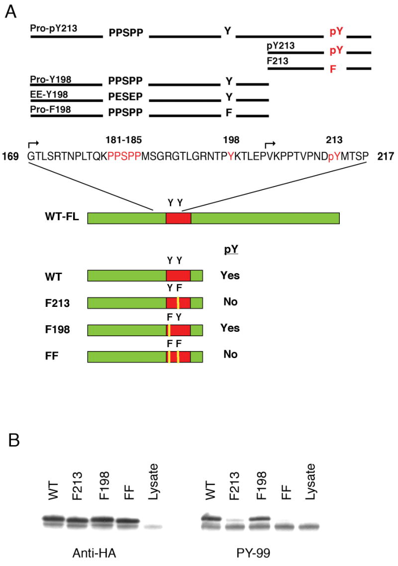
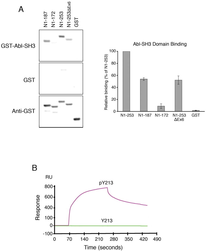
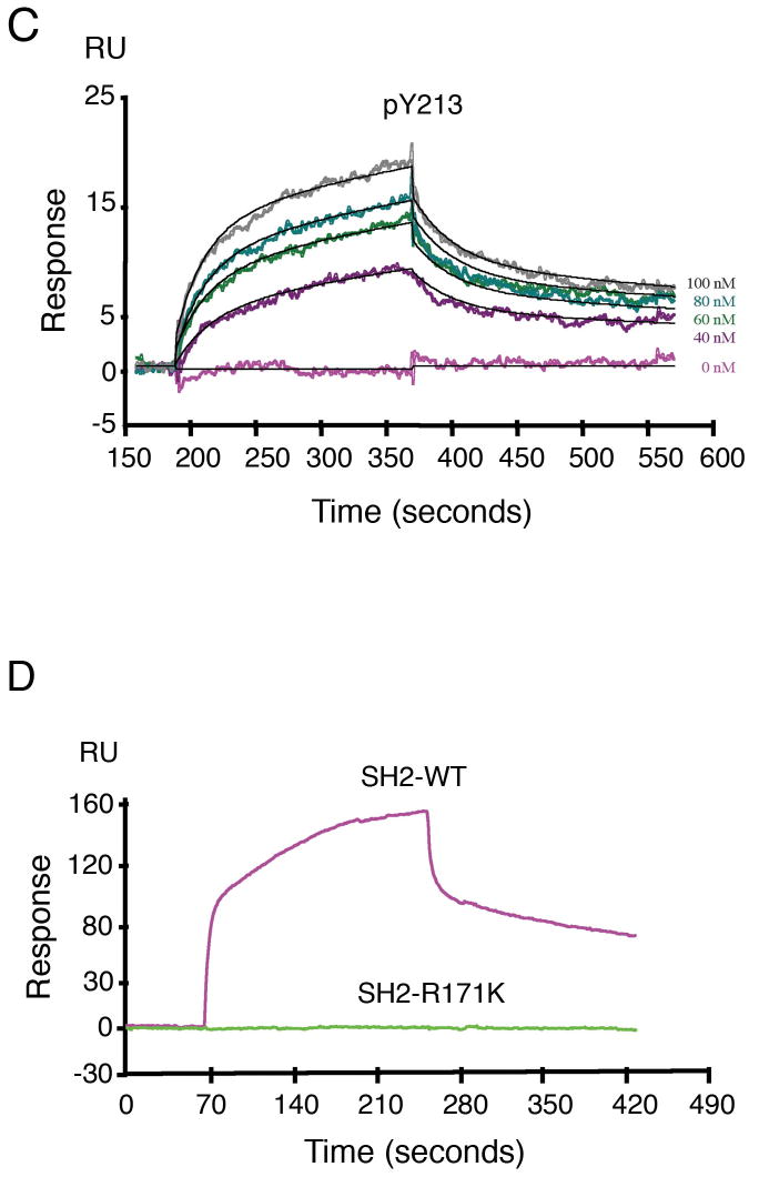
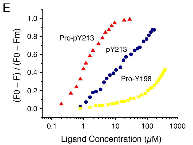
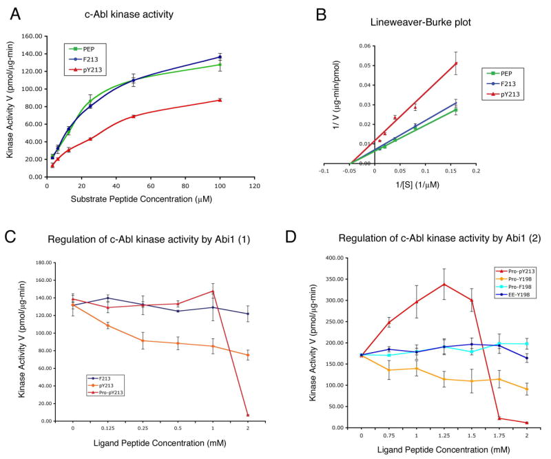
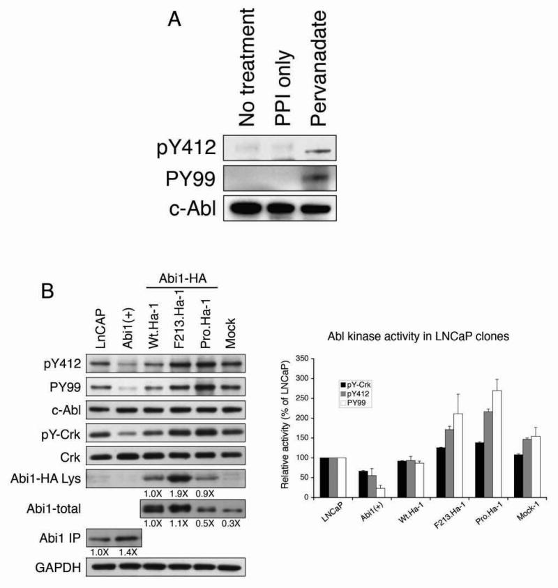
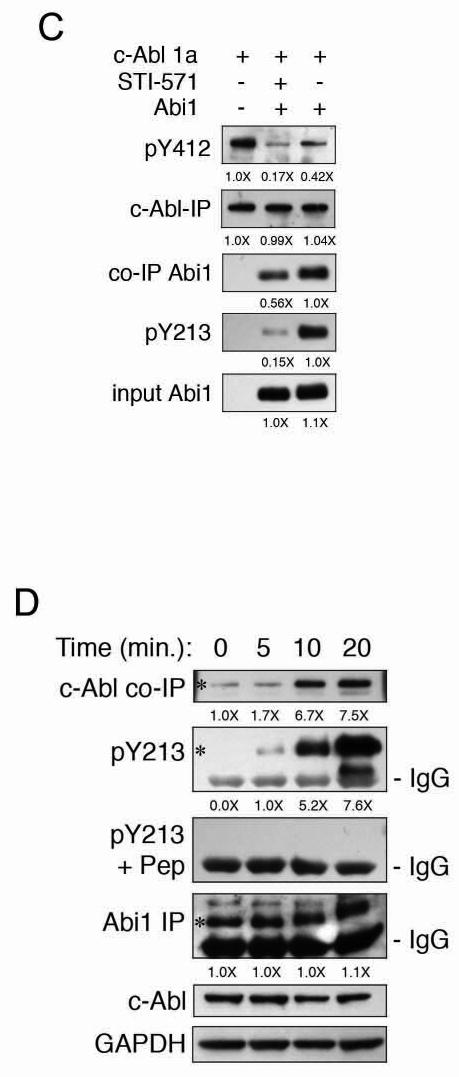
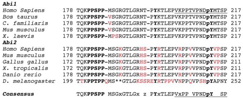
Here we report c-Abl kinase inhibition mediated by a phosphotyrosine located in trans in the c-Abl substrate, Abi1. The mechanism, which is pertinent to the nonmyristoylated c-Abl kinase, involves high affinity concurrent binding of the phosphotyrosine pY213 to the Abl SH2 domain and binding of a proximal PXXP motif to the Abl SH3 domain. Abi1 regulation of c-Abl in vivo appears to play a critical role, as demonstrated by inhibition of pY412 phosphorylation of the nonmyristoylated Abl by coexpression of Abi1. Pervanadate-induced c-Abl kinase activity was also reduced upon expression of the wild type Abi1 but not by expression of the Y213 to F213 mutant Abi1 in LNCaP cells, which are naturally deficient in the regulatory pY213. Our findings suggest a novel mechanism by which Abl kinase is regulated in cells.


Figures

Figure 1
Figure 1. Identification of c-Abl kinase phosphorylation site
A. Regulatory region of Abi1. Top: Diagram of peptides used in Abl kinase assays. Essential features of peptides are illustrated relative to Abi1 residues 169 to 217 (GenBank Accession No. U87166). Names of peptides are above lines depicting the specific peptide. Pro, indicates inclusion of the PXXP motif, PPSPP; Y, indicates tyrosine; pY, indicates phosphotyrosine; F, indicates tyrosine to phenylalanine replacement. The N-terminus of peptides Pro-pY213, pY213, and F213 peptides used in kinase assays is marked above the sequence with a hooked arrow. Peptides Pro-Y198 and Pro-F198 span residues 169-203; these peptides exclude pY213 sequence. Peptide EE-Y198 replaces the sequence PPSPP with PESEP resulting in the loss of PXXP SH3-binding motif. Bottom: Diagram depicting in vitro translated polypeptides used initially to characterize tyrosine phosphorylation of the N-terminus of Abi1. An HA epitope was introduced at the C-terminus of each polypeptide. Mutations replacing either or both tyrosine residues, Y198, and Y213, in the region of interest are indicated. Red box represents exon 6 of Abi1 that is lacking in LNCaP cells. B. Mapping of tyrosine phosphorylation of the in vitro translated N-terminus of Abi1 by c-Abl. Abi1 polypeptides containing the N-terminal half of the protein and indicated mutations of tyrosine residues were subjected to in vitro kinase reactions with c-Abl tyrosine kinase, followed by separation on SDS-Tricine polyacrylamide gels (7%) and blotting with anti-HA antibody (left) and with the anti-phosphotyrosine antibody, PY-99 (right). WT, wild type polypeptide; F213, polypeptide containing a phenylalanine replacement of tyrosine 213 (Y213F); F198, the polypeptide containing phenylalanine replacement of tyrosine 198 (Y198F); FF, the polypeptide containing Y213F and Y198F; Lysate, lysate with no Abi1 cDNA.
Figure 2
Figure 2. Identification of the minimal binding region of c-Abl SH3 and SH2 domains in the N-terminus of Abi1
A. Binding to c-Abl SH3 domain. Recombinant GST fusion polypeptides containing the N-terminal terminal region of Abi1/Hsshb3p1 were separated on SDS polyacrylamide gels and transferred onto the nitrocellulose membrane. The following Abi1 polypeptides were analyzed: N1-187, encoding residues 1-187; N1-172, encoding residues 1-172; N1-253, encoding residues 1-253; N1-253ΔEx6, encoding residues 1-253 but lacking exon 6 sequences of Abi1 (Macoska et al, 2001); GST, glutathione S-transferase. The bottom panel shows results when the blots were probed with the anti-GST monoclonal antibody (Anti-GST) to indicate level of protein expression. The top panel was incubated with biotinylated GST-Abl-SH3 domain (GST-Abl-SH3-B). No binding to biotinylated GST (GST-B) was observed (middle panel). The histogram shows relative binding of polypeptides, as percent of N1-253 binding, quantified from three independent experiments (n=3, ± s.d.). B-D. Binding to c-Abl SH2 domain. Surface plasmon resonance was used to determine binding between biotinylated 14-residue peptides containing either phosphorylated tyrosine 213 (pY213) or nonphosphorylated tyrosine 213 (Y213) to GST-tagged Abl SH2 domain. B. Biosensor chips coupled with pY213 or Y213 peptides were injected with the Abl SH2 domain (1 μM). C. Biosensor chip coupled with pY213 was injected with different concentrations of Abl SH2 domain, as indicated. D. Biosensor chip coupled with pY213 was injected with 0.5 μM of either the Abl SH2 domain or with the Abl SH2 domain R171K mutant. No binding to the GST protein alone was observed (not shown). RU, response unit. E. Representative fluorescence titration data using purified Abl dual SH3-SH2 domain. Titration curves for the consolidated dual SH3-SH2 domain ligand, Pro-pY213, and single domain ligands, Pro-Y198 for Abl SH3 domain, and pY213 for Abl SH2 domain, are indicated.
Figure 3
Figure 3. Phosphopeptides from Abi1 regulate c-Abl kinase activity in vitro
A. Kinase activities (vertical bars represent s.d.; n=3) of c-Abl after addition of indicated peptides at 2 mM. Activity was measured with 5-min kinase assays in the presence of increasing substrate peptide concentrations. B. Double reciprocal Lineweaver-Burke plots of the data in A. Vertical bars represent s.d. (n=3). See Table 2 for Vmax and Km data from these experiments. C and D. Regulation of c-Abl kinase activity by Abi1 peptides. Concentration of Abi1 peptides is indicated on X axis; substrate peptide, PEP, was used at 100 μM (see Kinase assay, Materials and Methods); vertical bars represent s.e.m. (n=3). See Figure 1A for sequence information of peptides used here.
Figure 4
Figure 4. Abi1 inhibits pervanadate-activated Abl kinase activity in LNCaP cells
A. Pervanadate treatment enhances c-Abl kinase activity in LNCaP cells. LNCaP cells were treated with 0.1 mM pervanadate for 10 min. prior to cell lysis. Following treatment c-Abl was immunoprecipitated with K12 antibody; proteins were separated on SDS-PAGE gels, and transferred and blotted with indicated antibodies. “pY412” shows level of phosphorylation of the activation loop tyrosine; “pY99” shows levels of total tyrosine phosphorylation of c-Abl. “c-Abl” indicates levels of c-Abl kinase in samples. “Pervanadate” indicates cells treated with pervanadate; cell lysis was performed with inclusion of phosphatase inhibitors (PPI) as described [25]; “PPI only” indicates untreated cells but lysed in the presence of phosphatase inhibitors. “No treatment” indicates untreated cells lysed without phosphatase inhibitors. B. Pervanadate-induced Abl kinase activity in stably transfected cell lines was evaluated by immunoprecipitation and examination of phosphorylation levels of the endogenous Abl substrate Crk (pY-Crk), as well as the c-Abl activation loop phosphotyrosine pY412 (pY412) and total tyrosine phosphorylation of c-Abl (PY99). Proteins were immunopreciptated from indicated stable cell lines: c-Abl with K-12, Crk with anti-Crk, Abi1 with 7B6. Expression of the recombinant Abi1 in cell lysates was evaluated with anti-HA antibody (Abi1-HA Lys); expression of total Abi1 in LNCaP clones expressing HA-tagged Abi1 (Abi1-total), and expression of total Abi1 in Abi1(+) and LnCAP cell lines was evaluated by blotting of the immunoprecipitated Abi1 with the polyclonal antibody Ab-2 (Abi1-IP). “GAPDH” indicates lysate input levels used for immunoprecipitation. Histogram on the right indicates quantitation of c-Abl kinase activity in the cell lines (n=3; ±s.d.). Crk phosphorylation is lower in Abi1(+) than in LnCAP cells or Mock cell line (p<0.001); and lower in Wt.Ha than in F213.Ha or Pro.Ha (p<0.001) or Mock (p<0.01). Differences in pY412 or PY99 phosphorylation among the groups are also significant (p<0.05). C. Co-expression of Abi1 or STI-571 treatment inhibits pY412 phosphorylation of nonmyristoylated c-Abl. STI-571 and pY213-dependent interaction of Abi1 with nonmyristoylated c-Abl. Cos 7 cells were co-transfected with HA tagged-Abi1 and GFP tagged-c-Abl (isoform 1a (nonmyristoylated) with the GFP tag at the C-teminus). Cells were treated with STI-571 (10 μM) for 30 min prior to immunoprecipitation as indicated. The level of immunoprecipitated c-Abl (c-Abl-IP) was evaluated with anti-GFP antibody; levels of pY412 (pY412) and pY213 (pY213) phosphorylation were evaluated with phosphospecific antibodies, as indicated. The level of co-immunoprecipitated recombinant Abi1 was evaluated with anti-HA antibody (co-IP Abi1). The level of Abi1 expression in transfected cultures is indicated by “input Abi1.” Numbers below blots shows density of specific bands. Note that treatment of cells with STI-571 reduces but does not completely abolish the association of Abl with the recombinant Abi1. D. pY213-dependent interaction of Abi1 with c-Abl in LNCaP. LNCaP cells were treated with pervanadate for increasing amounts of time as indicated. Abi1 was immunoprecipitated with monoclonal antibody 7B6 and the immunoprecipitated proteins were analyzed by Western blotting using specific antibodies: Abi1 with Ab-2 (Abi1-IP), c-Abl with K-12 (c-Abl co-IP). The level of Abi1 phosphorylation on Y213 in the samples was evaluated by blotting with pY213 (pY213); the level of pY213 immunoreactivity with addition of pY213 phosphopeptide (10 μM) is indicated (pY213+Pep). The level of c-Abl kinase in cell lysates following immunoprecipitation with K-12 antibody and blotting with 8E9 is indicated by “c-Abl.” “GAPDH” indicates lysate input levels used for immunoprecipitation. Numbers below blots shows density of bands indicated by asterisks. There is an apparent crossreactive band above the band with asterisk in the Abi1 IP panel; no crossreactivity of this band with pY213 antibody is observed. Note that binding of pY213 antibody to Abi1 can be blocked by inclusion of the phosphopeptide pY213 in the incubation mixture.
Figure 5
Figure 5
c-Abl kinase regulatory region of the Abi protein family. The region of Abi1 containing the Abl SH3 domain binding site containing the core PXXP consensus, PPSPP, Y198, and the SH2 domain binding site (underlined, with regulatory phosphotyrosine, pY) is highly conserved during evolution in the Abi protein family. Critical residues are in bold, non-conserved residues are in red. GenBank Accession numbers for the sequences are the following: for Abi1: Homo sapiens, U87166 and NM_005470; Bos taurus, XP_881074.1; Canis familiaris, XP_858231.1; Mus musculus, Q8CBW3; Xenopus laevis, AAH81178.1; Abi2: Homo sapiens, NP_005750.3; Mus musculus, AAH79646.1; Gallus gallus, XP_421962.1; Xenopus tropicalis, NP_001007488.1; Danio rerio, XP_685431.1, Drosophila melanogaster, AAD38382.1. ** denotes insertion of the sequence RAGNTGTLGKSVSNT in Drosophila melanogaster. Consensus is presented in the bottom, where “x” is K or R, and “z” is S or T, gaps indicate non-conserved residues.

Similar articles

Cited by

References

    1. Koleske AJ, Gifford AM, Scott ML, Nee M, Bronson RT, Miczek KA, Baltimore D. Essential roles for the Abl and Arg tyrosine kinases in neurulation. Neuron. 1998;21:1259–1272. - PubMed
    1. Woodring PJ, Hunter T, Wang JY. Regulation of F-actin-dependent processes by the Abl family of tyrosine kinases. J Cell Sci. 2003;116:2613–2626. - PubMed
    1. Druker BJ, Sawyers CL, Capdeville R, Ford JM, Baccarani M, Goldman JM. Chronic myelogenous leukemia. Hematology (Am Soc Hematol Educ Program) 2001:87–112. - PubMed
    1. Goldman JM, Melo JV. Chronic myeloid leukemia--advances in biology and new approaches to treatment. N Engl J Med. 2003;349:1451–1464. - PubMed
    1. Buchdunger E, Zimmermann J, Mett H, Meyer T, Muller M, Druker BJ, Lydon NB. Inhibition of the Abl protein-tyrosine kinase in vitro and in vivo by a 2-phenylaminopyrimidine derivative. Cancer Res. 1996;56:100–104. - PubMed

Publication types

MeSH terms