Comparative Study
. 2008 Mar 5;28(10):2320-31.
doi: 10.1523/JNEUROSCI.4760-07.2008.

A vicious cycle involving release of heat shock protein 60 from injured cells and activation of toll-like receptor 4 mediates neurodegeneration in the CNS

Affiliations
Comparative Study

A vicious cycle involving release of heat shock protein 60 from injured cells and activation of toll-like receptor 4 mediates neurodegeneration in the CNS

Seija Lehnardt et al. J Neurosci. .

Abstract

Infection, ischemia, trauma, and neoplasia elicit a similar inflammatory response in the CNS characterized by activation of microglia, the resident CNS monocyte. The molecular events leading from CNS injury to the activation of innate immunity is not well understood. We show here that the intracellular chaperone heat shock protein 60 (HSP60) serves as a signal of CNS injury by activating microglia through a toll-like receptor 4 (TLR4)-dependent and myeloid differentiation factor 88 (MyD88)-dependent pathway. HSP60 is released from CNS cells undergoing necrotic or apoptotic cell death and specifically binds to microglia. HSP60-induced synthesis of neurotoxic nitric oxide by microglia is dependent on TLR4. HSP60 induces extensive axonal loss and neuronal death in CNS cultures from wild-type but not TLR4 or MyD88 loss-of-function mutant mice. This is the first evidence of an endogenous molecular pathway common to many forms of neuronal injury that bidirectionally links CNS inflammation with neurodegeneration.


Figures

Figure 1.
Figure 1.
Mixed CNS cultures derived from lpsd mice are less vulnerable and contain a higher number of neurons compared with cultures derived from wild-type mice. Mixed CNS cultures from four pooled mouse forebrains derived from BALB/cJ or lpsd mice were prepared and cultured for 3 d. AC, Subsequent parallel (A) and individual (B, C) immunostaining with antibodies against MAP-2 and neurofilament marked neurons. Parallel DAPI staining revealed the total number of cells by staining all nuclei. Scale bars, 50 μm. For quantitation of DAPI- and MAP-2-positive cells in B, six high-power fields per coverslip were analyzed. For each condition, experiments were performed in triplicates. The data shown are representative of >10 individual experiments.
Figure 2.
Figure 2.
HSP60 induces neuronal cell death dependent on a functional TLR4 pathway. Mixed CNS cultures from forebrains of BALB/cJ and lpsd mice were incubated with 10 μg/ml HSP60 or PBS (control) for 2 d. A, C, Cultures were stained with antibodies against MAP-2 (A) and neurofilament (C) to mark neurons, whereas all nuclei were stained with DAPI. Scale bars, 50 μm. B, Quantitation of MAP-2-positive neurons from BALB/cJ and lpsd mice in the presence or absence of HSP60. Six high-power fields per coverslip were analyzed. For each condition, experiments were performed in triplicates. The data shown are representative of four individual experiments. The results are presented as mean ± SD (p < 0.001, Student's t test) for the comparison of BALB/cJ cells with or without HSP60. D, Fifteen micrograms of the HSP60 preparation were analyzed by SDS-PAGE and subsequent Coomassie staining. PS, Protein standards.
Figure 3.
Figure 3.
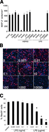
HSP60-induced neuronal cell death is not attributable to LPS contamination. Cortical neurons were prepared from BALB/cJ mice forebrains and were supplemented with purified microglia from BALB/cJ mice. A, Cells were incubated with 10 μg/ml HSP60 or 1 μg/ml LPS. Neurotoxic effects of HSP60 were abrogated by pretreatment of samples with trypsin and proteinase K (PK) or by boiling for 20 min before incubation. Neurotoxic effects of HSP60 were not abrogated by combined treatment of cultures with PMBS. In parallel, LPS and PBS (control) were also treated under the same conditions before incubation. After 2 d, cultures were fixed and stained both with MAP-2 antibody and with DAPI to mark neurons and nuclei, respectively. The results are presented as mean ± SD. Cells were incubated with various doses of LPS, as indicated, for 2 d. B, Staining with the NeuN antibody and DAPI (units are in nanograms per milliliter). C, Quantitation of NeuN-positive neurons as a percentage of all DAPI-positive cells in cultures supplemented with BALB/cJ microglia. Scale bar, 50 μm. Results are presented as mean ± SD (*p < 0.001, Student's t test) for comparison of the indicated group to the control group. All experiments were repeated three times.
Figure 4.
Figure 4.
Neurons treated with HSP60 undergo apoptotic cell death dependent on a TLR4-MyD88 pathway in microglia. Cortical neurons were prepared from BALB/cJ mice forebrains. Purified neurons alone as well as neurons cocultured with purified microglia from BALB/cJ (wt) or lpsd mice were incubated with 10 μg/ml HSP60 or PBS. A, After 2 d, cultures were fixed and stained both with NeuN antibody and with IB4 to mark neurons and microglia, respectively. B, Quantitation of NeuN-positive neurons as a percentage of all DAPI-positive cells in purified cultures and cultures supplemented with BALB/cJ (WT)- or lpsd-derived microglia in the presence or absence of HSP60. Results are presented as mean ± SD (p < 0.001, Student's t test) for comparison of the group, in which wild-type microglia and HSP60 are added, with other groups. C, Cells were incubated with 10 μg/ml HSP60 or Camptothecin and analyzed by a TUNEL assay. D, Cortical neurons were prepared from C57Bl/6J mice forebrains. Purified neurons alone as well as neurons cocultured with purified microglia from C57Bl/6J (wt) or MyD88−/− mice were incubated with 10 μg/ml HSP60 or PBS. After 2 d, cultures were fixed and stained both with MAP-2 antibody and with IB4 to mark neurons and microglia, respectively. E, Quantitation of MAP-2-positive neurons as a percentage of all DAPI-positive cells in purified and C57Bl/6J (wt) or MyD88−/− microglia-enriched cultures in the presence or absence of HSP60. Results are presented as mean ± SD (p < 0.001, Student's t test) for comparison of the group, in which wild-type microglia and HSP60 are added, with other groups. For all quantitations (B, E), six high-power fields per coverslip were analyzed. For each condition, experiments were performed in triplicates. The data shown are representative of four individual experiments each. Scale bars, 50 μm.
Figure 5.
Figure 5.
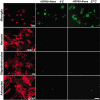
Microglia, but not neurons, oligodendrocytes, or astrocytes, bind HSP60-Alexa. Primary cultures of rat microglia, neurons, oligodendrocytes, and astrocytes were incubated with 1 μg/ml HSP60-Alexa at 4°C or 37°C and analyzed by immunofluorescence. Parallel cultures were immunostained for microglia (IB4), neurons (MAP-2), oligodendrocytes (O4), and astrocytes (GFAP). Scale bar, 50 μm.
Figure 6.
Figure 6.
HSP60 is released by apoptotic and necrotic CNS cells and causes neuronal injury. Mixed CNS cultures derived from wild-type mice were forced to undergo apoptosis or necrosis. A, Untreated and drug-treated cells were stained with DAPI. B, Culture supernatants (Sup) of treated cells were analyzed by SDS-PAGE and subsequent immunoblotting with an HSP60 antibody. Cell lysate derived from A431 cells served as a positive control. C, Culture supernatants of treated cells were immunoprecipitated with an HSP60 antibody or normal mouse IgG. Neuronal-enriched mixed CNS cultures derived from wild-type mice were treated with either plain or immunoprecipitated (with an antibody against HSP60 or with normal mouse IgG) culture supernatants of live, apoptotic, and necrotic cells. Cells were then immunostained with NeuN to mark neurons and with DAPI. D, Quantitation of NeuN-positive neurons in CNS cultures treated with supernatants (S/N) from live, apoptotic, or necrotic CNS cells with or without immunoprecipitation (IP) against HSP60. Six high-power fields per coverslip were analyzed. For each condition, experiments were performed in triplicates. The data shown are representative of three individual experiments. The results are presented as mean ± SD (**p < 0.01, ***p < 0.001; Student's t test) for the comparison of S/N from apoptotic or necrotic cells versus S/N from live cells and for the comparison of S/N treated with anti-HSP60 antibody versus isotype control antibody or no treatment. E, Immunoblot of wild-type mouse-derived mixed CNS cells and heat-shocked HeLa cells (positive control) probed with the SPA-807 antibody. Scale bars, 50 μm.
Figure 7.
Figure 7.
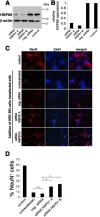
Silencing of HSP60 reduces neurotoxicity of HEK293 cell lysates added to cocultures of neurons and microglia. HEK293 cells were transfected with siRNA A and B targeting HSP60. Untransfected cells (control) and cells transfected with nonsilencing siRNA (neg. siRNA) served as controls. A, Immunoblot of lysed cells probed with the SPA-807 antibody directed against HSP60. β-Actin served as a loading control. B, Densitometric analysis of the immunoblot signals after detection by chemiluminescence. C, Cocultures of neurons and microglia derived from wild-type mice were treated with cell lysates of transfected and untransfected HEK293 cells and were subsequently immunostained with NeuN to mark neurons, and with DAPI. Scale bar, 50 μm. D, Quantitation of NeuN-positive neurons as a percentage of all DAPI-positive cells in cocultures treated with HEK293 cell lysates. Six high-power fields per coverslip were analyzed. For each condition, experiments were performed in duplicates. The data shown are representative of four individual experiments. The results are presented as mean ± SD (*p < 0.001) for the comparison of neg. siRNA with siRNA HSPD1A and for the comparison of neg. siRNA with siRNA HSPD1B (one-way ANOVA and post hoc Bonferroni's test).
Figure 8.
Figure 8.
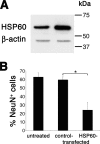
Overexpression of HSP60 increases neurotoxicity of HEK293 cell lysates added to cocultures of neurons and microglia. HEK293 cells were transfected with plasmids encoding hHSP60 or GFP (control). A, Immunoblot of lysed cells probed with the SPA-807 antibody directed against HSP60. Left lane, Control-transfected; right lane, HSP60-transfected. β-Actin served as a loading control. B, Quantitation of NeuN-positive neurons as a percentage of all DAPI-positive cells in cocultures of neurons and microglia derived from wild-type mice treated with cell lysates of hHSP60- or control-transfected HEK293 cells. Cell lysates were diluted 1:15. Quantitation of NeuN-positive neurons was performed by analyzing four high-power fields per coverslip. For each condition, experiments were performed in duplicates. The data shown are pooled from three individual experiments with three different preparations of transfected cells. The results are presented as mean ± SD (*p < 0.001) for the comparison of control-transfected cells with hHSP60-transfected cells (one-way ANOVA and post hoc Bonferroni's test).
Figure 9.
Figure 9.
HSP60 induces release of neurotoxic NO from microglia through a TLR4-dependent pathway. A, Purified microglia derived from BALB/cJ mice were incubated with supernatants of apoptotic and necrotic mixed CNS cells for the indicated duration. The amount of nitrite in the culture media was determined by the Griess reaction. B, Quantitation of NeuN-positive neurons as a percentage of all DAPI-positive cells in cocultures of neurons and microglia incubated with either 10 μg/ml HSP60 alone or in combination with the iNOS inhibitor aminoguanidine (200 μm). Cultures were fixed after 72 h and immunostained with NeuN and DAPI, and neurons were quantified. Results are presented as mean ± SD (*p < 0.001, Student's t test) for the comparison of the control group with HSP60 alone or with HSP60 plus AG and for the comparison of HSP60 alone with HSP60 and AG. C, D, Purified microglia derived from BALB/cJ and lpsd mice were incubated for 48 h with increasing concentrations of HSP60 (C) or were treated with 10 μg/ml HSP60 for various incubation times (D). The amount of nitrite in the culture supernatant was determined by the Griess reaction. For each condition, experiments were performed in duplicates. The data shown are representative of three individual experiments. Results are presented as mean ± SD.

Similar articles

Cited by

References

    1. Akira S, Uematsu S, Takeuchi O. Pathogen recognition and innate immunity. Cell. 2006;124:783–801. - PubMed
    1. Anderson KV, Bokla L, Nusslein-Volhard C. Establishment of dorsal-ventral polarity in the Drosophila embryo: the induction of polarity by the Toll gene product. Cell. 1985;42:791–798. - PubMed
    1. Asea A, Rehli M, Kabingu E, Boch JA, Bare O, Auron PE, Stevenson MA, Calderwood SK. Novel signal transduction pathway utilized by extracellular HSP70: role of toll-like receptor (TLR) 2 and TLR4. J Biol Chem. 2002;277:15028–15034. - PubMed
    1. Babcock AA, Wirenfeldt M, Holm T, Nielsen HH, Dissing-Olesen L, Toft-Hansen H, Millward JM, Landmann R, Rivest S, Finsen B, Owens T. Toll-like receptor 2 signaling in response to brain injury: an innate bridge to neuroinflammation. J Neurosci. 2006;26:12826–12837. - PMC - PubMed
    1. Basu S, Binder RJ, Suto R, Anderson KM, Srivastava PK. Necrotic but not apoptotic cell death releases heat shock proteins, which deliver a partial maturation signal to dendritic cells and activate the NF-kappa B pathway. Int Immunol. 2000;12:1539–1546. - PubMed

Publication types

MeSH terms