

The recognition and retrotranslocation of misfolded proteins from the endoplasmic reticulum
- PMID: 18315532
- PMCID: PMC2754126
- DOI: 10.1111/j.1600-0854.2008.00729.x
The recognition and retrotranslocation of misfolded proteins from the endoplasmic reticulum
Abstract
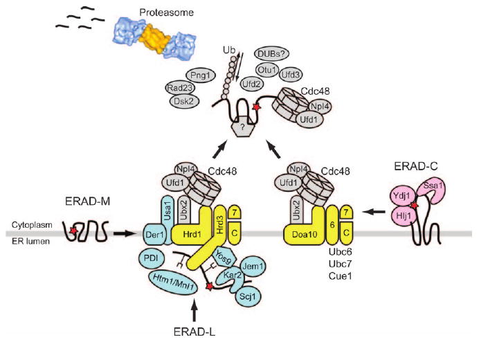
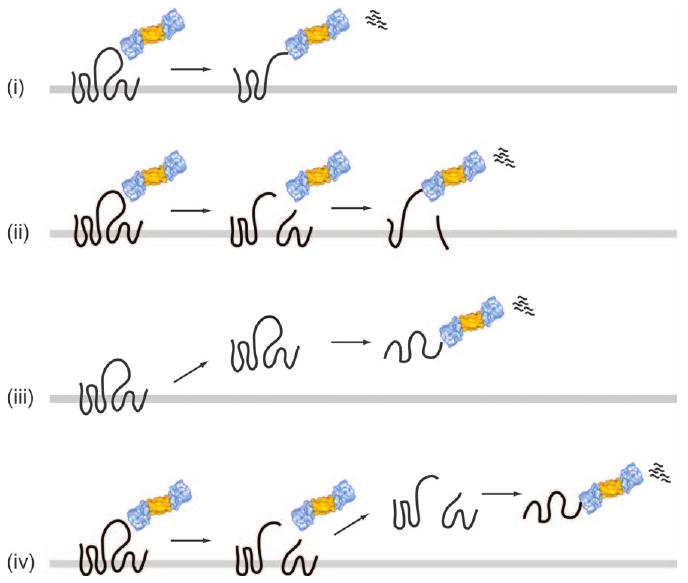
Secretory and membrane proteins that fail to fold in the endoplasmic reticulum (ER) are retained and may be sorted for ER-associated degradation (ERAD). During ERAD, ER-associated components such as molecular chaperones and lectins recognize folding intermediates and specific oligosaccharyl modifications on ERAD substrates. Substrates selected for ERAD are then targeted for ubiquitin- and proteasome-mediated degradation. Because the catalytic steps of the ubiquitin-proteasome system reside in the cytoplasm, soluble ERAD substrates that reside in the ER lumen must be retrotranslocated back to the cytoplasm prior to degradation. In contrast, it has been less clear how polytopic, integral membrane substrates are delivered to enzymes required for ubiquitin conjugation and to the proteasome. In this review, we discuss recent studies addressing how ERAD substrates are recognized, ubiquitinated and delivered to the proteasome and then survey current views of how soluble and integral membrane substrates may be retrotranslocated.
Figures
Similar articles
-
A Cdc48 "Retrochaperone" Function Is Required for the Solubility of Retrotranslocated, Integral Membrane Endoplasmic Reticulum-associated Degradation (ERAD-M) Substrates.J Biol Chem. 2017 Feb 24;292(8):3112-3128. doi: 10.1074/jbc.M116.770610. Epub 2017 Jan 11. J Biol Chem. 2017. PMID: 28077573 Free PMC article.
-
One step at a time: endoplasmic reticulum-associated degradation.Nat Rev Mol Cell Biol. 2008 Dec;9(12):944-57. doi: 10.1038/nrm2546. Epub 2008 Nov 12. Nat Rev Mol Cell Biol. 2008. PMID: 19002207 Free PMC article. Review.
-
The evolving role of ubiquitin modification in endoplasmic reticulum-associated degradation.Biochem J. 2017 Feb 15;474(4):445-469. doi: 10.1042/BCJ20160582. Biochem J. 2017. PMID: 28159894 Free PMC article. Review.
-
Recognition and delivery of ERAD substrates to the proteasome and alternative paths for cell survival.Curr Top Microbiol Immunol. 2005;300:17-40. doi: 10.1007/3-540-28007-3_2. Curr Top Microbiol Immunol. 2005. PMID: 16573235 Review.
-
Transmembrane helix hydrophobicity is an energetic barrier during the retrotranslocation of integral membrane ERAD substrates.Mol Biol Cell. 2017 Jul 15;28(15):2076-2090. doi: 10.1091/mbc.E17-03-0184. Epub 2017 May 24. Mol Biol Cell. 2017. PMID: 28539401 Free PMC article.
Cited by
-
Molecular chaperones: providing a safe place to weather a midlife protein-folding crisis.Nat Struct Mol Biol. 2016 Jul 6;23(7):621-3. doi: 10.1038/nsmb.3255. Nat Struct Mol Biol. 2016. PMID: 27384188 No abstract available.
-
Endoplasmic reticulum stress, the unfolded protein response, autophagy, and the integrated regulation of breast cancer cell fate.Cancer Res. 2012 Mar 15;72(6):1321-31. doi: 10.1158/0008-5472.CAN-11-3213. Cancer Res. 2012. PMID: 22422988 Free PMC article. Review.
-
Membrane proteases in the bacterial protein secretion and quality control pathway.Microbiol Mol Biol Rev. 2012 Jun;76(2):311-30. doi: 10.1128/MMBR.05019-11. Microbiol Mol Biol Rev. 2012. PMID: 22688815 Free PMC article. Review.
-
Life and death of a BiP substrate.Semin Cell Dev Biol. 2010 Jul;21(5):472-8. doi: 10.1016/j.semcdb.2009.12.008. Epub 2009 Dec 21. Semin Cell Dev Biol. 2010. PMID: 20026282 Free PMC article. Review.
-
Sec61p is part of the endoplasmic reticulum-associated degradation machinery.EMBO J. 2009 Oct 7;28(19):2874-84. doi: 10.1038/emboj.2009.231. Epub 2009 Aug 20. EMBO J. 2009. PMID: 19696741 Free PMC article.
References
-
- Ghaemmaghami S, Huh WK, Bower K, Howson RW, Belle A, Dephoure N, O'Shea EK, Weissman JS. Global analysis of protein expression in yeast. Nature. 2003;425:737–741. - PubMed
-
- Ellgaard L, Helenius A. Quality control in the endoplasmic reticulum. Nat Rev Mol Cell Biol. 2003;4:181–191. - PubMed
-
- Meusser B, Hirsch C, Jarosch E, Sommer T. ERAD: the long road to destruction. Nat Cell Biol. 2005;7:766–772. - PubMed
-
- Sayeed A, Ng DT. Search and destroy: ER quality control and ER-associated protein degradation. Crit Rev Biochem Mol Biol. 2005;40:75–91. - PubMed
Publication types
MeSH terms
Substances
Grants and funding
LinkOut - more resources
Full Text Sources
Molecular Biology Databases
