

doi: 10.1021/pr070451j.
Epub 2008 Feb 14.
Rapid isolation and identification of bacteriophage T4-encoded modifications of Escherichia coli RNA polymerase: a generic method to study bacteriophage/host interactions
Affiliations
- PMID: 18271525
- PMCID: PMC2612130
- DOI: 10.1021/pr070451j
Item in Clipboard
Rapid isolation and identification of bacteriophage T4-encoded modifications of Escherichia coli RNA polymerase: a generic method to study bacteriophage/host interactions
J Proteome Res.
2008 Mar.
Abstract
Bacteriophages are bacterial viruses that infect bacterial cells, and they have developed ingenious mechanisms to modify the bacterial RNA polymerase. Using a rapid, specific, single-step affinity isolation procedure to purify Escherichia coli RNA polymerase from bacteriophage T4-infected cells, we have identified bacteriophage T4-dependent modifications of the host RNA polymerase. We suggest that this methodology is broadly applicable for the identification of bacteriophage-dependent alterations of the host synthesis machinery.
Figures
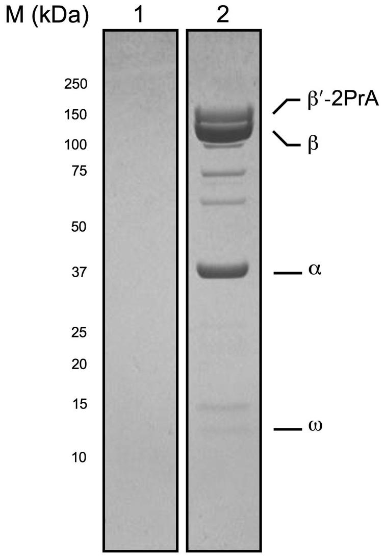
Immunoisolation of β′-2PrA and co-isolating proteins from T4-infected Ec cells. Complexes were isolated via a PrA tag under conditions that co-isolated interacting proteins. Proteins were resolved by denaturing SDS-PAGE, visualized by Coomassie blue staining, and analyzed by MALDI MS. Lanes are loaded as follows: lane 1, Immunoisolated proteins from Ec wild-type cells infected with T4; lane 2, immunoisolated proteins from EcrpoC::2PrA cells infected with T4. RNAP subunits in the 2PrA-tagged sample (lane 2) are labeled.
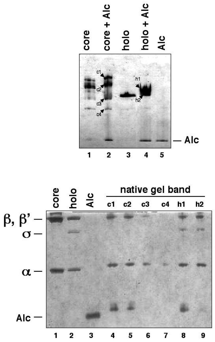
Non-denaturing PAGE and denaturing SDS-PAGE were used to probe the associations of Alc with the core enzyme and the σ70-holoenzyme. (A) Different combinations of Alc, core, and σ70-holoenzyme were incubated together and resolved under non-denaturing conditions. (B) Bands due to the proteins separated by non-denaturing PAGE, labeled in (A), were excised and their composition revealed by denaturing SDS-PAGE.
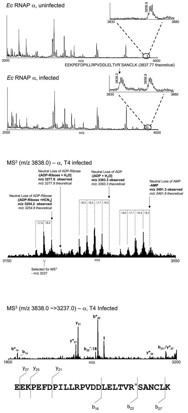
Identification of T4-dependent ADP-ribosylation of RNAP α subunit Arg 265. Bands due to the RNAP α subunit isolated from uninfected and T4-infected cells were excised, digested with trypsin, and analysed by MALDI MS. The resulting mass spectra were aligned and scanned for peptides present in the T4 infected sample, but absent from the uninfected sample. (A) Mass spectrum of tryptic peptides due to the RNAP α subunit isolated from uninfected Ec rpoC::2PrA cells (top panel) and Ec rpoC::2PrA cells infected with T4 (bottom panel). The appearance of a peak at 3838.0 m/z, with a mass that corresponds to a singly protonated ADP-ribsoylated peptide derived from α residues 244 to 271 in the spectrum of the infected phage that is absent in the uninfected sample spectrum suggests that this peptide is ADP-ribosylated in vivo in a T4 dependent manner. (B) The mass spectrum of the MS2 analysis of the species with an m/z of 3838.0 described in (A) illustrates neutral loss of AMP, ADP and ADP-ribose, together with various water/ammonia neutral losses. This fragmentation is consistent with the interpretation that the peptide ion at 3838.0 m/z is indeed an ADP-ribsoylated species. (C) MS3 analysis of the ions in the MS2 spectrum with a nominal m/z of 3237.0 (corresponding to the neutral loss of ADP-Ribose +HCN2 +water/ammonia) yielded backbone fragmentation providing partial sequence of the peptide. The fragmentation pattern is consistent with the interpretation that the original peptide ion at 3838.0 m/z is indeed derived from α residues 244 to 271. Observed are fragments corresponding to the preferential cleavage of the singly-protonated peptide ion carboxy-terminal to acidic residues and amino-terminal to Pro residues. Together, the MS, MS2, and MS3 spectra in panels (A), (B) and (C) provide strong evidence that α is ADP-ribosylated on an internal Arg residue within the tryptic peptide encoding residues 244-271.
Similar articles
-
Transcription regulation by bacteriophage T4 AsiA.Protein Expr Purif. 2005 May;41(1):1-8. doi: 10.1016/j.pep.2004.09.019. Protein Expr Purif. 2005. PMID: 15802215 Review.
-
The bacteriophage T4 late-transcription coactivator gp33 binds the flap domain of Escherichia coli RNA polymerase.Proc Natl Acad Sci U S A. 2004 Dec 14;101(50):17365-70. doi: 10.1073/pnas.0408028101. Epub 2004 Dec 1. Proc Natl Acad Sci U S A. 2004. PMID: 15574501 Free PMC article.
-
A mutation within the β subunit of Escherichia coli RNA polymerase impairs transcription from bacteriophage T4 middle promoters.J Bacteriol. 2010 Nov;192(21):5580-7. doi: 10.1128/JB.00338-10. Epub 2010 Aug 20. J Bacteriol. 2010. PMID: 20729353 Free PMC article.
-
T4 early promoter strength probed in vivo with unribosylated and ADP-ribosylated Escherichia coli RNA polymerase: a mutation analysis.Microbiology (Reading). 2000 Oct;146 ( Pt 10):2643-2653. doi: 10.1099/00221287-146-10-2643. Microbiology (Reading). 2000. PMID: 11021939
-
Xenogeneic Regulation of the Bacterial Transcription Machinery.J Mol Biol. 2019 Sep 20;431(20):4078-4092. doi: 10.1016/j.jmb.2019.02.008. Epub 2019 Feb 15. J Mol Biol. 2019. PMID: 30776429 Review.
Cited by
-
Improved methodology for the affinity isolation of human protein complexes expressed at near endogenous levels.Biotechniques. 2012 May;0(0):1-6. doi: 10.2144/000113864. Biotechniques. 2012. PMID: 22668517 Free PMC article.
-
Improved native isolation of endogenous Protein A-tagged protein complexes.Biotechniques. 2013 Apr;54(4):213-6. doi: 10.2144/000114012. Biotechniques. 2013. PMID: 23581468 Free PMC article.
-
Affinity isolation and I-DIRT mass spectrometric analysis of the Escherichia coli O157:H7 Sakai RNA polymerase complex.J Bacteriol. 2008 Feb;190(4):1284-9. doi: 10.1128/JB.01599-07. Epub 2007 Dec 14. J Bacteriol. 2008. PMID: 18083804 Free PMC article.
-
Temporal regulation of gene expression of the Thermus thermophilus bacteriophage P23-45.J Mol Biol. 2011 Jan 7;405(1):125-42. doi: 10.1016/j.jmb.2010.10.049. Epub 2010 Nov 2. J Mol Biol. 2011. PMID: 21050864 Free PMC article.
-
SepL resembles an aberrant effector in binding to a class 1 type III secretion chaperone and carrying an N-terminal secretion signal.J Bacteriol. 2010 Nov;192(22):6093-8. doi: 10.1128/JB.00760-10. Epub 2010 Sep 10. J Bacteriol. 2010. PMID: 20833800 Free PMC article.
References
-
- Matsuzaki S, Rashel M, Uchiyama J, Sakurai S, Ujihara T, Kuroda M, Ikeuchi M, Tani T, Fujieda M, Wakiguchi H, Imai S. Bacteriophage therapy: a revitalized therapy against bacterial infectious diseases. J. Infect. Chemother. 2005;11:211–9. - PubMed
-
- Nechaev S, Severinov K. Bacteriophage-induced modifications of host RNA polymerase. Annu. Rev. Microbiol. 2003;57:301–22. - PubMed
-
- Severinov K, Kashlev M, Severinova E, Bass I, McWilliams K, Kutter E, Nikiforov V, Snyder L, Goldfarb A. A non-essential domain of Escherichia coli RNA polymerase required for the action of the termination factor Alc. J. Biol. Chem. 1994;269:14254–9. - PubMed
-
- Kashlev M, Nudler E, Goldfarb A, White T, Kutter E. Bacteriophage T4 Alc protein: a transcription termination factor sensing local modification of DNA. Cell. 1993;75:147–54. - PubMed
Publication types
MeSH terms
Substances
Grants and funding
- P41 RR000862-35/RR/NCRR NIH HHS/United States
- U54 RR022220-046885/RR/NCRR NIH HHS/United States
- U54 RR022220/RR/NCRR NIH HHS/United States
- R01 GM059295/GM/NIGMS NIH HHS/United States
- GM59295/GM/NIGMS NIH HHS/United States
- R01 GM061898/GM/NIGMS NIH HHS/United States
- RR00862/RR/NCRR NIH HHS/United States
- R01 GM062427/GM/NIGMS NIH HHS/United States
- R01 GM062427-08/GM/NIGMS NIH HHS/United States
- R01 GM059295-08/GM/NIGMS NIH HHS/United States
- P41 RR000862/RR/NCRR NIH HHS/United States
- GM61898/GM/NIGMS NIH HHS/United States
- R01 GM061898-08/GM/NIGMS NIH HHS/United States
LinkOut - more resources
Full Text Sources
