Comparative Study
. 2008 May;105(4):1536-49.
doi: 10.1111/j.1471-4159.2008.05283.x. Epub 2008 Feb 5.

Lovastatin inhibits amyloid precursor protein (APP) beta-cleavage through reduction of APP distribution in Lubrol WX extractable low density lipid rafts

Affiliations
Comparative Study

Lovastatin inhibits amyloid precursor protein (APP) beta-cleavage through reduction of APP distribution in Lubrol WX extractable low density lipid rafts

Je-Seong Won et al. J Neurochem. 2008 May.

Abstract

Previous studies have described that statins (inhibitors of cholesterol and isoprenoid biosynthesis) inhibit the output of amyloid-beta (Abeta) in the animal model and thus decrease risk of Alzheimer's disease. However, their action mechanism(s) in Abeta precursor protein (APP) processing and Abeta generation is not fully understood. In this study, we report that lovastatin treatment reduced Abeta output in cultured hippocampal neurons as a result of reduced APP levels and beta-secretase activities in low density Lubrol WX (non-ionic detergent) extractable lipid rafts (LDLR). Rather than altering cholesterol levels in lipid raft fractions and thus disrupting lipid raft structure, lovastatin decreased Abeta generation through down-regulating geranylgeranyl-pyrophosphate dependent endocytosis pathway. The inhibition of APP endocytosis by treatment with lovastatin and reduction of APP levels in LDLR fractions by treatment with phenylarsine oxide (a general endocytosis inhibitor) support the involvement of APP endocytosis in APP distribution in LDLR fractions and subsequent APP beta-cleavage. Moreover, lovastatin-mediated down-regulation of endocytosis regulators, such as early endosomal antigen 1, dynamin-1, and phosphatidylinositol 3-kinase activity, indicates that lovastatin modulates APP endocytosis possibly through its pleiotropic effects on endocytic regulators. Collectively, these data report that lovastatin mediates inhibition of LDLR distribution and beta-cleavage of APP in a geranylgeranyl-pyrophosphate and endocytosis-dependent manner.


Figures

Fig. 1
Fig. 1
Lovastatin reduces Aβ generation but increases cellular APP levels in primary hippocampal neurons. The primary rat hippocampal neurons were treated with lovastatin (LOVA) for 36hrs, and secreted Aβ40 and Aβ42 levels (A), cell viability (B) and protein levels of APP695, C99 (β-secretase processed C-terminal fragment of APP; βCTF), BACE1 and ADAM10 (C) were measured. All experiments were performed at least three times and showed the same tendency. The vertical bar on each group indicates the standard error of mean (* P < 0.05, ** P < 0.01, *** p < 0.001 compared to control group).
Fig. 2
Fig. 2
Lovastatin reduces APP levels in low-density lipid raft fractions. A. To determine the distribution of APP in membrane microdomains, the lipid raft fractions from rat hippocampal neurons were extracted by using 0.5% Triton X-100 or 0.5% Lubrol WX and the distribution of APP in the lipid raft fractions was determined by comparing protein levels of APP and non-lipid raft markers (CD71 and clathrin), caveolae marker (flotillin-1) or marker for glycosylphosphatidylinositol-anchored protein containing lipid rafts (prion protein, PrP) by Western immunoblot analysis. B. To examine the effect of lovastatin on APP levels in lipid rafts, the neuron cells were cultured for 36 hrs in the presence or absence of lovastatin (5 µM). Following the extraction of lipid raft fractions in 0.5% Lubrol WX, the protein levels of APP in each fraction were analyzed by Western immunoblot analysis. As loading control of density gradient centrifugation, β-actin levels were measured from post nuclear lysates (lysate). In addition, the lack of correlation between protein levels of APP and flotillin-1 in low density lipid raft fractions (fraction# 2 and 3) in lovastatin treated and untreated control gradient fractions indicate that the observed alterations in APP levels are not due to the differences of protein amounts in each group of fractions. All experiments were performed at least three times and showed the same tendency.
Fig. 3
Fig. 3
Lovastatin reduces β-secretase activity without altering α-secretase activity. To examine the effect of lovastatin on α- and β-secretases, the α- and β-secretase activities were measured from primary cultured hippocampal neuron cells after treatment with lovastatin (5µM/36hrs) (A). For the lipid raft distribution of β-secretase, the lipid raft fractions were extracted from post nuclear fractions by using 0.5% Lubrol WX and enzyme levels of α- and β-secretases (B) and β-secretase enzyme activity in low density lipid raft fraction (fraction # 2) (C) were measured. All experiments were done at least three times and showed same tendency. The vertical bar indicates the standard error of mean (* P < 0.05 compared to control group).
Fig. 4
Fig. 4
Mevalonate reverses lovastatin-mediated reduction of APP levels in low density lipid raft fractions, β-secretase activity and Aβ generation. To define the role of metabolites of the mevalonate pathway in lovastatin-mediated anti-amyloidogenic effects, primary cultured hippocampal neuron cells were treated with lovastatin (5µM) in the presence or absence of mevalonate (250µM) for 36 hrs and then APP levels in lipid rafts (A), Aβ40 levels in media (B) and β-secretase activity in post nuclear cell lysate (C) were measured as described in materials and methods. For the lipid raft distribution of APP, the lipid raft fractions were extracted from post nuclear fractions by using 0.5% Lubrol WX and APP levels in lipid raft fraction were measured (A). As loading control of density gradient centrifugation, β-actin levels were measured from post nuclear lysates (lysate) by Western immunoblot analysis (A-ii). All experiments were performed at least three times. VHC (vehicle) represents dimethylsulfoxide treatment as control. The vertical bar indicates the standard error of mean (* P < 0.05, ** P < 0.01, *** p < 0.001 compared to control group; + P < 0.05, +++ p < 0.001 compared to lovastatin treated group).
Fig. 5
Fig. 5
Cellular cholesterol levels were not altered by lovastatin treatment. To examine the involvement of cholesterol in lovastatin-mediated anti-amyloidogenic effect, the effect of lovastatin (5µM/36hrs) on the levels of cholesterol in post nuclear whole cell extracts (A) and in lipid raft fractions (B) were measured. To analyze half life of cholesterol under lovastatin treatment, the neurons were labeled with [14C]-cholesterol and treated with lovastatin (5µM). Thirty six hr after, the levels of [14C]-cholesterol were measured as described in Experimental Procedure (C).
Fig. 6
Fig. 6
Lovastatin may exert its anti-amyloidogenic effect through inhibition of geranylgeranyl-pyrophosphate synthesis. To examine the involvement of isoprenoids in lovastatin-mediated anti-amyloidogenic activity, hippocampal neuron cells were treated with farnesyl-pyrophosphate (FPP) or geranylgeranyl-pyrophosphate (GGPP) in the presence or absence of lovastatin (LOVA; 5µM/36hrs), followed by measurement of level of APP in membrane microdomain fractions (A) and Aβ40 levels in culture media (B) as described in materials and methods. To examine the involvement of farnesylation and geranylgeranylation in lovastatin-mediated anti-amyloidogenic activity, the cells were treated with FTI-276 (FTI; a farnesyl-transferase inhibitor; 5 µM/36hrs) or GGTI-298 (GGTI; a geranylgeranyl-transferase inhibitor; 1 µM), followed by measurement of Aβ40 levels in culture media (C) and β-secretase activity (D) and APP levels in lipid raft fractions (E). For confirmation of equal amount protein loading in the process of lipid raft extraction, β-actin levels were measured from post nuclear lysates (lysate) by Western immunoblot analysis (A and E). All experiments were done at least three times and showed the same tendency. VHC (vehicle) represents dimethylsulfoxide treatment as control. The vertical bar indicates the standard error of mean (* P < 0.05, ** P < 0.01, *** p < 0.001 compared to control group).
Fig. 7
Fig. 7
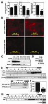
Anti-amyloidogenic activity of lovastatin is mediated via inhibition of APP endocytosis. To examine the effect of lovastatin on APP endocytosis, hippocampal neuronal cells were treated with lovastatin (LOVA; 5µM/36hrs) and cell surface and internalized APP levels were measured by fluorometric (A) or fluoromicroscopic (B) methods. For this the cells were incubated with APP antibody (22C11) at 4°C and fixed for staining of cell surface APP or further incubated at 37°C and washed with acid PBS and fixed for staining of internalized APP. To examine the effect of lovastatin on endosomal APP levels, APP and Rab5 levels in post nuclear (S1), light mitochondrial (P15), post-light mitochondrial (S15), microsomal (P100) and post microsomal (S100) fractions were measured by Western immunoblot analysis (C). For analysis of APP association with Rab5 in the microsomes, the P100 fractions were immunoprecipitated (IP) by using anti-Rab5 polyclonal antibody and co-precipitated APP levels were quantified by Western immunoblot using 22C11 APP antibody (D). For analysis of subcellular distribution of APP, subcellular organelles were purified by Optiprep® density gradient centrifugation (E). As loading control of density gradient centrifugation, β-actin levels were measured from post nuclear lysates (lysate) by Western immunoblot analysis. In addition, no correlation between protein levels of APP or EEA1 and β-actin in each subcellular fraction indicated that the observed alteration in APP and EEA1 levels may not due to protein amounts in each fractions. To examine the involvement of APP endocytosis in the regulation of Aβ40 generation (F) and APP distribution in lipid raft fractions (G), the cells were treated with phenylarsine oxide (PAO; 200nM/18hrs). For confirmation of equal amount protein loading in the process of lipid raft extraction, β-actin levels were measured from post nuclear lysates (lysate) by Western immunoblot analysis. All experiments were performed at least three times. The vertical bar indicates the standard error of mean (** P < 0.01, *** p < 0.001 compared to control group; CON).
Fig. 8
Fig. 8
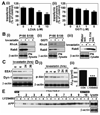
Pleiotropic roles of lovastatin in the down-regulation of endocytosis may be involved in the reduction of APP endocytosis. A. To examine the effect of lovastatin on clathrin-mediated transferrin endocytosis, hippocampal neuron cells were treated with lovastatin (5µM/36hrs) (i) or GGTI-298 (GGTI; 1µM/36hrs) (ii) and incubated with fluorescent transferrin. The endocytosis of fluorescent transferrin was measured as described in materials and methods. B. To characterize the possible involvement of geranylgeranylation small GTPases in lovastatin-mediated antiamyloidogenesis, hippocampal neuron cells were treated with lovastatin (i) or GGTI-298 (GGTI) (ii), and the levels of membrane (P100) or cytoplasm (S100) associated RhoA and Rab5 were measured. For analysis of geranylgeranylated Rab5 protein levels, post-nuclear cell lysates were extracted by using Triton X-114 (iii). Rab5 level in water soluble (Water sol.) or Triton X-114 soluble (TX-114 sol.) represent ungeranylgeranylated or geranylgeranylated Rab5, respectively. The β-actin levels in post-nuclear cell lysates (PN-lysate) were analyzed for loading controls of differential centrifugation and TX-114 extraction. C. To characterize the possible involvement of EEA1 (an early endosomal antigen 1) and dynamin-I in lovastatin-mediated anti-amyloidogenesis, the levels of these proteins from post-nuclear fractions were measured after treatment with lovastatin (5µM/36 hrs). To characterize the possible involvement of phosphatidylinositol 3-kinase (PI3-K)/Akt pathway in lovastatin-mediated anti-amyloidogenesis, the effect of lovastatin on Akt phosphorylation (D-i) and the effect of LY294002 (a PI3-K inhibitor; 20µM/24hr) on Aβ40 levels in culture media (D-ii) or distribution of APP in lipid raft fractions (E) were analyzed. For confirmation of equal amount protein loading in the process of lipid raft extraction, β-actin levels were measured from post nuclear lysates (lysate) by Western immunoblot analysis. All experiments were done at least three times and showed the same tendency. The vertical bar indicates the standard error of mean (* P < 0.05, ** P < 0.01, *** p < 0.001 compared to control group).

Similar articles

Cited by

References

    1. Andersson M, Elmberger PG, Edlund C, Kristensson K, Dallner G. Rates of cholesterol, ubiquinone, dolichol and dolichyl-P biosynthesis in rat brain slices. FEBS Lett. 1990;269:15–18. - PubMed
    1. Antonny B. SNARE filtering by dynamin. Cell. 2004;119:581–582. - PubMed
    1. Bamberger ME, Harris ME, McDonald DR, Husemann J, Landreth GE. A cell surface receptor complex for fibrillar beta-amyloid mediates microglial activation. J Neurosci. 2003;23:2665–2674. - PMC - PubMed
    1. Bonifacino JS, Traub LM. Signals for sorting of transmembrane proteins to endosomes and lysosomes. Annu Rev Biochem. 2003;72:395–447. - PubMed
    1. Brown DA, Rose JK. Sorting of GPI-anchored proteins to glycolipid-enriched membrane subdomains during transport to the apical cell surface. Cell. 1992;68:533–544. - PubMed

Publication types

MeSH terms