. 2008 Feb;20(2):337-52.
doi: 10.1105/tpc.107.052142. Epub 2008 Feb 5.

The Arabidopsis phytochrome-interacting factor PIF7, together with PIF3 and PIF4, regulates responses to prolonged red light by modulating phyB levels

Affiliations

The Arabidopsis phytochrome-interacting factor PIF7, together with PIF3 and PIF4, regulates responses to prolonged red light by modulating phyB levels

Pablo Leivar et al. Plant Cell. 2008 Feb.

Abstract

We show that a previously uncharacterized Arabidopsis thaliana basic helix-loop-helix (bHLH) phytochrome interacting factor (PIF), designated PIF7, interacts specifically with the far-red light-absorbing Pfr form of phyB through a conserved domain called the active phyB binding motif. Similar to PIF3, upon light exposure, PIF7 rapidly migrates to intranuclear speckles, where it colocalizes with phyB. However, in striking contrast to PIF3, this process is not accompanied by detectable light-induced phosphorylation or degradation of PIF7, suggesting that the consequences of interaction with photoactivated phyB may differ among PIFs. Nevertheless, PIF7 acts similarly to PIF3 in prolonged red light as a weak negative regulator of phyB-mediated seedling deetiolation. Examination of pif3, pif4, and pif7 double mutant combinations shows that their moderate hypersensitivity to extended red light is additive. We provide evidence that the mechanism by which these PIFs operate on the phyB signaling pathway under prolonged red light is through maintaining low phyB protein levels, in an additive or synergistic manner, via a process likely involving the proteasome pathway. These data suggest that the role of these phyB-interacting bHLH factors in modulating seedling deetiolation in prolonged red light may not be as phy-activated signaling intermediates, as proposed previously, but as direct modulators of the abundance of the photoreceptor.


Figures
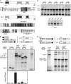
Figure 1.
Figure 1.
bHLH072 Is a phy-Interacting Protein, Now Designated PIF7. (A) Alignment of PIF7, PIF3, PIF4, and bHLH016 full-length proteins. Two different aligned regions are shown: amino acids 1 to 26 of PIF7, including the APB domain (top); and amino acids 152 to 270 of PIF7, including the nuclear localization signal (NLS), the bHLH region, and the Q-rich motif present in PIF7 (bottom). Schematic diagrams of the PIF7 protein domains are also shown. (B) PIF7 binds conformer-specifically to phyB Pfr. The GAL4 transcriptional activation domain (GAD) alone or fusions of GAD to full-length PIF7 (PIF7:GAD) or PIF3 (GAD:PIF3) were used as bait in in vitro coimmunoprecipitation assays. The Pfr and Pr forms of phyB were used as prey. All proteins were synthesized as [35S]Met-labeled products in TnT reactions. Schematic diagrams at top show the design of the experiment, and the SDS-PAGE separations of the pellet fractions and the phyB input (5%) are shown in the center. A quantification of the binding, expressed as percentage of the total phyB input recovered as phyB bound to the bait, is shown at bottom. Data from a representative experiment are shown. (C) PIF7 binds selectively to phyB. A GAD:full-length PIF7 fusion protein was used as bait in in vitro coimmunoprecipitation assays. The Pfr and Pr forms of phy (A to E) were used as prey. All proteins were synthesized as [35S]Met-labeled products in TnT reactions. SDS-PAGE separations of the pellet fractions and the inputs (5%) are shown. (D) The APB motif mediates the binding of PIF7 to phyB Pfr. GAD fusions with full-length wild-type or mutated PIF7 were used as bait in in vitro coimmunoprecipitation assays. Mutated PIF7 carries E8A and G14A substitution mutations in the APB motif. Pfr and Pr forms of phyB were used as prey. All proteins were synthesized as [35S]Met-labeled products in TnT reactions. Schematic diagrams at top show the design of the experiment, and the SDS-PAGE separations of the pellet fractions and the phyB input (5%) are shown at bottom.
Figure 2.
Figure 2.
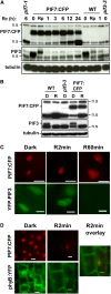
PIF7 Is a Light-Stable Nuclear Protein That Colocalizes with phyB in Rc-Induced Early Speckles. (A) Immunoblot of protein extracts from T2 segregating 35S:PIF7:CFP seedlings. Seedlings were grown in the dark for 2 d (0), transferred to Rc for a 30-s pulse, followed by 10 min of dark (Rp), or transferred to Rc for 1, 3, 6, 12, or 24 h. PIF7-specific polyclonal antibody (top) and a PIF3-specific polyclonal antibody (middle) were used as probes. As controls, protein extracts from the wild type, pif7-1, and pif3-3 are included. Tubulin was used as a loading control (bottom). n.s., nonspecific, cross-reacting bands. (B) Immunoblot of protein extracts from 35S:PIF7:CFP seedlings. Seedlings were grown for 4 d in the dark (D) or in Rc at 6.2 μmol·m−2·s−1 (R). PIF7-specific polyclonal antibody (top) and a PIF3-specific polyclonal antibody (middle) were used as probes. As controls, protein extracts from the wild type and pif3-3 are included. Tubulin was used as a loading control (bottom). (C) Epifluorescence imaging of YFP (displayed in green) and CFP (displayed in red) fluorescence in nuclei of Arabidopsis transgenic seedlings expressing PIF7:CFP (top panels) or YFP:PIF3 (bottom panels). Seedlings were grown in the dark for 4 d and then either maintained in darkness (Dark), exposed to Rc for 2 min (R2min) provided by passing the microscope light through a red filter (140 μmol·m−2·s−1), or exposed to Rc for 60 min of 7 μmol·m−2·s−1 (R60min). Images were recorded within 2 min of termination of the Rc treatments. Bars = 10 μm. (D) Epifluorescence imaging of YFP (displayed in green) and CFP (displayed in red) fluorescence in nuclei of Arabidopsis transgenic seedlings coexpressing PIF7:CFP (top panels) and phyB:YFP (bottom panels) in the phyB-9 background. Seedlings were grown in the dark for 4 d and then either maintained in darkness (Dark) or exposed to Rc for 2 min (R2min) provided by passing the microscope light through a red filter (140 μmol·m−2·s−1). Images were recorded within 2 min of termination of the Rc treatments. The overlay of the PIF7:YFP and the phyB:CFP signals after a Rc irradiation is indicated in yellow in the merged image (right panel). Bars = 10 μm.
Figure 3.
Figure 3.
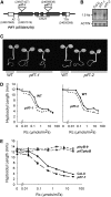
T-DNA Insertional pif7 Mutants Are Hypersensitive to Rc. (A) Mutations identified in the Arabidopsis PIF7 gene (At5g61270). T-DNA insertions in pif7-1 and pif7-2 are indicated at positions 24,657,427 and 24,656,638, respectively, on chromosome V. The location of the bHLH domain is indicated. The coding region is represented by shaded boxes, and the untranslated regions are represented by open boxes (exons), with introns shown as short lines connecting the boxes. (B) RNA gel blots of 4-d-old Rc-grown (10 μmol·m−2·s−1) wild-type Columbia (Col-0) and pif7 mutant seedlings probed with a PIF7-specific probe. As a loading control, the blot was reprobed with an ACTIN-specific probe. (C) Visual phenotype of 4-d-old Rc-grown (10 μmol·m−2·s−1) wild-type and pif7-1 and pif7-2 mutant seedlings. (D) Fluence response curves for hypocotyl length in 4-d-old wild-type and pif7-1 (left) and pif7-2 (right) seedlings grown in Rc. Mean and se values are representative of at least 25 seedlings for each light treatment. (E) Fluence response curves for hypocotyl length in 4-d-old wild-type (Col-0), phyB-9, pif7-1, and pif7 phyB seedlings grown in Rc. Mean and se values are representative of at least 25 seedlings for each light treatment.
Figure 4.
Figure 4.
Hypersensitivity of pif3, pif4, and pif7 to Prolonged Rc Is Additive. (A) Visual phenotypes of 4-d-old Rc-grown (10 μmol·m−2·s−1) wild-type and pif seedlings. (B) Hypocotyl length in 4-d-old Rc-grown wild-type and pif seedlings. Rc fluence rate was 0.8 μmol·m−2·s−1 for seedlings in all panels except for the middle right panel, in which it was 0.3 μmol·m−2·s−1. Mean and se values are representative of at least 25 seedlings.
Figure 5.
Figure 5.
phyB Levels Are Elevated in pif3, pif4, and pif7 Single and Double Mutant Seedlings Grown under Prolonged Rc. phyA and phyB levels in 4-d-old wild-type Col-0 and pif mutant seedlings. Set 1 and set 2 correspond to two sets of seeds that were harvested at different times. (A) Representative immunoblots of protein extracts of wild-type and pif seedlings grown in Rc (0.9 μmol·m−2·s−1) and probed with phyB-specific monoclonal antibodies (top) and phyA-specific monoclonal antibodies (bottom). Tubulin was used as a loading control (middle). (B) phyA and phyB protein levels in protein extracts of 4-d-old wild-type and pif seedlings grown in Rc (0.9 μmol·m−2·s−1). phyA and phyB signal normalized to the tubulin was quantified using NIH Image software in at least three independent biological replicates. To ensure the linearity of phyA, phyB, and tubulin chemiluminescence signals, a protein extract dilution curve was always run and exposed in parallel. Fold increase for each set was calculated considering the corresponding wild-type value as 1. Mean fold increase value were calculated from three (set 1), four (set 2 except for pif7), or six (wild type and pif7) independently grown replicates. Two additional technical replicates quantified in the Odyssey infrared imaging system were included in the analysis of phyB levels of set 1 seeds. Immunoblots corresponding to all of the replicates analyzed are shown in (A) and Supplemental Figure 10A online. Error bars indicate se, based on biological replicates. (C) Representative immunoblots of protein extracts of 4-d-old wild-type and pif seedlings. Seedlings were grown in the dark, and extracts were probed with phyB-specific monoclonal antibodies (top). Tubulin was used as a loading control (bottom). (D) Correlation of the level of phyB protein with the hypocotyl length displayed by wild-type and pif seedlings grown in Rc (0.9 μmol·m−2·s−1). Plotted hypocotyl and phyB values are shown as mean fold increase over the wild type. The best-fit curve by eye is shown. Error bars indicate se, based on biological replicates.
Figure 6.
Figure 6.
Changes of phyB Levels in pif3 Mutant Seedlings Precede the Changes in Hypocotyl Elongation. (A) Time course of hypocotyl length and phyB level in wild-type Col-0 and pif3-3 mutant seedlings. Seedlings were germinated and grown as described in Methods (Monte et al., 2004). Germination was induced for 3 h in white light followed by 21 h of darkness, and the seedlings were then moved to Rc (7.7 μmol·m−2·s−1) for up to 36, 48, 60, or 96 h after inducing germination before measuring hypocotyl length (top) and phyB levels (bottom). Mean values of hypocotyl length are representative of at least 25 seedlings. phyB normalized to tubulin (phyB/tub) was quantified as in Figure 5B, and mean values from three biological replicates are shown (bottom). Bars represent se. P values from a t test analysis for differences between wild-type and pif3-3 mutant seedlings are shown. (B) Immunoblots corresponding to all of the replicates (rep.) analyzed in (A), bottom panel, are shown.

Similar articles

Cited by

References

    1. Alonso, J.M., et al. (2003). Genome-wide insertional mutagenesis of Arabidopsis thaliana. Science 301 653–657. - PubMed
    1. Al-Sady, B., Kikis, E.A., Monte, E., and Quail, P.H. (2008). Mechanistic duality of transcription factor function in phytochrome signaling. Proc. Natl. Acad. Sci. USA, in press. - PMC - PubMed
    1. Al-Sady, B., Ni, W., Kircher, S., Schafer, E., and Quail, P.H. (2006). Photoactivated phytochrome induces rapid PIF3 phosphorylation as a prelude to proteasome-mediated degradation. Mol. Cell 23 439–446. - PubMed
    1. Aukerman, M.J., Hirschfeld, M., Wester, L., Weaver, M., Clack, T., Amasino, R.M., and Sharrock, R.A. (1997). A deletion in the PHYD gene of the Arabidopsis Wassilewskija ecotype defines a role for phytochrome D in red/far-red light sensing. Plant Cell 9 1317–1322. - PMC - PubMed
    1. Bailey, P.C., Martin, C., Toledo-Ortiz, G., Quail, P.H., Huq, E., Heim, M.A., Jakoby, M., Werber, M., and Weisshaar, B. (2003). Update on the basic helix-loop-helix transcription factor gene family in Arabidopsis thaliana. Plant Cell 15 2497–2502. - PMC - PubMed

Publication types

MeSH terms

LinkOut - more resources