

STIM1 is a MT-plus-end-tracking protein involved in remodeling of the ER
- PMID: 18249114
- PMCID: PMC2600655
- DOI: 10.1016/j.cub.2007.12.050
STIM1 is a MT-plus-end-tracking protein involved in remodeling of the ER
Abstract
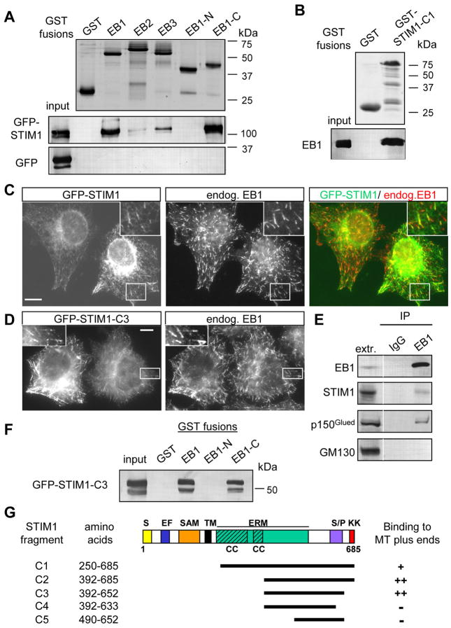
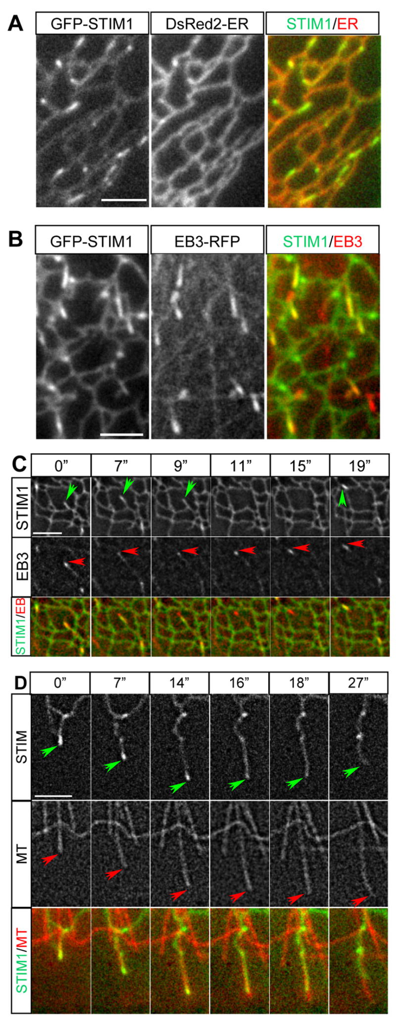
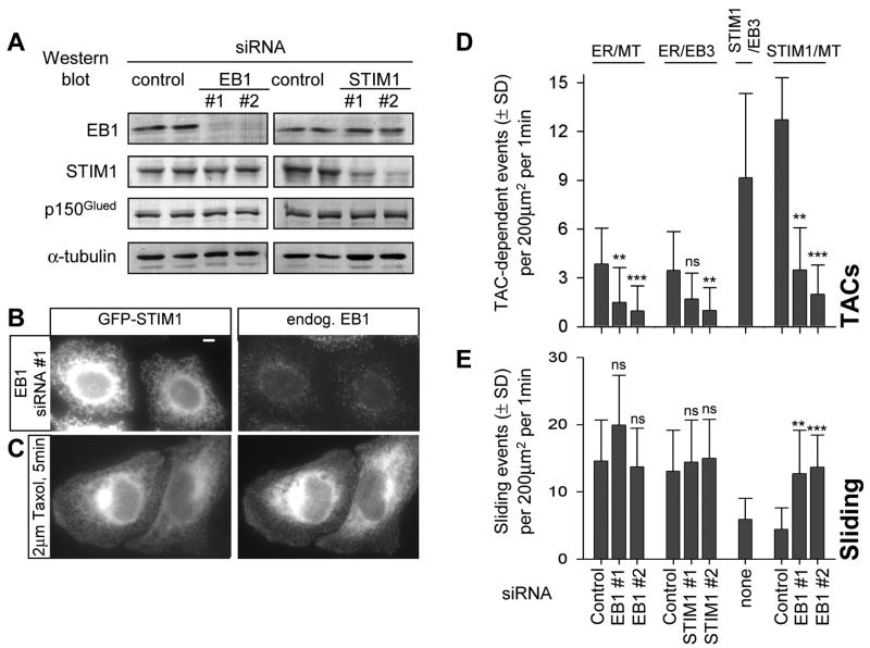
Stromal interaction molecule 1 (STIM1) is a transmembrane protein that is essential for store-operated Ca(2+) entry, a process of extracellular Ca(2+) influx in response to the depletion of Ca(2+) stores in the endoplasmic reticulum (ER) (reviewed in [1-4]). STIM1 localizes predominantly to the ER; upon Ca(2+) release from the ER, STIM1 translocates to the ER-plasma membrane junctions and activates Ca(2+) channels (reviewed in [1-4]). Here, we show that STIM1 directly binds to the microtubule-plus-end-tracking protein EB1 and forms EB1-dependent comet-like accumulations at the sites where polymerizing microtubule ends come in contact with the ER network. Therefore, the previously observed tubulovesicular motility of GFP-STIM1 [5] is not a motor-based movement but a traveling wave of diffusion-dependent STIM1 concentration in the ER membrane. STIM1 overexpression strongly stimulates ER extension occurring through the microtubule "tip attachment complex" (TAC) mechanism [6, 7], a process whereby an ER tubule attaches to and elongates together with the EB1-positive end of a growing microtubule. Depletion of STIM1 and EB1 decreases TAC-dependent ER protrusion, indicating that microtubule growth-dependent concentration of STIM1 in the ER membrane plays a role in ER remodeling.
Figures

Similar articles
-
Phosphorylation of STIM1 at ERK1/2 target sites regulates interaction with the microtubule plus-end binding protein EB1.J Cell Sci. 2013 Jul 15;126(Pt 14):3170-80. doi: 10.1242/jcs.125054. Epub 2013 May 17. J Cell Sci. 2013. PMID: 23687376
-
EB1 binding restricts STIM1 translocation to ER-PM junctions and regulates store-operated Ca2+ entry.J Cell Biol. 2018 Jun 4;217(6):2047-2058. doi: 10.1083/jcb.201711151. Epub 2018 Mar 21. J Cell Biol. 2018. PMID: 29563214 Free PMC article.
-
STIM1 Is Required for Remodeling of the Endoplasmic Reticulum and Microtubule Cytoskeleton in Steering Growth Cones.J Neurosci. 2019 Jun 26;39(26):5095-5114. doi: 10.1523/JNEUROSCI.2496-18.2019. Epub 2019 Apr 25. J Neurosci. 2019. PMID: 31023836 Free PMC article.
-
Ca2+ signaling and STIM1.Prog Biophys Mol Biol. 2010 Sep;103(1):51-8. doi: 10.1016/j.pbiomolbio.2010.02.004. Epub 2010 Mar 11. Prog Biophys Mol Biol. 2010. PMID: 20226808 Review.
-
Molecular physiology and pathophysiology of stromal interaction molecules.Exp Biol Med (Maywood). 2018 Mar;243(5):451-472. doi: 10.1177/1535370218754524. Epub 2018 Jan 24. Exp Biol Med (Maywood). 2018. PMID: 29363328 Free PMC article. Review.
Cited by
-
How filopodia respond to calcium in the absence of a calcium-binding structural protein: non-channel functions of TRP.Cell Commun Signal. 2022 Aug 26;20(1):130. doi: 10.1186/s12964-022-00927-y. Cell Commun Signal. 2022. PMID: 36028898 Free PMC article.
-
L-type Ca2+ channel activation of STIM1-Orai1 signaling remodels the dendritic spine ER to maintain long-term structural plasticity.Proc Natl Acad Sci U S A. 2024 Aug 27;121(35):e2407324121. doi: 10.1073/pnas.2407324121. Epub 2024 Aug 23. Proc Natl Acad Sci U S A. 2024. PMID: 39178228
-
Kif18A and chromokinesins confine centromere movements via microtubule growth suppression and spatial control of kinetochore tension.Dev Cell. 2012 May 15;22(5):1017-29. doi: 10.1016/j.devcel.2012.02.013. Dev Cell. 2012. PMID: 22595673 Free PMC article.
-
The intracellular transport and secretion of calumenin-1/2 in living cells.PLoS One. 2012;7(4):e35344. doi: 10.1371/journal.pone.0035344. Epub 2012 Apr 13. PLoS One. 2012. PMID: 22514732 Free PMC article.
-
Endoplasmic Reticulum-Plasma Membrane Contact Sites as an Organizing Principle for Compartmentalized Calcium and cAMP Signaling.Int J Mol Sci. 2021 Apr 29;22(9):4703. doi: 10.3390/ijms22094703. Int J Mol Sci. 2021. PMID: 33946838 Free PMC article. Review.
References
Publication types
MeSH terms
Substances
Grants and funding
LinkOut - more resources
Full Text Sources
Other Literature Sources
Miscellaneous
