

doi: 10.1186/1476-4598-7-8.
An integrative model for recurrence in ovarian cancer
Affiliations
- PMID: 18211683
- PMCID: PMC2248209
- DOI: 10.1186/1476-4598-7-8
Item in Clipboard
An integrative model for recurrence in ovarian cancer
Mol Cancer.
.
No abstract available
Figures
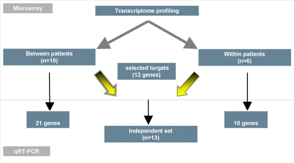
Flow chart of our study design. 2 cohorts were used in this study: In the first one, we selected a homogeneous series of primary and recurrent serous papillary adenocarcinomas from different patients(Between patient cohort). The second cohort consisted of 3 paired ovarian cancers (primary and recurrent samples coming from the same patient) but of different histology (Within patient cohort). Selected genes identified from microarray experiments were validated for both cohorts and a subset of these genes (n = 12) were validated in an independent set (test set) of 13 serous papillary adenocarcinomas using TaqMan® PCR.
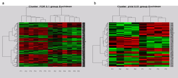
Hierarchical cluster heatmaps demonstrating distinct patterns of gene expression between primary and recurrent ovarian tumors. (a) Heatmap of the ovarian tumors in cohort 1 based on the FDR0.1 list with the primary clustering on the left and the recurrent samples on the right. Vertical bars represent the samples and the horizontal bars represent the genes. Green bars reflect downregulated genes and red bars upregulated genes. (b) Heat map discriminating recurrent (left) and primary (right) ovarian tumours in cohort 2 based on the p0.01 list. P, primary tumours; R, recurrent tumors.
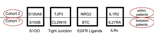
Gene families involved in the molecular regulation of recurrence in ovarian cancer. Some of the upregulated genes in recurrent compared to primary ovarian carcinomas that we validated in cohort 2 belong in the same gene families with some of the upregulated genes validated in cohort 1. Upregulation of tight junction proteins and EGFR ligands, development of a cytokine response via interleukin receptors and intracellular signaling via calcium binding S100 proteins seem to contribute to the "recurrent" signature and possibly have a role in drug resistance.
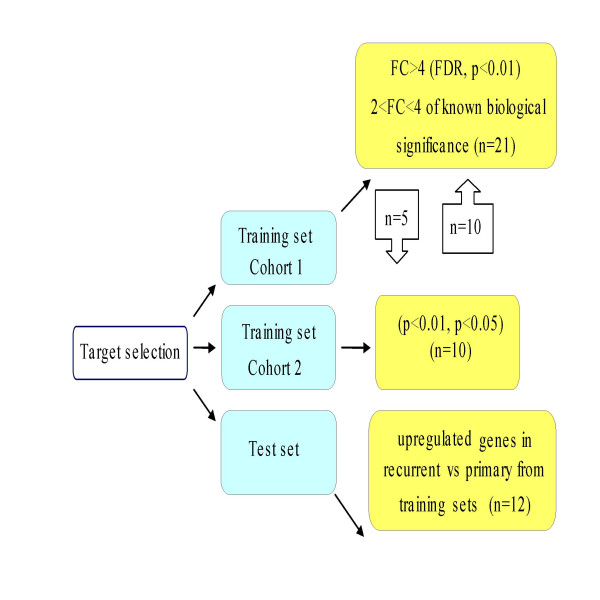
TaqMan® PCR validation of target genes identified in both training and test sets. Gene selection for TaqMan® validation was based on the most differentially expressed genes from the p and FDR value list with a fold change > 4 but also included genes that had a 2–4 fold change and also some genes involved in the most differentially expressed pathways. Priority was given to selection of genes upregulated in recurrent compared to primary samples, which might provide "recurrence" signatures in ovarian cancer. Upregulated genes validated in both cohorts were alternatively interrogated (external validation) and further advanced for validation in the test set. Independent validation on a test set refers to completely distinct samples of serous histology that were not previously employed in marker development (n = number of gene targets selected for validation).
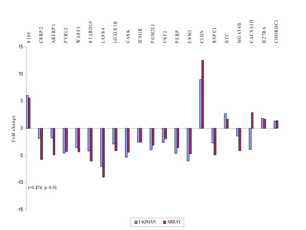
TaqMan® PCR validation of microarray experiments in cohort 1. The fold changes in the arrays were plotted against the relative quantitation from TaqMan® in recurrent vs primary tumours. The TaqMan® values are displayed in blue and the array results in red. Spearman correlation r showed high concordance between the 2 experiments.
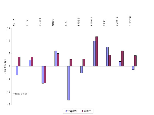
TaqMan® PCR validation of microarray experiments in cohort 2. A similar concordance was observed as in cohort 1.
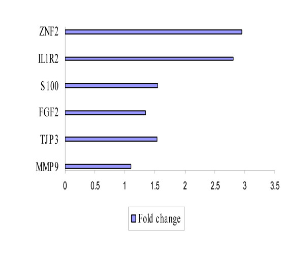
External validation of a subset of upregulated genes in cohort 2 that validated in cohort 1. Bars indicate the relative overexpression of target genes in recurrent vs primary tumors. IL1R2 and ZNF218 gave the best distinction between recurrent and primary tumors with greater than twofold changes.
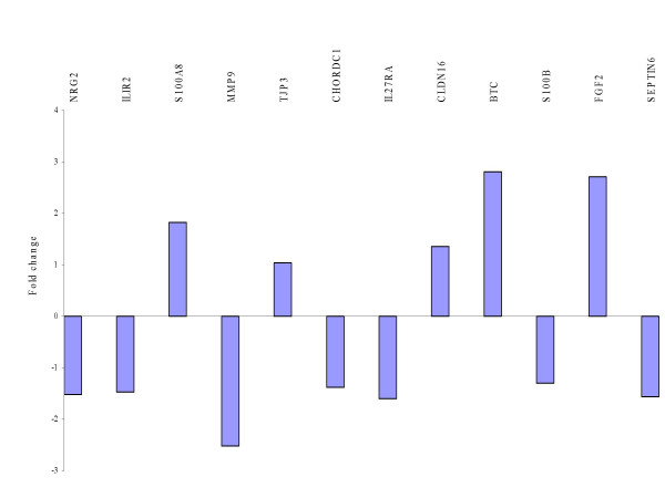
Independent TaqMan® PCR validation of a set of selected genes from both cohorts in a test set of serous papillary adenocarcinomas of varying grade and stage. BTC and FGF2 provided the best distinction between recurrent and primary tumours with fold changes of 2.8 and 2.71 respectively.
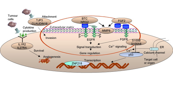
An integrative model for recurrence in ovarian cancer. Schematic representation of putative genes and gene families involved in the recurrence of ovarian carcinomas. According to our current working concept, tumour cells during relapse produce adhesion molecules to mediate attachment and invasion via co-overexpression of matrix metalloproteinases, cytokines and inflammatory mediators to stimulate survival and a variety of growth factors bound to their cognate receptors to fully proliferate in order to confront and modulate their immediate environment, which they must eventually overtake.
Similar articles
-
Gene expression profiling of epithelial ovarian tumours correlated with malignant potential.Mol Cancer. 2004 Oct 7;3:27. doi: 10.1186/1476-4598-3-27. Mol Cancer. 2004. PMID: 15471544 Free PMC article.
-
Independent test set performance in the prediction of early relapse in ovarian cancer with gene expression profiles.Clin Cancer Res. 2005 Nov 1;11(21):7958-9; author reply 7959. doi: 10.1158/1078-0432.CCR-05-1216. Clin Cancer Res. 2005. PMID: 16278422 No abstract available.
-
Potential role of miR-9 and miR-223 in recurrent ovarian cancer.Mol Cancer. 2008 Apr 28;7:35. doi: 10.1186/1476-4598-7-35. Mol Cancer. 2008. PMID: 18442408 Free PMC article.
-
Modelling genetic and clinical heterogeneity in epithelial ovarian cancers.Carcinogenesis. 2011 Oct;32(10):1540-9. doi: 10.1093/carcin/bgr140. Epub 2011 Aug 22. Carcinogenesis. 2011. PMID: 21859834
-
Microarray-based gene expression studies in ovarian cancer.Cancer Control. 2011 Jan;18(1):8-15. doi: 10.1177/107327481101800102. Cancer Control. 2011. PMID: 21273975 Review.
Cited by
-
Interleukin-1 Receptor Type 2 Acts with c-Fos to Enhance the Expression of Interleukin-6 and Vascular Endothelial Growth Factor A in Colon Cancer Cells and Induce Angiogenesis.J Biol Chem. 2015 Sep 4;290(36):22212-24. doi: 10.1074/jbc.M115.644823. Epub 2015 Jul 24. J Biol Chem. 2015. PMID: 26209639 Free PMC article.
-
Glomus tumors in neurofibromatosis type 1: genetic, functional, and clinical evidence of a novel association.Cancer Res. 2009 Sep 15;69(18):7393-401. doi: 10.1158/0008-5472.CAN-09-1752. Epub 2009 Sep 8. Cancer Res. 2009. PMID: 19738042 Free PMC article.
-
Targeting AMPK signaling in combating ovarian cancers: opportunities and challenges.Acta Biochim Biophys Sin (Shanghai). 2016 Apr;48(4):301-17. doi: 10.1093/abbs/gmv128. Epub 2016 Jan 12. Acta Biochim Biophys Sin (Shanghai). 2016. PMID: 26764240 Free PMC article. Review.
-
Immune-Related Genes' Prognostic, Therapeutic and Diagnostic Value in Ovarian Cancer Immune-Related Gene Biomarker in Ovarian Cancer.Cancer Control. 2023 Jan-Dec;30:10732748231168756. doi: 10.1177/10732748231168756. Cancer Control. 2023. PMID: 37078136 Free PMC article.
-
Pre-Treatment of platinum resistant ovarian cancer cells with an MMP-9/MMP-2 inhibitor prior to cisplatin enhances cytotoxicity as determined by high content screening.Int J Mol Sci. 2013 Jan 22;14(1):2085-103. doi: 10.3390/ijms14012085. Int J Mol Sci. 2013. PMID: 23340649 Free PMC article.
References
-
- Parkin DM, Bray F, Ferlay J, Pisani P. Global cancer statistics, 2002. CA Cancer J Clin. 2005;55:74–108. - PubMed
-
- The National Cancer Registry Ireland http://www.ncri.ie
-
- DiSaia PJ, Creasman WT, eds . Epithelial ovarian cancer. Clinical gynecologic oncology. St. Louis: Mosby Year Book; 1997. pp. 282–350.
-
- Kikkawa F, Nawa A, Ino K, Shibata K, Kajiyama H, Nomura S. Advances in treatment of epithelial ovarian cancer. Nagoya J Med Sci. 2006;68:19–26. - PubMed
MeSH terms
LinkOut - more resources
Full Text Sources
Medical
