. 1991 Apr;1(1):41-53.

Expression of histone-U1 snRNA chimeric genes: U1 promoters are compatible with histone 3' end formation

Affiliations

Expression of histone-U1 snRNA chimeric genes: U1 promoters are compatible with histone 3' end formation

D R Pilch et al. Gene Expr. 1991 Apr.

Abstract

Chimeric genes which fuse the mouse histone H2a gene and the mouse U1b gene were constructed and introduced into CHO cells by cotransfection. In the UH genes, the U1b gene promoter and the start of the U1b gene were fused to the H2a gene in the 5' untranslated region. In the HU genes, the U1b 3' end was inserted into the 3' untranslated region of the H2a gene replacing the normal histone 3' end. Transcripts from the UH genes initiated at the start of the U1 gene and ended at the normal histone 3' end. Transcripts from the HU chimeric genes did not end at the U1 3' end but extended at least 80 nucleotides further and had heterogeneous 3' ends. Placing both a U1 snRNA promoter and a U1 snRNA 3' end around a histone coding region resulted in transcripts which initiate and terminate at the appropriate U1 ends. These results are consistent with previous reports that formation of the U1 3' ends require U1 promoters, but indicate that the histone 3' end can be formed on transcripts initiating at U1 promoters. The transcripts initiated at the U1 start site and ending at the histone 3' end are present on polyribosomes and show proper posttranscriptional regulation.


Figures
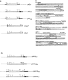
Figure 1
Figure 1
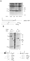
The histone-Ul chimeric genes. A. The parent histone H2a-614 gene is shown (pH2a), with the 5′ and 3′ untranslated regions and the coding region indicated. The U5H and U50H gene have 226 nucleotides of 5′ flanking sequence and 5 or 50 nucleotides of the mouse Ulb coding region. The HU117 and HU156 genes have the Ul 3′ end starting at nucleotides 156 and 117 respectively, attached in the 3′ untranslated region of the histone H2a614 gene. B. The UHU156 and UHU117 genes have both the Ul promoter and Ul 3′ end flanking the histone gene. The UHHU genes have the Ul 3′ end starting at nucleotide 156 or 117 of the coding region attached 25 nucleotides 3′ of the end of the U5H gene. C. The sequences of the Ul-histone junctions in the U5H, U50H, HU117, HU156 genes are given. The coding region sequences of the Ul genes are underlined, as are the distal sequence element (DSE), proximal sequence element (PSE) of the Ul promoter, the ATG codon of the H2a-614 gene, and the 3′ end of the histone mRNA. The polylinker sequences are indicated. The restriction sites used are: formula image AvaI; formula image NarI.
Figure 2
Figure 2
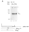
Expression of the UH genes. A. 5 μg RNA from duplicate samples of CHO cells transfected with the U5H, U50H or H2a-614 genes were analyzed by SI nuclease mapping using the H2a-614 gene labeled at the 3′ end of the Aval site at codon 20 as a probe. The protected fragments are: H2aH–protection of the endogenous hamster H2a mRNAs. H2aM–protection of the RNA expressed from the transfected genes. Band P is the probe band. The RNA samples are indicated under each lane. In the lanes labeled CHO, RNAs from untransfected CHO cells were analyzed. The marker (last lane on the right) is pUC18 digested with HpaII. B. The same RNA samples as in A were analyzed by S1 nuclease mapping using either the U50H gene (lanes 1–4), or the U5H gene (lanes 5–8) labeled at the 5′ end of the Aval site as a probe. Lanes 1, 2, and 7 are analyses of RNAs from cells transfected with the U50H gene, Lanes 3, 5, 6 are analyses of RNAs from cells transfected with the U5H gene. Lanes 4 and 8 are analyses of RNAs from cells transfected with the H2a-614 gene. Lanes 2 and 7 and lanes 3 and 6 contain the same RNA samples. The protected fragments are: H2an-protection of the endogenous hamster H2a mRNAs; H2aM - protection of the mouse H2a-614 mRNA; U15–protection of the RNA from the U5H gene starting at the U1 RNA start site; U115–protection of RNA from the U150H gene starting at the U1 start site. P indicates the undigested probe fragment. In lanes 3 and 7, the RNAs are mapped to the point of divergence between the U15H and U150H genes. Lane M is marker pUC18 digested with HpaII.
Figure 3
Figure 3
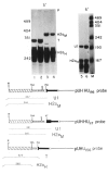
A. Localization of the U5H transcripts. CHO cells transfected with the U5H gene were fractionated into nuclear (N), polysomal (P) and cytosol (S) fractions and equal cell equivalents of each fraction assayed by S1 nuclease mapping using the H2a-614 gene labeled at the 3′ end of the Aval site at codon 20 as a probe as in Figure 2A. The protected fragments are: H2aH–protection of the endogenous hamster H2a mRNAs; H2aM–protection of the RNA expressed from the U5H gene. B. Effect of hydroxyurea on U5H RNA. Duplicate cultures of CHO cells transfected with the U5H gene were treated with 5mM hydroxyurea for one hour and RNA prepared from control (lanes 1 and 3) and treated cultures (lanes 2 and 4). 5 μg total cell RNA was assayed by SI nuclease mapping using the U5H gene labeled at the 5′ end of the Narl site at codon 43 as a probe. Lane M is marker pUC18 digested with HpaII. The protected fragments are: H2aH–protection to the ATG codon by the endogenous hamster H2a mRNAs; U1–protection to the first nucleotide in the U1 sequence of the U5H gene.
Figure 4
Figure 4
Expression of the HU genes. A. Duplicate preparations of RNA from cells transfected with the HU117 genes (lanes 1 and 2) or the HU156 genes (lanes 3 and 4) were analyzed by S1 nuclease mapping using the H2a-614 gene labeled at the 3′ end of the Aval site as a probe. This probe will map all transcripts from the HU genes which extend more than 60 nucleotides past the 3′ end of the histone coding region as a single protected fragment (labeled T). The other protected fragment (H2aH) is due to protection of the endogenous hamster H2a mRNAs; B. Duplicate preparations of RNA (5 μg) from cells transfected with the HU117 (lanes 1 and 2) or the HU156 genes (lanes 5 and 6) were analyzed by S1 nuclease mapping using the HU117 (lanes 1–4) or HU156 (lanes 5–8) genes labeled at the 3′ end of the Narl site at codon 43 as probes. RNA from cells transfected with the H2a-614 gene was analyzed in lanes 3, 4, 7, and 8. The arrows indicate the expected positions of transcripts ending at the U1 3′ end (332 and 376 nucleotides respectively). Band P is undigested probe. C. RNAs from cells transfected with the HU156; or the HU117 were analyzed by SI nuclease mapping using the probes UHU117PL or UHUnvPL respectively. The RNAs used are indicated beneath the lanes. These probes have a polylinker insert either 112 or 82 nucleotides after the U1 3′ end, which allows the mapping of longer heterogenous read-through transcripts from these genes as a single protected fragment (labeled RT). The position of the protected fragment which would correspond to the U1 3′ end is indicated. The protected fragment labeled H2aH corresponds to protection of the endogenous hamster H2a mRNAs. Lane M is marker pUC18 digested with HpaII. P is the position of the undigested probe.
Figure 4
Figure 4
Expression of the HU genes. A. Duplicate preparations of RNA from cells transfected with the HU117 genes (lanes 1 and 2) or the HU156 genes (lanes 3 and 4) were analyzed by S1 nuclease mapping using the H2a-614 gene labeled at the 3′ end of the Aval site as a probe. This probe will map all transcripts from the HU genes which extend more than 60 nucleotides past the 3′ end of the histone coding region as a single protected fragment (labeled T). The other protected fragment (H2aH) is due to protection of the endogenous hamster H2a mRNAs; B. Duplicate preparations of RNA (5 μg) from cells transfected with the HU117 (lanes 1 and 2) or the HU156 genes (lanes 5 and 6) were analyzed by S1 nuclease mapping using the HU117 (lanes 1–4) or HU156 (lanes 5–8) genes labeled at the 3′ end of the Narl site at codon 43 as probes. RNA from cells transfected with the H2a-614 gene was analyzed in lanes 3, 4, 7, and 8. The arrows indicate the expected positions of transcripts ending at the U1 3′ end (332 and 376 nucleotides respectively). Band P is undigested probe. C. RNAs from cells transfected with the HU156; or the HU117 were analyzed by SI nuclease mapping using the probes UHU117PL or UHUnvPL respectively. The RNAs used are indicated beneath the lanes. These probes have a polylinker insert either 112 or 82 nucleotides after the U1 3′ end, which allows the mapping of longer heterogenous read-through transcripts from these genes as a single protected fragment (labeled RT). The position of the protected fragment which would correspond to the U1 3′ end is indicated. The protected fragment labeled H2aH corresponds to protection of the endogenous hamster H2a mRNAs. Lane M is marker pUC18 digested with HpaII. P is the position of the undigested probe.
Figure 5
Figure 5
Expression of the UHU genes. A. 10 μg RNA from CHO cells transfected with the UHU156 genes (lanes 1–3) or the UHU117 genes (lanes 4 and 5) was analyzed by S1 nuclease mapping using the appropriate homologous gene labeled at the 3′ end of the Narl site at codon 43 as a probe. The protected fragments are: H2aH- protection to the TAA codon by the endogenous hamster H2a mRNAs; U1–protection by transcripts from the UHU genes extending to the normal U1 3′ end. P is the undigested probe band. The positions of the marker fragments, pUC18 digested with HpaII, are indicated. B. 10 μg total cell RNA from CHO cells transfected with the UHU156 genes (lane 2) or the UHU117 genes (lanes 3 and 4) was analyzed by S1 nuclease mapping using the UHU156PL (lanes 1 and 2) or UHU117PL (lanes 3 and 4) probes described in Fig. 4C labeled at the 3′ end of the Narl site at codon 43. These probes diverge from the transfected gene after the U1 3′ end as in Figure 4B, allowing detection of all longer transcripts as a single protected fragment (RT). The protected fragments are: H2aH–protection to the TAA codon by the endogenous hamster H2a mRNAs; U1–protection by transcripts from the UHU genes extending to the normal U1 3′ end; RT–protection by transcripts which extend past the U1 3′ end. Lane 1 is 1 μg RNA from untransfected CHO cells. C. 5 μg total cell RNA, cells transfected with the UHU117 gene (lane 1), the UHU156 gene (lane 2), or untransfected CHO cells (lane 3) were analyzed by S1 nuclease mapping using the U5H gene labeled at the 5′ end of the NarI site (codon 43) as a probe. The protected fragments are: H2aH–protection to the ATG codon by the endogenous hamster H2a mRNAs; U1–protection by transcripts from the UHU genes extending to the normal U1 5′ end. Lane M is marker pUC18 digested with HpaII. D. 10 μg total cell RNA from cells transfected with the UHU117 genes (lanes 1 and 2) or the CHO-156C cells transfected with the UHU156 gene (lanes 3 and 4) were analyzed by S1 nuclease mapping using the UHU117 gene (lanes 1–3) or the UHU156 gene (lane 5) labeled at the 3′ end of the NarI site (codon 43) as a probe. The protected fragments are labeled as in Figure 5B. The fragment labeled T is due to protection of all transcripts from the UHU156 gene to the point of divergence of the UHU156 and the UHU117 genes. Band P is the undigested probe. Lane 4 is marker pUC18 digested with HpaII. The same CHO-156C [UHU156] (lane 6) and UHU117 RNAs (lane 7) were analyzed by S1 nuclease mapping using the U5H gene labeled at the 5′ end of the NarI site as a probe. The protected fragments are labeled as in Figure 5C. The fragment labeled U1RT results from protection by transcripts which initiated upstream from the normal U1 start site. Band P is undigested probe DNA. The position of the marker fragments is shown. The hamster histone RNAs were not detected efficiently in lane 7 for unknown reasons.
Figure 5
Figure 5
Expression of the UHU genes. A. 10 μg RNA from CHO cells transfected with the UHU156 genes (lanes 1–3) or the UHU117 genes (lanes 4 and 5) was analyzed by S1 nuclease mapping using the appropriate homologous gene labeled at the 3′ end of the Narl site at codon 43 as a probe. The protected fragments are: H2aH- protection to the TAA codon by the endogenous hamster H2a mRNAs; U1–protection by transcripts from the UHU genes extending to the normal U1 3′ end. P is the undigested probe band. The positions of the marker fragments, pUC18 digested with HpaII, are indicated. B. 10 μg total cell RNA from CHO cells transfected with the UHU156 genes (lane 2) or the UHU117 genes (lanes 3 and 4) was analyzed by S1 nuclease mapping using the UHU156PL (lanes 1 and 2) or UHU117PL (lanes 3 and 4) probes described in Fig. 4C labeled at the 3′ end of the Narl site at codon 43. These probes diverge from the transfected gene after the U1 3′ end as in Figure 4B, allowing detection of all longer transcripts as a single protected fragment (RT). The protected fragments are: H2aH–protection to the TAA codon by the endogenous hamster H2a mRNAs; U1–protection by transcripts from the UHU genes extending to the normal U1 3′ end; RT–protection by transcripts which extend past the U1 3′ end. Lane 1 is 1 μg RNA from untransfected CHO cells. C. 5 μg total cell RNA, cells transfected with the UHU117 gene (lane 1), the UHU156 gene (lane 2), or untransfected CHO cells (lane 3) were analyzed by S1 nuclease mapping using the U5H gene labeled at the 5′ end of the NarI site (codon 43) as a probe. The protected fragments are: H2aH–protection to the ATG codon by the endogenous hamster H2a mRNAs; U1–protection by transcripts from the UHU genes extending to the normal U1 5′ end. Lane M is marker pUC18 digested with HpaII. D. 10 μg total cell RNA from cells transfected with the UHU117 genes (lanes 1 and 2) or the CHO-156C cells transfected with the UHU156 gene (lanes 3 and 4) were analyzed by S1 nuclease mapping using the UHU117 gene (lanes 1–3) or the UHU156 gene (lane 5) labeled at the 3′ end of the NarI site (codon 43) as a probe. The protected fragments are labeled as in Figure 5B. The fragment labeled T is due to protection of all transcripts from the UHU156 gene to the point of divergence of the UHU156 and the UHU117 genes. Band P is the undigested probe. Lane 4 is marker pUC18 digested with HpaII. The same CHO-156C [UHU156] (lane 6) and UHU117 RNAs (lane 7) were analyzed by S1 nuclease mapping using the U5H gene labeled at the 5′ end of the NarI site as a probe. The protected fragments are labeled as in Figure 5C. The fragment labeled U1RT results from protection by transcripts which initiated upstream from the normal U1 start site. Band P is undigested probe DNA. The position of the marker fragments is shown. The hamster histone RNAs were not detected efficiently in lane 7 for unknown reasons.
Figure 6
Figure 6
Expression of the UHHU genes. 10 μg total cell RNA from cells transfected with the UHH156 gene lanes 1, 4, and 5) or the UHHU117 gene (lanes 2, 3, and 6) were analyzed by S1 nuclease mapping. The UHU156 gene labeled at the 3′ end of the Narl site as a probe in lanes 1 and 2. The UHHU117 or UHHU156 gene labeled at the Narl site was used as a probe in lanes 3 and 4 respectively. The 5′ ends of the genes were analyzed in lanes 5 and 6 using the U5H gene labeled at the 5′ end of the NarI site as a probe as in Figure 3B. Lane M is marker pUC18 digested with HpaII. The protected fragments are: H2aH-protection of the endogenous hamster H2a mRNAs; H2aM–protection by transcripts extending to the 3′ end of the mouse H2a–614 gene; T–protection by all transcripts from the UHHU genes to the point of divergence between the UHHU gene and the UHU156 gene; U1–protection by transcripts initiating at the U1 start site. Band P is unprotected probe DNA. A schematic of the S1 nuclease assays is shown below the figure.

Similar articles

Cited by

References

    1. Ach R. A. and Weiner A. M. (1987), Mol Cell Biol 7, 2070–2079. - PMC - PubMed
    1. Birchmeier C., Schümperli D., Sconzo G., and Birnstiel M. L. (1984), Proc Nad Acad Sci USA 81, 1057–1061. - PMC - PubMed
    1. Birnstiel M. L., Busslinger M., and Strub K. (1985), Cell 41, 349–359. - PubMed
    1. Chaney W. G., Howard D. R., Pollard J. W., Sallustio S., and Stanley P. (1986), Somatic Cell Mol Genet 12, 237–244. - PubMed
    1. Chodchoy N., Levine B. J., Sprecher C., Skoultchi A. I., and Marzluff W. F. (1987), Mol Cell Biol 7, 1039–1047. - PMC - PubMed

Publication types

LinkOut - more resources