. 2008 Jan 9;3(1):e1414.
doi: 10.1371/journal.pone.0001414.

The Drosophila cytosine-5 methyltransferase Dnmt2 is associated with the nuclear matrix and can access DNA during mitosis

Affiliations

The Drosophila cytosine-5 methyltransferase Dnmt2 is associated with the nuclear matrix and can access DNA during mitosis

Matthias Schaefer et al. PLoS One. .

Abstract

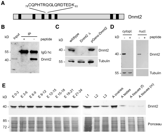
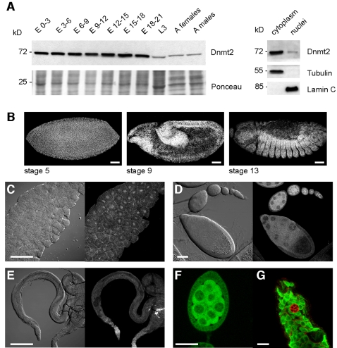
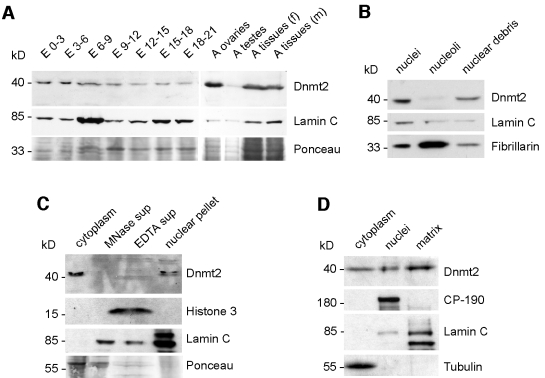
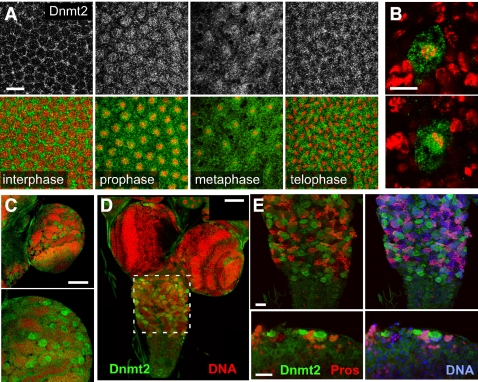
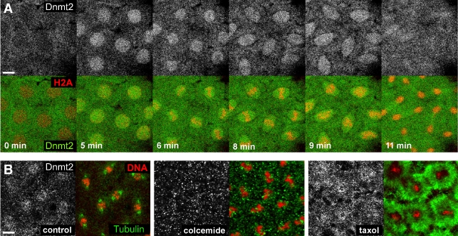
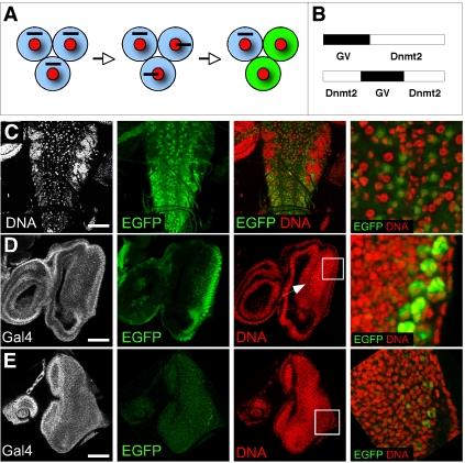
Cytosine-5 methyltransferases of the Dnmt2 family are highly conserved in evolution and their biological function is being studied in several organisms. Although all structural DNA methyltransferase motifs are present in Dnmt2, these enzymes show a strong tRNA methyltransferase activity. In line with an enzymatic activity towards substrates other than DNA, Dnmt2 has been described to localize to the cytoplasm. Using molecular and biochemical approaches we show here that Dnmt2 is both a cytoplasmic and a nuclear protein. Sub-cellular fractionation shows that a significant amount of Dnmt2 is bound to the nuclear matrix. Sub-cellular localization analysis reveals that Dnmt2 proteins are enriched in actively dividing cells. Dnmt2 localization is highly dynamic during the cell cycle. Using live imaging we observed that Dnmt2-EGFP enters prophase nuclei and shows a spindle-like localization pattern during mitotic divisions. Additional experiments suggest that this localization is microtubule dependent and that Dnmt2 can access DNA during mitotic cell divisions. Our results represent the first comprehensive characterization of Dnmt2 proteins on the cellular level and have important implications for our understanding of the molecular activities of Dnmt2.


Conflict of interest statement

Competing Interests: The authors have declared that no competing interests exist.

Figures

Figure 1
Figure 1. Establishment of Dnmt2-specific antibodies and characterization of Dnmt2 expression during Drosophila development.
(A) Sequence of the epitope recognized by polyclonal antibodies against Dnmt2 peptide spanning amino acids 78–93. (B) Affinity-purified anti-Dnmt2 antibodies efficiently immunoprecipitate Dnmt2 from whole cell protein extracts. (C) Anti-Dnmt2 specifically detects a 40 kDa protein. Whole-cell protein extracts from wildtype, homozygous mutants (Dnmt2Δ) and rescued mutants (pGenoDnmt2) were blotted and probed with anti-Dnmt2. (D) Early embryonic tissue (0–6 hr AEL) was fractionated, blotted as cytoplasmic and nuclear fractions and probed with anti-Dnmt2 and anti-〈-tubulin as control. (E) Developmental Western blot. Wildtype cytoplasmic protein extract from various embryonic, larval and adult stages were blotted and probed with anti-Dnmt2. Ponceau staining of blot is shown as loading control. hc-heavy chain, cytopl.-cytoplasmic, nucl.-nuclear, E-embryo, L1-first instar larvae, L2-second instar larvae, L3-third instar larvae, A-adult, f-female, m-males.
Figure 2
Figure 2. Tissue specific expression of Dnmt2.
(A) Left panel: developmental Western blot of pGeno-Dnmt2-EGFP animals. Whole protein extract from embryonic, larval and adult stages were blotted and probed with anti-Dnmt2. Ponceau staining is shown as a loading control. Right panel: Dnmt2-EGFP is expressed both in the cytoplasm and in nuclei. Fractionation efficiency was confirmed by probing the blot for cytoplasmic 〈-tubulin and nuclear Lamin C. Antibodies against EGFP reveal Dnmt2 in developing embryos (B), third instar larval salivary glands (C), ovaries (D) and testes (E). (F) Dnmt2-EGFP in a developing cyst of the female germline is mostly cytoplasmic. Nuclear staining can be observed in nurse cell nuclei. (G) Dnmt2-EGFP in the male germline is ubiquitous in all cells with the exception of somatic hub cells (marked by Armadillo, red). Scale bars: (B) 100 µm; (C) 10 µm; (D) 50 µm; (E) 100 µm; (F) 20 µm; (G) 10 µm.
Figure 3
Figure 3. Dnmt2 is present in nuclei and resides in an insoluble part of the nuclear matrix.
(A) Developmental Western blot. Purified nuclei from embryonic and adult tissues were urea extracted, blotted and probed with anti-Dnmt2. Ponceau staining is shown as a loading control, lamin C staining is shown as a control for the enrichment of nuclear proteins. (B) Dnmt2 is not enriched in nucleoli. Sucrose density fractionation of nucleoli followed by blotting and tracing of nucleolar material using anti-Fibrillarin shows that Dnmt2 is part of other nuclear structures. (C) Dnmt2 is not enriched in nucleosomes. Micrococcal nuclease digest of purified nuclei releases histone H3 (EDTA supernatant), but not Dnmt2. The EDTA extracted nuclei still contain Dnmt2 suggesting association of Dnmt2 with non-chromatin structures. (D) Dnmt2 is associated with the nuclear matrix. Nuclear Dnmt2 is insoluble after DNAse digest, followed by high salt extraction and can only be solublilzed using urea extraction. CP-190 is included as a peripheral and lamin C as an integral nuclear matrix interacting protein. E-embryo, A-adult, f-female, m-males, MNase-micrococcal nuclease, sup-supernatant.
Figure 4
Figure 4. Dnmt2 localizes to mitotically active cells.
(A) pGeno-Dnmt2-EGFP localizes to mitotic nuclei during prophase and localizes to midbody structures in telophase as visualized by anti-EGFP staining. Pictures show the distribution of Dnmt2-EGFP (gray, upper panel) and merged images (lower panel) with Dnmt2-EGFP in green and DNA in red. (B) Ectopic Dnmt2-EGFP localizes around metaphase chromosomes. Cell-type specific expression of UAS-Dnmt2-EGFP using asense-GAL4 causes Dnmt2 to localize to nuclear structures during mitosis. Ectopic protein localizes to prophase chromatin of CNS neuroblasts (upper panel) and aggregates densely around the mitotic metaphase plate (lower panel). (C) pGeno-Dnmt2-EGFP localizes to mitotic nuclei during larval development (upper panel). Frequently dividing optic lobe neuroblasts are enriched for Dnmt2-EGFP expression as visualized by anti-EGFP staining (lower panel). (D–E) Ventral ganglion neuroblasts express high levels of Dnmt2-EGFP, whereas ganglion mother cells (marked by Prospero, red, in E), which give rise to differentiated neurons and glia, do not express Dnmt2-EGFP. Scale bars: (A) 10 µm; (B) 10 µm; (C, D) 50 µm; (E, F) 20 µm; (G) 10 µm.
Figure 5
Figure 5. Dnmt2 localization to mitotic nuclei is dynamic and microtubuli-dependent.
(A) Nuclear division cycle 10 embryos expressing pUbq-Dnmt2-EGFP and His2Av-mRFP1 were mounted on an open coverslip and viewed by dual wavelength time-lapse LSCM. Selected frames are shown for Dnmt2-EGFP (gray, upper panel) and as merged images (lower panel) with Dnmt2-EGFP in green and His2A-mRFP1 in red. A movie of the sequence is available as supporting material. (B) 1–3 h old embryos expressing pGeno-Dnmt2-EGFP were collected and fixed immediately (left panel), treated with colcemide (middle panel) or with taxol (right panel) in order to disrupt or stabilize microtubuli, respectively. Metaphase chromosomes are shown with Dnmt2 (gray), DNA (red) and 〈-tubulin (green). While Dnmt2-EGFP is maintained around metaphase plates in taxol-treated embryos, the protein is delocalized in colcemide-treated embryos. Scale bars: (A) 10 µm; (B) 5 µm.
Figure 6
Figure 6. Nuclear access reporter assay using GAL4:VP16-Dnmt2 fusion proteins.
(A) Schematic representation of the nuclear access reporter assay. Ubiquitous expression of the GAL4:VP16 (GV) fusion proteins (black bar) causes the expression of EGFP only in some cells (green) due to selective nuclear access of the fusion protein. (B) GV-fusion proteins. GV-Dnmt2 (upper panel), Dnmt2-GV-Dnmt2 fusion expressing GV in the context of a split Dnmt2 protein (lower panel) which served as control. (C) GAL4:VP16-Dnmt2 expression in third instar larval brains causes widespread UAS-EGFP expression in the ventral ganglion. The magnification shows that regularly spaced cells (neuroblasts) express EGFP (green). (D) Ubiquitous GAL4:VP16-Dnmt2 in third instar larval eye discs causes specific UAS-EGFP expression behind the morphogentic furrow (arrow). Magnification of a field of cells shows robust expression in differentiated photoreceptor cells. (E) The control construct (GAL4:VP16 in a split Dnmt2 protein) causes background levels of UAS-EGFP expression. GAL4 (gray), EGFP (green), DNA (red). Scale bars: (C) 100 µm; (D, E) 70 µm.

Similar articles

Cited by

References

    1. Rozenski J, Crain PF, McCloskey JA. The RNA Modification Database: 1999 update. Nucleic Acids Res. 1999;27:196–197. - PMC - PubMed
    1. Klose RJ, Bird AP. Genomic DNA methylation: the mark and its mediators. Trends Biochem Sci. 2006;31:89–97. - PubMed
    1. Jones PA, Baylin SB. The epigenomics of cancer. Cell. 2007;128:683–692. - PMC - PubMed
    1. Yoder JA, Bestor TH. A candidate mammalian DNA methyltransferase related to pmt1p of fission yeast. Hum Mol Genet. 1998;7:279–284. - PubMed
    1. Okano M, Xie S, Li E. Dnmt2 is not required for de novo and maintenance methylation of viral DNA in embryonic stem cells. Nucleic Acids Res. 1998;26:2536–2540. - PMC - PubMed

Publication types

LinkOut - more resources