

Peptide mimic of the HIV envelope gp120-gp41 interface
- PMID: 18178220
- PMCID: PMC2265733
- DOI: 10.1016/j.jmb.2007.12.001
Peptide mimic of the HIV envelope gp120-gp41 interface
Abstract
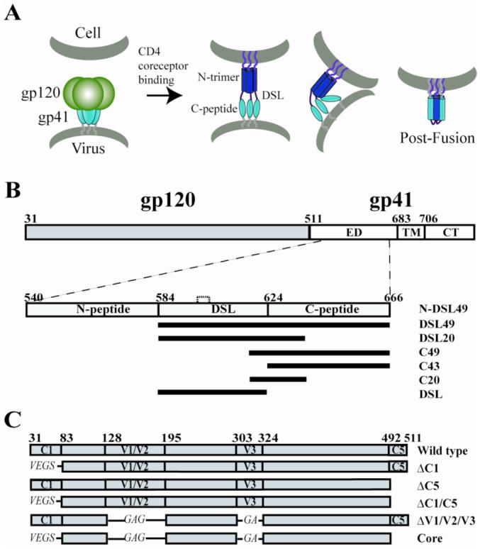
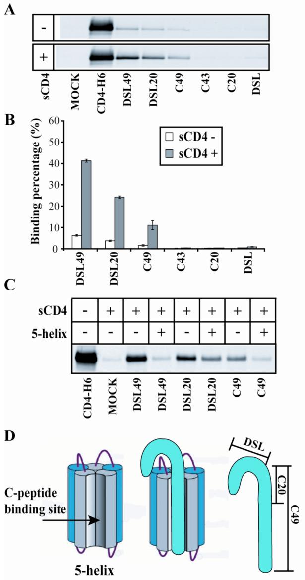
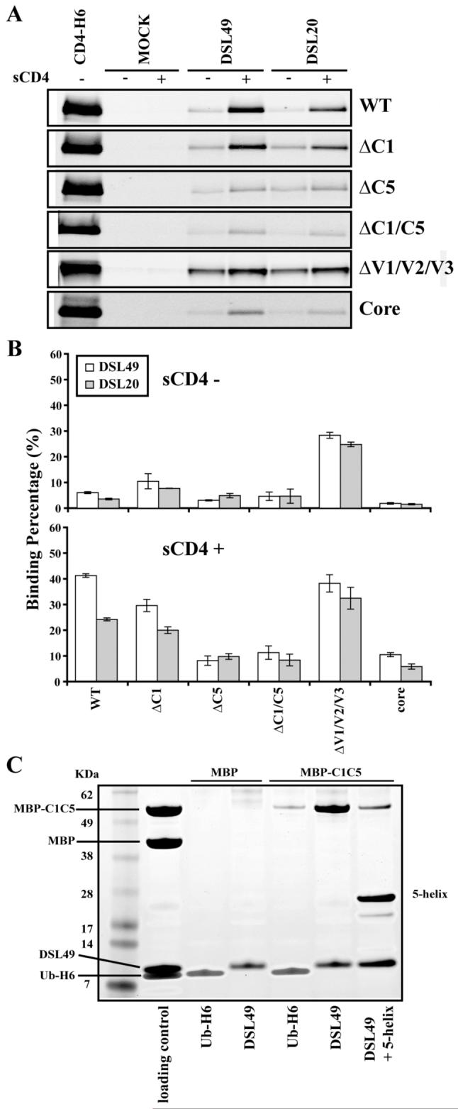
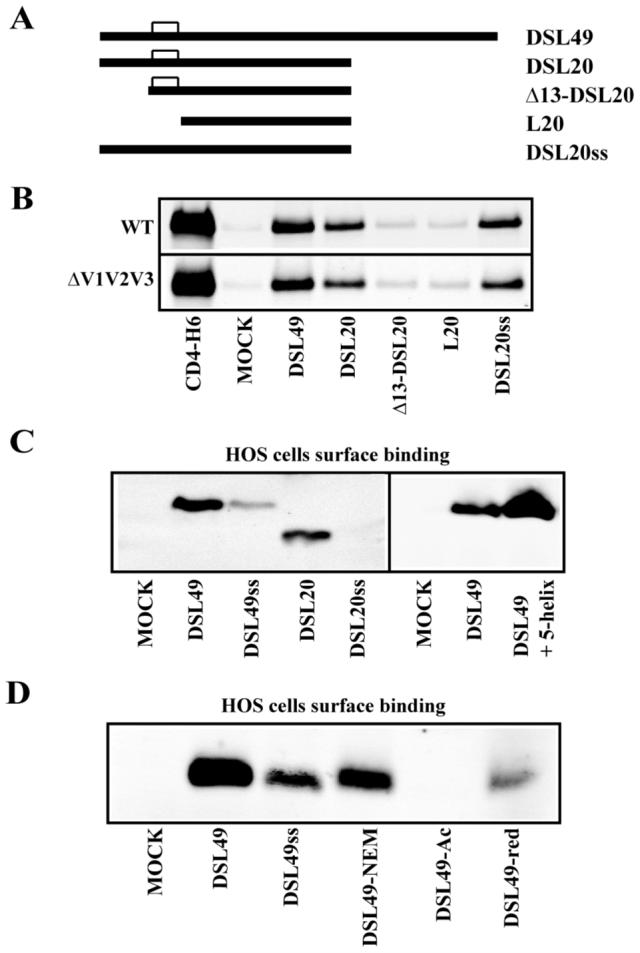
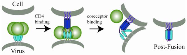
The human immunodeficiency virus envelope glycoprotein (Env) is composed of surface (gp120) and transmembrane (gp41) subunits, which are noncovalently associated on the viral surface. Human immunodeficiency virus Env mediates viral entry after undergoing a complex series of conformational changes induced by interaction with cellular CD4 and a chemokine coreceptor. These changes propagate from gp120 to gp41 via the gp120-gp41 interface, ultimately exposing gp41 and allowing it to form the trimer-of-hairpins structure that provides the driving force for membrane fusion. Key unresolved questions about the gp120-gp41 interface include the specific regions of gp41 and gp120 involved, the mechanism by which receptor and coreceptor-binding-induced conformational changes in gp120 are communicated to gp41, how trimer-of-hairpins formation is prevented in the prefusogenic gp120-gp41 complex, and, ultimately, the structure of the prefusion gp120-gp41 complex. Here, we develop a biochemical model system that mimics a key portion of the gp120-gp41 interface in the prefusogenic state. We find that a gp41 fragment containing the disulfide bond loop and C-peptide region binds primarily to the gp120 C5 region and that this interaction is incompatible with trimer-of-hairpins formation. Based on these data, we propose that in prefusogenic Env, gp120 sequesters the gp41 C-peptide region away from the N-trimer region, preventing trimer-of-hairpins formation until coreceptor binding disrupts this interface. This model system is a valuable tool for studying the gp120-gp41 complex, conformational changes induced by CD4 and coreceptor binding, and the mechanism of membrane fusion.
Figures
Similar articles
-
Cryo-EM structure of a CD4-bound open HIV-1 envelope trimer reveals structural rearrangements of the gp120 V1V2 loop.Proc Natl Acad Sci U S A. 2016 Nov 15;113(46):E7151-E7158. doi: 10.1073/pnas.1615939113. Epub 2016 Oct 31. Proc Natl Acad Sci U S A. 2016. PMID: 27799557 Free PMC article.
-
HIV-1 gp41 Residues Modulate CD4-Induced Conformational Changes in the Envelope Glycoprotein and Evolution of a Relaxed Conformation of gp120.J Virol. 2018 Jul 31;92(16):e00583-18. doi: 10.1128/JVI.00583-18. Print 2018 Aug 15. J Virol. 2018. PMID: 29875245 Free PMC article.
-
Effects of the I559P gp41 change on the conformation and function of the human immunodeficiency virus (HIV-1) membrane envelope glycoprotein trimer.PLoS One. 2015 Apr 7;10(4):e0122111. doi: 10.1371/journal.pone.0122111. eCollection 2015. PLoS One. 2015. PMID: 25849367 Free PMC article.
-
CD4 activation of HIV fusion.Int J Cell Cloning. 1992 Nov;10(6):323-32. doi: 10.1002/stem.5530100603. Int J Cell Cloning. 1992. PMID: 1281202 Review.
-
Progress in targeting HIV-1 entry.Drug Discov Today. 2005 Aug 15;10(16):1085-94. doi: 10.1016/S1359-6446(05)03550-6. Drug Discov Today. 2005. PMID: 16182193 Review.
Cited by
-
Novel ring structure in the gp41 trimer of human immunodeficiency virus type 1 that modulates sensitivity and resistance to broadly neutralizing antibodies.J Virol. 2009 Aug;83(15):7728-38. doi: 10.1128/JVI.00688-09. Epub 2009 May 27. J Virol. 2009. PMID: 19474108 Free PMC article.
-
Membrane-anchored HIV-1 N-heptad repeat peptides are highly potent cell fusion inhibitors via an altered mode of action.PLoS Pathog. 2009 Jul;5(7):e1000509. doi: 10.1371/journal.ppat.1000509. Epub 2009 Jul 10. PLoS Pathog. 2009. PMID: 19593361 Free PMC article.
-
HIV-1 fusion is blocked through binding of GB Virus C E2-derived peptides to the HIV-1 gp41 disulfide loop [corrected].PLoS One. 2013;8(1):e54452. doi: 10.1371/journal.pone.0054452. Epub 2013 Jan 22. PLoS One. 2013. PMID: 23349893 Free PMC article.
-
Structure based antibody-like peptidomimetics.Pharmaceuticals (Basel). 2012 Feb 16;5(2):209-35. doi: 10.3390/ph5020209. Pharmaceuticals (Basel). 2012. PMID: 24288089 Free PMC article.
-
Asymmetric deactivation of HIV-1 gp41 following fusion inhibitor binding.PLoS Pathog. 2009 Nov;5(11):e1000674. doi: 10.1371/journal.ppat.1000674. Epub 2009 Nov 26. PLoS Pathog. 2009. PMID: 19956769 Free PMC article.
References
-
- Wyatt R, Sodroski J. The HIV-1 envelope glycoproteins: fusogens, antigens, and immunogens. Science. 1998;280:1884–8. - PubMed
-
- Eckert DM, Kim PS. Mechanisms of viral membrane fusion and its inhibition. Annu Rev Biochem. 2001;70:777–810. - PubMed
-
- Wu L, Gerard NP, Wyatt R, Choe H, Parolin C, Ruffing N, Borsetti A, Cardoso AA, Desjardin E, Newman W, Gerard C, Sodroski J. CD4-induced interaction of primary HIV-1 gp120 glycoproteins with the chemokine receptor CCR-5. Nature. 1996;384:179–83. - PubMed
-
- Chen B, Vogan EM, Gong H, Skehel JJ, Wiley DC, Harrison SC. Structure of an unliganded simian immunodeficiency virus gp120 core. Nature. 2005;433:834–41. - PubMed
Publication types
MeSH terms
Substances
Grants and funding
LinkOut - more resources
Full Text Sources
Other Literature Sources
Research Materials
Miscellaneous
