. 2007 Dec 31;179(7):1385-98.
doi: 10.1083/jcb.200708106. Epub 2007 Dec 24.

DNA-activated protein kinase functions in a newly observed S phase checkpoint that links histone mRNA abundance with DNA replication

Affiliations

DNA-activated protein kinase functions in a newly observed S phase checkpoint that links histone mRNA abundance with DNA replication

Berndt Müller et al. J Cell Biol. .

Erratum in

  • J Cell Biol. 2008 Feb 25;180(4):843

Abstract

DNA and histone synthesis are coupled and ongoing replication is required to maintain histone gene expression. Here, we expose S phase-arrested cells to the kinase inhibitors caffeine and LY294002. This uncouples DNA replication from histone messenger RNA (mRNA) abundance, altering the efficiency of replication stress-induced histone mRNA down-regulation. Interference with caffeine-sensitive checkpoint kinases ataxia telangiectasia and Rad3 related (ATR)/ataxia telangiectasia mutated (ATM) does not affect histone mRNA down- regulation, which indicates that ATR/ATM alone cannot account for such coupling. LY294002 potentiates caffeine's ability to uncouple histone mRNA stabilization from replication only in cells containing functional DNA-activated protein kinase (DNA-PK), which indicates that DNA-PK is the target of LY294002. DNA-PK is activated during replication stress and DNA-PK signaling is enhanced when ATR/ATM signaling is abrogated. Histone mRNA decay does not require Chk1/Chk2. Replication stress induces phosphorylation of UPF1 but not hairpin[corrected]-binding protein/stem-loop binding protein at S/TQ sites, which are preferred substrate recognition motifs of phosphatidylinositol 3-kinase-like kinases, which indicates that histone mRNA stability may be directly controlled by ATR/ATM- and DNA-PK-mediated phosphorylation of UPF1.


Figures

Figure 1.
Figure 1.
Caffeine abrogates the replication checkpoint and inhibits histone mRNA decay induced by replication stress in HeLa cells. (A) Caffeine abrogates the replication checkpoint. (top) Asynchronous HeLa cells were pulsed with CldU for 20 min and incubated without drugs (mock), in the presence of APH alone, or in the presence of both APH and caffeine. At different times (typically 6–24 h) after the CldU pulse, cells were washed free of drugs and pulsed with IdU for 20 min. CldU or IdU incorporation was visualized by immunofluorescence confocal microscopy. Data are single optical sections showing the initial CldU pulse (green) incorporated into an early replication pattern and, at times thereafter, IdU pulses (red) either colocalized (yellow) with the CldU pulse in the presence of APH or being incorporated into progressively later replication patterns in mock-treated cells and in cells where the replication checkpoint has been abrogated by caffeine addition. Bar, 10 μm. (middle) Graph summarizes typical data and shows the fraction of those cells with an early replication pattern at the first pulse proceeding into the indicated pattern (early, early/mid, mid, or late) visualized by the second pulse after each treatment. (bottom) Nocodazole-arrested HeLa cells were released into drug-free medium for 14 h. The cells were either mock treated (control) or treated with 5 mM caffeine alone (+caffeine) for 1 h before being treated for a further 2 h either with (+HU and +caffeine +HU) or without (control and +caffeine) HU to induce replication stress. Cell lysates were then immunoblotted for Chk1. (B) Caffeine inhibits histone mRNA decay. Asynchronous HeLa cells were treated with the indicated caffeine concentration or mock treated. After 1 h, 2 mM HU or water was added and incubation was continued for 30 min followed by isolation of RNA. RNA was analyzed by Northern blotting using probes detecting histone H2B mRNA, H3 mRNA, or GAPDH mRNA. Histone mRNA levels were standardized with respect to GAPDH mRNA levels and the graph shows histone mRNA levels in HU-treated cells expressed as a percentage of the level in untreated cells.
Figure 2.
Figure 2.
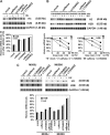
Inhibition of ATR signaling does not interfere with replication stress–induced histone mRNA decay. (A) Time course of HU-induced decay of histone mRNA in cells expressing kd-ATR. Expression of wt-ATR or kd-ATR was either induced (+Dox) or not (−Dox) by treatment of U2OS/ATR and U2OS/kd-ATR cells with doxycycline. Cells were treated with 2 mM HU and RNA was isolated at indicated times after HU addition. Histone RNA was analyzed as in Fig. 1. Changes in histone H2A, H2B, and H3 mRNA levels were compared with the level at 0 min. (B) Inactivation of ATR signaling using cells expressing kd-ATR suppresses phosphorylation and activation of Chk1 in response to replication stress. Expression of kd-ATR was either induced (+kd-ATR) or not (−kd-ATR) by treatment of U2OS/kd-ATR cells with doxycycline or buffer. Cells were then treated with 50 μg/ml APH for 16 h to induce replication stress. Subsequently, cell lysates were subjected to a Chk1 immunoprecipitation kinase assay (top) or immunoblotted using Chk1 antibodies (bottom, anti–phospho-Ser345 and total Chk1 antibody). (C) Ablation of ATR by RNAi does not interfere with HU-induced histone mRNA decay. Asynchronous HeLa cells were transfected with siRNA targeting ATR, control siRNA 1 (targeting luciferase), control siRNA 2 (targeting a nonrelevant gene), or mock transfected. Cell lysates were immunoblotted for ATR and nucleolin (top). 32 h after transfection, cells were treated with or without 2 mM HU for 0, 20, or 60 min or left untreated for 60 min, and then lysed for RNA analysis (middle). Histone H3 mRNA levels were analyzed as in Fig. 1. Graph (bottom) shows changes in H3 mRNA levels compared with the level at 0 min. In parallel, cell lysates were immunoblotted for ATR (to confirm knockdown) and nucleolin (loading; left). Data are expressed as mean ± range for duplicate experiments.
Figure 3.
Figure 3.
Replication stress induces efficient, caffeine-sensitive histone mRNA decay in cells lacking ATM. (A) ATM is not required for histone mRNA decay induced by replication stress. AT22IJE-T/pEBS (AT/pEBS) cells lacking ATM and AT22IJE-T/pEBS-YZ5 (AT/pEBS-YZ5) cells complemented with ATM were treated with 2 mM HU for the times indicated, and RNA was isolated and analyzed by Northern blotting as in Fig. 1. Histone H3 mRNA levels were normalized to GAPDH mRNA levels and compared with the level at 0 min. (B) Caffeine inhibits histone mRNA decay in AT cells. AT/pEBS cells were treated with caffeine or mock treated. After 1 h, 2 mM HU or water was added and cells were incubated for the indicated times before RNA isolation and analysis. Graph shows changes in histone H2B and H3 mRNA levels compared with the level at 0 min.
Figure 4.
Figure 4.
Wortmannin induces a delay in replication stress–induced histone mRNA decay. (A) Concentration dependence of delay in histone mRNA decay induced by wortmannin. Asynchronous HeLa cells were treated with the indicated concentration of wortmannin or mock treated with DMSO. After 1 h, cells were treated with or without 2 mM HU and RNA was isolated after a further 30 min for analysis by Northern blotting. Histogram shows histone mRNA levels in HU-treated cells compared with levels in corresponding non–HU-treated cells. (B) Kinetic analysis of delay on histone mRNA decay induced by wortmannin. Asynchronous HeLa cells were treated with 100 μM wortmannin or mock treated with DMSO. After 1 h, cells were treated with or without 2 mM HU. RNA was isolated at the indicated times after HU addition and isolated from untreated cells after 50 min. Graph shows the changes in histone mRNA levels compared with the level at 0 min.
Figure 5.
Figure 5.
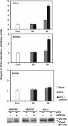
Inhibition of ATR/ATM signaling by caffeine results in a DNA-PK–mediated activation of Chk2. HeLa, MO59J (DNA-PK), or MO59K (DNA-PK+) cells were either untreated (Con) or synchronized in metaphase by treatment with nocodazole for 14 h. Mitotic cells were removed and plated in fresh medium containing APH for 24 h (0 h). Caffeine or buffer was added and cells were incubated for an additional 5 h. Cell lysates were subjected to Chk2 immunoprecipitation kinase assay (top) or immunoblotted with Chk2 antibody (bottom). Chk2 band shifting (pChk2) is indicative of phosphorylation (Feijoo et al., 2001). Data are expressed as mean ± range for duplicate determinations.
Figure 6.
Figure 6.
DNA-PK is a regulator of histone mRNA stability. (A) Asynchronous HeLa cells were treated with 5 mM caffeine alone, 200 μM LY294002 alone or 5 mM caffeine and 200 μM LY294002 together or mock treated with DMSO. After 1 h, cells were treated with or without 2 mM HU and RNA was isolated after an additional 30 min for analysis as in Fig. 1. The graph shows histone mRNA levels in HU-treated cells, with 100% defined as histone mRNA levels in non–HU-treated cells. (B) Asynchronous HeLa cells were treated with 5 mM caffeine alone, 200 μM LY294002 alone, or 5 mM caffeine and 200 μM LY294002 together or mock treated with DMSO. After 1 h, cells were treated with or without 2 mM HU, RNA was isolated at the indicated times, and all samples were analyzed by Northern blotting as in Fig. 1. Graphs show changes in histone mRNA levels compared with the level at 0 min. (C) M059J (DNA-PK) and M059K (DNA-PK+) cells were treated with 5 mM caffeine alone, 200 μM LY294002 alone, or 5 mM caffeine and 200 μM LY294002 together or mock treated with DMSO. After 1 h, cells were treated with or without 2 mM HU and RNA was isolated after a further 1-h incubation and analyzed as in Fig. 1. The graph shows histone mRNA levels in HU-treated cells compared with histone mRNA levels in non–HU-treated cells.
Figure 7.
Figure 7.
Inhibition of Chk1 function results in increased phosphorylation of Chk2. Chk1 and 2 are not functionally limiting for the control of replication stress–induced histone mRNA decay. (A) Asynchronous HeLa cells were transfected with siRNA targeting Chk1 or control siRNA 1 (targeting luciferase) or mock transfected. Cell lysates were immunoblotted for Chk1 to confirm knockdown and actin (loading control). (B, left) Asynchronous HeLa cells were transfected with siRNA targeting Chk1, control siRNA 1 (targeting luciferase) or siRNA 2 (targeting a nonrelevant gene) or mock transfected. After 32 h, cells were treated with or without 2 mM HU for 0, 20, and 60 min or left untreated for 60 min and then lysed for RNA analysis. Histone H3 mRNA levels were analyzed as in Fig. 1. The graph shows changes in H3 mRNA levels compared with the level at 0 min. (B, right) Asynchronous HeLa cells were incubated with or without 300 nM of Chk1-selective inhibitor UCN-01. 1 h later, cells were incubated with or without 2 mM HU for the indicated times and lysed for RNA analysis as before. The graph shows changes in histone mRNA levels compared with the level at 0 min. (C) HeLa cells were synchronized in metaphase by treatment with nocodazole for 14 h and then released. After 14 h, cells were treated with or without 300 nM UCN-01 for 1 h, then 2 mM HU was added for a further 2 h. Cell lysates were immunoblotted with a phosphospecific Chk2 antibody (top) and a total anti-Chk2 antibody (bottom). (D) Asynchronous DLD-1 cells were incubated with or without 300 nM of Chk1-selective inhibitor UCN-01 or with or without caffeine. 1 h later, cells were incubated with or without 2 mM HU for the indicated times and lysed for RNA analysis as before. The graphs show changes in histone mRNA levels compared with the level at 0 min.
Figure 8.
Figure 8.
Replication stress results in increased phosphorylation of UPF1 but not HBP/SLBP on PIKK substrate motifs. Asynchronous HeLa cells were incubated with or without 2 mM HU for 16 h. (A) Cell lysates were subjected to immunoprecipitation using goat anti-UPF1 antibodies or IgG control, and immunoprecipitates were treated with AP or buffer before SDS-PAGE and immunoblotting with anti–phospho-S/TQ (top) and anti-UPF1 antibodies (bottom). (B) Cell lysates were subjected to immunoprecipitation using anti-HBP antisera and immunoprecipitates were immunoblotted with anti–phospho-S/TQ (top) and anti-HBP antiserum (bottom).
Figure 9.
Figure 9.
Model for the coordination of DNA replication and histone production in mammalian cells. Exposure of cells to replication stress induces histone mRNA decay via a currently poorly understood pathway (top right arrow). Replication stress results in stabilization of slowed or stalled replication forks via a pathway involving ATR and Chk1 (top left arrow). Replication restart from stalled replication forks occurs predominantly via a pathway involving homologous recombination. At some frequency, replication forks either encounter complex DNA damage or fail to be stabilized by the ATR–Chk1 pathway, which results in replication fork collapse, generating DNA double-strand breaks. These may be repaired either via ATM-dependent homologous recombination–induced replication restart or via NHEJ mediated by DNA-PK. Failure of ATR signaling (such as occurs in the presence of caffeine) will result in higher levels of fork collapse, generating an increased level of substrate for DNA-PK–mediated NHEJ. Coordinated regulation of histone mRNA decay by both ATR/ATM and DNA-PK during replication stress ensures that, irrespective of the extent to which each pathway operates in any given circumstance, the supply of histones will remain closely coupled to the demand required for the assembly of newly synthesized chromosomes. The RNA helicase UPF1 involved in histone mRNA decay (Kaygun and Marzluff, 2005) may act as an effector of ATR/DNA-PK signaling and induce histone mRNA decay. Upon relief from replication stress, histone mRNA stability is restored to normal levels presumably by a mechanism linked to the restart of replication forks and involving changes in UPF1 phosphorylation.

Similar articles

Cited by

References

    1. Abraham, R.T. 2001. Cell cycle checkpoint signaling through the ATM and ATR kinases. Genes Dev. 15:2177–2196. - PubMed
    1. Arnaudeau, C., C. Lundin, and T. Helleday. 2001. DNA double-strand breaks associated with replication forks are predominantly repaired by homologous recombination involving an exchange mechanism in mammalian cells. J. Mol. Biol. 307:1235–1245. - PubMed
    1. Azzalin, C.M., and J. Lingner. 2006. The human RNA surveillance factor UPF1 is required for S phase progression and genome stability. Curr. Biol. 16:433–439. - PubMed
    1. Baumbach, L.L., G.S. Stein, and J.L. Stein. 1987. Regulation of human histone gene expression: transcriptional and posttranscriptional control in the coupling of histone messenger RNA stability with DNA replication. Biochemistry. 26:6178–6187. - PubMed
    1. Bhattacharyya, N.P., A. Ganesh, G. Phear, B. Richards, A. Skandalis, and M. Meuth. 1995. Molecular analysis of mutations in mutator colorectal carcinoma cell lines. Hum. Mol. Genet. 4:2057–2064. - PubMed

Publication types

MeSH terms